. 2019 Sep;44(3):939-948.
doi: 10.3892/ijmm.2019.4273. Epub 2019 Jul 10.

Expression changes of CX3CL1 and CX3CR1 proteins in the hippocampal CA1 field of the gerbil following transient global cerebral ischemia

Affiliations

Expression changes of CX3CL1 and CX3CR1 proteins in the hippocampal CA1 field of the gerbil following transient global cerebral ischemia

Ji Hyeon Ahn et al. Int J Mol Med. 2019 Sep.

Abstract

Chemokine C‑X3‑C motif ligand 1 (CX3CL1) and its sole receptor, CX3CR1, are known to be involved in neuronal damage/death following brain ischemia. In the present study, time‑dependent expression changes of CX3CL1 and CX3CR1 proteins were investigated in the hippocampal CA1 field following 5 min of transient global cerebral ischemia (tgCI) in gerbils. To induce tgCI in gerbils, bilateral common carotid arteries were occluded for 5 min using aneurysm clips. Expression changes of CX3CL1 and CX3CR1 proteins were assessed at 1, 2 and 5 days after tgCI using western blotting and immunohistochemistry. CX3CL1 immunoreactivity was strong in the CA1 pyramidal cells of animals in the sham operation group. Weak CX3CL1 immunoreactivity was detected at 6 h after tgCI, recovered at 1 day after tgCI and disappeared from 5 days after tgCI. CX3CR1 immunoreactivity was very weak in CA1 pyramidal cells of the sham animals. CX3CR1 immunoreactivity in CA1 pyramidal cells was significantly increased at 1 days after tgCI and gradually decreased thereafter. On the other hand, CX3CR1 immunoreactivity was significantly increased in microglia from 5 days after tgCI. These results showed that CX3CL1 and CX3CR1 protein expression levels in pyramidal cells and microglia in the hippocampal CA1 field following tgCI were changed, indicating that tgCI‑induced expression changes of CX3CL1 and CX3CR1 proteins might be closely associated with tgCI‑induced delayed neuronal death and microglial activation.


Figures
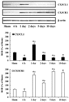
Figure 1
Figure 1
Western blot analyses of CX3CL1 and CX3CR1 in the hippocampal CA1 field of sham operated and ischemia operated gerbils. ROD as % value of immunoblot band is represented (n=7 per group). *P<0.05 vs. the sham operated gerbils; P<0.05 vs. the pre-time-point group. CX3CL1, C-X3-C motif ligand 1; CX3CR1, CX3C chemokine receptor 1; ROD, relative optical density.
Figure 2
Figure 2
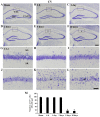
CV staining. Low magnification of the hippocampus of the (A) sham group and ischemia operated groups at (B) 6 h, (C) 1 day, (D) 2 days, (E) 5 days and (F) 10 days after tgCI. High magnification of the CA1 field of the (G) sham group and ischemia operated groups at (H) 6 h, (I) 1 day, (J) 2 days, (K) 5 days, and (L) 10 days after tgCI. CV positive cells are well observed in the SP in the CA1-3 field in the sham operated group. In the ischemia operated group, CV positive cells are significantly decreased only in the SP (asterisks) of the CA1 field at 5 and 10 days after tgCI. Scale bars=400 µm (A-F) and 50 µm (G-L). (M) Numbers of CV positive cells in the CA1 field (n=7 at each point in time). *P<0.05 vs. the sham operated group. The bars indicate the mean ± standard error of the mean. CV, cresyl violet; SP, stratum pyramidale; SO, stratum oriens; SR, stratum radiatum; tgCI, transient global cerebral ischemia.
Figure 3
Figure 3
FJB histofluorescence staining. FJB histofluorescence staining in the CA1 field of the (A) sham group and ischemia operated groups at (B) 6 h, (C) 1 day, (D) 2 days, (E) 5 days and (F) 10 days after tgCI. FJB positive cells are not detected in the SP until 2 days after tgCI. However, FJB positive cells (indicated by asterisks) are detected in the SP at 5 and 10 days after tgCI. Scale bar, 50 µm. (G) Numbers of FJB positive cells in the CA1 field (n=7 at each point in time). *P<0.05 vs. the sham operated gerbils. The bars indicate the mean ± standard error of the mean. SP, stratum pyramidale; FJB, Fluoro-Jade B; tgCI, transient global cerebral ischemia.
Figure 4
Figure 4
Iba-1 immunohistochemistry. (A-F) Iba1 immunohistochemistry in the CA1 field of the (A) sham group and ischemia operated groups at (B) 6 h, (C) 1 day, (D) 2 days, (E) 5 days and (F) 10 days after tgCI. In the ischemia operated gerbils, Iba1 immunoreactivity is gradually increased after tgCI. In particular, Iba1 immunoreactive microglia are aggregated in the SP (asterisks) at 5 and 10 days after tgCI. Scale bar, 50 µm. (G) ROD as % of Iba1 immunoreactive structures after tgCI (n=7 at each point in time). *P<0.05 vs. the sham operated gerbils; P<0.05 vs. the pre-time-point gerbils. Bars indicate the mean ± standard error of the mean. SP, stratum pyramidale; tgCI, transient global cerebral ischemia; ROD, relative optical density; Iba1, ionized calcium-binding adapter molecule 1.
Figure 5
Figure 5
CX3CL1 immunohistochemistry. (A-F) CX3CL1 immunohistochemistry in the CA1 field of the (A) sham group and ischemia operated groups at (B) 6 h, (C) 1 day, (D) 2 days, (E) 5 days and (F) 10 days after tgCI. Strong CX3CL1 immunoreactivity is shown in CA1 pyramidal cells of the SP at sham and 1 day after tgCI. CX3CL1 immunoreactivity is very weak in the CA1 field at 6 h (asterisk), 5 days and 10 days after tgCI. Scale bar, 50 µm. (G) ROD as % of CX3CL1 immunoreactive structures in the CA1 field after tgCI (n=7 at each point in time) *P<0.05 vs. the sham operated gerbils; P<0.05 vs. the pre-time-point group. Bars indicate the means ± standard error of the mean. SP, stratum pyramidale; ROD, relative optical density; tgCI, transient global cerebral ischemia; CX3CL1, C-X3-C motif ligand 1; CX3CR1, CX3C chemokine receptor 1.
Figure 6
Figure 6
CX3CR1 immunohistochemistry. (A-F) CX3CR1 immunohistochemistry in the CA1 field of the (A) sham group and ischemia operated groups at (B) 6 h, (C) 1 day, (D) 2 days, (E) 5 days and (F) 10 days after tgCI. Weak CX3CR1 immunoreactivity is detected in the SP (asterisk) of the sham operated gerbils and CX3CR1 immunoreactivity in the SP (asterisk) at 1 day after tgCI is strong. At 5 and 10 days after tgCI, strong CX3CR1 immunoreactivity is shown in cells (arrows) in the SO and SR. Scale bar, 50 µm. (G) ROD as % of CX3CR1-immunoreactive structures in the CA1 field after tgCI (n=7 at each point in time). *P<0.05 vs. the sham operated gerbils; P<0.05 vs. the pre-time-point group. Bars indicate the mean ± standard error of the mean. SO, stratum oriens; SP, stratum pyramidale; SR, stratum radiatum; tgCI, transient global cerebral ischemia; CX3CL1, C-X3-C motif ligand 1; CX3CR1, CX3C chemokine receptor 1; ROD, relative optical density.
Figure 7
Figure 7
Double immunofluorescence staining for (A) CX3CR1 (green), (B) Iba1 (red) and (C) merged images in the CA1 field at 10 days after tgCI. CX3CR1 immunoreactivity is identified as Iba1-immunoreactive microglia (arrows). Scale bar, 50 µm. CX3CL1, C-X3-C motif ligand 1; CX3CR1, CX3C chemokine receptor 1; tgCI, transient global cerebral ischemia; Iba1, ionized calcium-binding adapter molecule 1.

References

    1. Arundine M, Tymianski M. Molecular mechanisms of glutamate-dependent neurodegeneration in ischemia and traumatic brain injury. Cell Mol Life Sci. 2004;61:657–668. doi: 10.1007/s00018-003-3319-x. - DOI - PMC - PubMed
    1. Lee JC, Kim IH, Park JH, Ahn JH, Cho JH, Cho GS, Tae HJ, Chen BH, Yan BC, Yoo KY, et al. Ischemic preconditioning protects hippocampal pyramidal neurons from transient ischemic injury via the attenuation of oxidative damage through upregulating heme oxygenase-1. Free Radic Biol Med. 2015;79:78–90. doi: 10.1016/j.freeradbiomed.2014.11.022. - DOI - PubMed
    1. Park JH, Kim YH, Ahn JH, Choi SY, Hong S, Kim SK, Kang IJ, Kim YM, Lee TK, Won MH, Lee CH. Atomoxetine protects against NMDA receptor-mediated hippocampal neuronal death following transient global cerebral ischemia. Curr Neurovasc Res. 2017;14:158–168. doi: 10.2174/1567202614666170328094042. - DOI - PubMed
    1. Rastogi L, Godbole MM, Ray M, Rathore P, Rathore P, Pradhan S, Gupta SK, Pandey CM. Reduction in oxidative stress and cell death explains hypothyroidism induced neuroprotection subsequent to ischemia/reperfusion insult. Exp Neurol. 2006;200:290–300. doi: 10.1016/j.expneurol.2006.02.013. - DOI - PubMed
    1. Barone FC, Feuerstein GZ. Inflammatory mediators and stroke: New opportunities for novel therapeutics. J Cereb Blood Flow Metab. 1999;19:819–834. doi: 10.1097/00004647-199908000-00001. - DOI - PubMed