. 2019 Nov;76(11-12):549-561.
doi: 10.1002/cm.21562. Epub 2019 Oct 11.

Computational model demonstrates that Ndc80-associated proteins strengthen kinetochore-microtubule attachments in metaphase

Affiliations

Computational model demonstrates that Ndc80-associated proteins strengthen kinetochore-microtubule attachments in metaphase

Samuel Campbell et al. Cytoskeleton (Hoboken). 2019 Nov.

Abstract

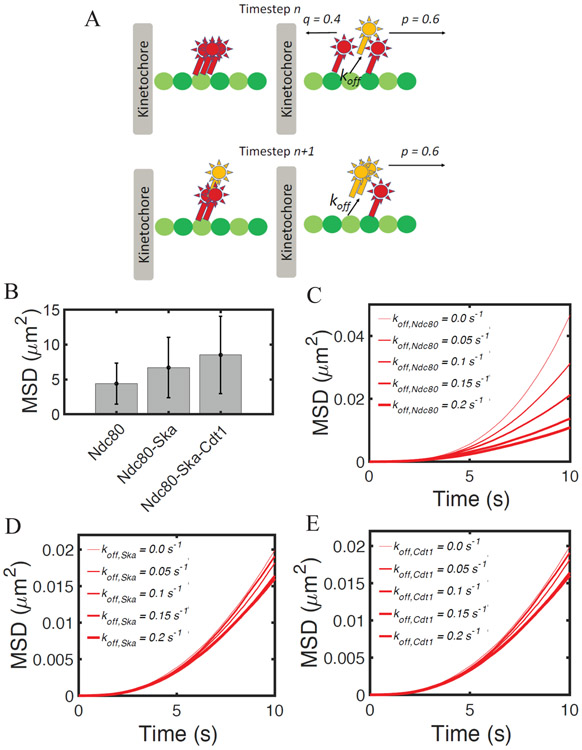
Chromosome segregation is mediated by spindle microtubules that attach to the kinetochore via dynamic protein complexes, such as Ndc80, Ska, Cdt1 and ch-TOG during mitotic metaphase. While experimental studies have previously shown that these proteins and protein complexes are all essential for maintaining a stable kinetochore-microtubule (kMT) interface, their exact roles in the mitotic metaphase remains elusive. In this study, we employed experimental and computational methods in order to characterize how these proteins can strengthen kMT attachments in both nonload-bearing and load-bearing conditions, typical of prometaphase and metaphase, respectively. Immunofluorescence staining of HeLa cells showed that the levels of Ska and Cdt1 significantly increased from prometaphase to metaphase, while levels of the Ndc80 complex remained unchanged. Our new computational model showed that by incorporating binding and unbinding of each protein complex coupled with a biased diffusion mechanism, the displacement of a possible complex formed by Ndc80-Ska-Cdt1 is significantly higher than that of Ndc80 alone or Ndc80-Ska. In addition, when we incorporate Ndc80/ch-TOG in the model, rupture force and time of attachment of the kMT interface increases. These results support the hypothesis that Ndc80-associated proteins strengthen kMT attachments, and that the interplay between kMT protein complexes in metaphase ensures stable attachments.

Keywords: Ndc80; kinetochore; microtubule; mitosis; modeling.


Figures

Figure 1:
Figure 1:
Kinetochore localization of microtubule-binding proteins that are required for stabilizing end-on kinetochore-microtubule attachments in fixed mitotic HeLa cells. (A) Immunofluorescence staining of mitotic cells for the Hec1 subunit of the Ndc80 complex shown alongside another kinetochore marker, Zwint1 in either prometaphase (top panel) or metaphase (bottom panel). (B) Comparative quantification of Hec1 intensity at kinetochores in prometaphase vs metaphase cells from A. n = 75 from 5 cells. (C) Immunofluorescence staining of mitotic cells for the Ska3 subunit of the Ska complex shown alongside kinetochore markers, anti-CREST antiserum (ACA) in prometaphase (top panel) or Hec1 in metaphase (bottom panel), as indicated. (D) Comparative quantification of Ska3 intensity in prometaphase vs metaphase cells from C. n=120 from 5 cells. (E) Immunofluorescence staining of mitotic cells for Cdt1 shown alongside kinetochore markers, anti-CREST antiserum (ACA) in prometaphase (top panel) or Hec1 in metaphase (bottom panel), as indicated. (F) Comparative quantification of Cdt1 intensity in prometaphase vs metaphase cells from E. n=120 from 5 cells.
Figure 2.
Figure 2.. Flow chart of the computational model.
The model implements a Kinetic Monte Carlo algorithm, where state and position of each particle are updated at each time step. All particles are initially in the unbound state and at position 0. At each time step: their binding probability is evaluated; then, for the bound particles, unbinding probability is updated; bound particles can move; last, particles’ positions are updated. In the flow chart, black boxes indicate updates in particles’ states and green boxes refer to changes in position. Red is for conditional statements
Figure 3.
Figure 3.. Computational model of kMT attachments incorporating the Ndc80-accessory proteins
(A) Schematics of the computational model, incorporating binding, unbinding and biased diffusion of Ndc80-associated proteins Ska and Cdt1. (B) Heatmap showing the diffusion coefficient of a protein complex undergoing biased diffusion as a function of kon and koff. Parameter values of kon and koff for Ndc80, Ska and Cdt1 are evaluated based on previous in vitro characterization of the complex dynamics on immobilized microtubules. Simulations are run for 300 s and data are averages between 1000 independent runs. (C) Mean squared displacement of Ndc80 at 1 s of simulation using different values of koff, in order to mimic the effect of phosphomimetic Ndc80 mutants. Results are computed as averages from 10,000 independent runs. (D) Heat map of Mean Square displacement (MSD) of the Ndc80-Ska complex at 10 s of simulation, varying the number of Ndc80 and by systematically changing Ska kon. Results are computed as averages from 200 independent runs.
Figure 4.
Figure 4.. The interplay between Ndc80, Ska and Cdt1 ensures stable kinetochore-microtubule attachment in non-load-bering conditions.
(A) Schematics of the model with a complex of multiple proteins. At each time step, the position of the proteins is calculated as the average position of the bound proteins. Assuming that different proteins move in opposite directions at timestep n (upper panels), their position is updated at timestep n+1 (lower panels). Only after this update in position, the model evaluates binding, unbinding and diffusion. (B) Average Mean square displacement (MSD) of the Ndc80 complex at 300 seconds of simulations. Data are computed from 1,000 independent runs. Error bars denote standard deviation from the mean. MSD of the Ndc80 complex, including Ska and Cdt1, at 10 s of simulations, for different values of (C) koff,Ndc80, (D) koff,ska or (E) koff,cdt1. Data are computed from 5,000 independent runs.
Figure 5.
Figure 5.. Ndc80, Ska and Cdt1 increase kinetochore-microtubule rupture force and attachment time in load-bearing conditions.
(A) Spindle microtubules localization of the Ska3 subunit of the Ska complex (red, top panel) or Cdt1 (red, bottom panel) in methanol-fixed (bottom) metaphase HeLa cells. Microtubules are in green while DAPI/chromosomes are in blue. (B) Implemented bond lifetimes for Ndc80, Ska, and Cdt1. (C) Average MSD of the Ndc80 complex at 300 seconds of simulations. Error bars denote standard deviation from the mean. Data are computed from 100 independent runs. (D) Average rupture force (force at which all proteins become unbound) evaluated for Ndc80, Ndc80-Ska, and Ndc80-Ska-Cdt1 versus the number of MT protofilaments. (E) Average attachment lifetime for Ndc80, Ndc80-Ska, and Ndc80-SkaCdt1 versus the number of MT protofilaments. Data are averages from 1,000 independent runs.
Figure 6.
Figure 6.. ch-TOG significantly enhances rupture force and attachment time via tension-dependent mechanisms.
(A) Localization of GFP-tagged ch-TOG to kinetochores (green, top panel) in paraformaldehyde-fixed and to spindle microtubules (green, bottom panel) in methanol-fixed metaphase HeLa cells. (B) Average attachment time for Ndc80 and Ndc80/ch-TOG versus number of MT protofilaments. Data are extracted from 1,000 independent runs. (C) Average rupture force for Ndc80 and Ndc80/ch-TOG versus the number of tubulin protofilaments. Results are computed from 1,000 independent runs. (D) Implemented bond lifetimes for ch-TOG. Bond lifetime versus force relation for varying τMAX are tested. (E) Heatmap of average attachment time by systematically varying the number of microtubules and τMAX. SE is below 1 min for all τMAX. (F) Heatmap of average rupture force by systematically varying number of microtubules and τMAX. SE is below 0.3 pN for all τMAX.

References

    1. Agarwal S, Smith KP, Zhou Y, Suzuki A, McKenney RJ, & Varma D (2018). Cdt1 stabilizes kinetochore-microtubule attachments via an Aurora B kinase-dependent mechanism. The Journal of Cell Biology. doi:10.1083/jcb.201705127 - DOI - PMC - PubMed
    1. Alushin GM, Ramey VH, Pasqualato S, Ball DA, Grigorieff N, Musacchio A, & Nogales E (2010). The Ndc80 kinetochore complex forms oligomeric arrays along microtubules. Nature. doi:10.1038/nature09423 - DOI - PMC - PubMed
    1. Alushin G, & Nogales E (2011). Visualizing kinetochore architecture. Current Opinion in Structural Biology. doi:10.1016/j.sbi.2011.07.009 - DOI - PMC - PubMed
    1. Chan YW, Jeyaprakash AA, Nigg EA, & Santamaria A (2012). Aurora B controls kinetochore-microtubule attachments by inhibiting Ska complex-KMN network interaction. Journal of Cell Biology. doi:10.1083/jcb.201109001 - DOI - PMC - PubMed
    1. Cheerambathur DK, Prevo B, Hattersley N, Lewellyn L, Corbett KD, Oegema K, & Desai A (2017). Dephosphorylation of the Ndc80 Tail Stabilizes Kinetochore-Microtubule Attachments via the Ska Complex. Developmental Cell. doi:10.1016/j.devcel.2017.04.013 - DOI - PMC - PubMed

Publication types

LinkOut - more resources