

Preactivation of Notch1 in remote ischemic preconditioning reduces cerebral ischemia-reperfusion injury through crosstalk with the NF-κB pathway
- PMID: 31526384
- PMCID: PMC6747758
- DOI: 10.1186/s12974-019-1570-9
Preactivation of Notch1 in remote ischemic preconditioning reduces cerebral ischemia-reperfusion injury through crosstalk with the NF-κB pathway
Abstract
Background: Remote ischemic preconditioning (RIPC) initiates endogenous protective pathways in the brain from a distance and represents a new, promising paradigm in neuroprotection against cerebral ischemia-reperfusion (I/R) injury. However, the underlying mechanism of RIPC-mediated cerebral ischemia tolerance is complicated and not well understood. We reported previously that preactivation of Notch1 mediated the neuroprotective effects of cerebral ischemic preconditioning in rats subjected to cerebral I/R injury. The present study seeks to further explore the role of crosstalk between the Notch1 and NF-κB signaling pathways in the process of RIPC-induced neuroprotection.
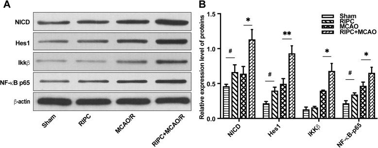
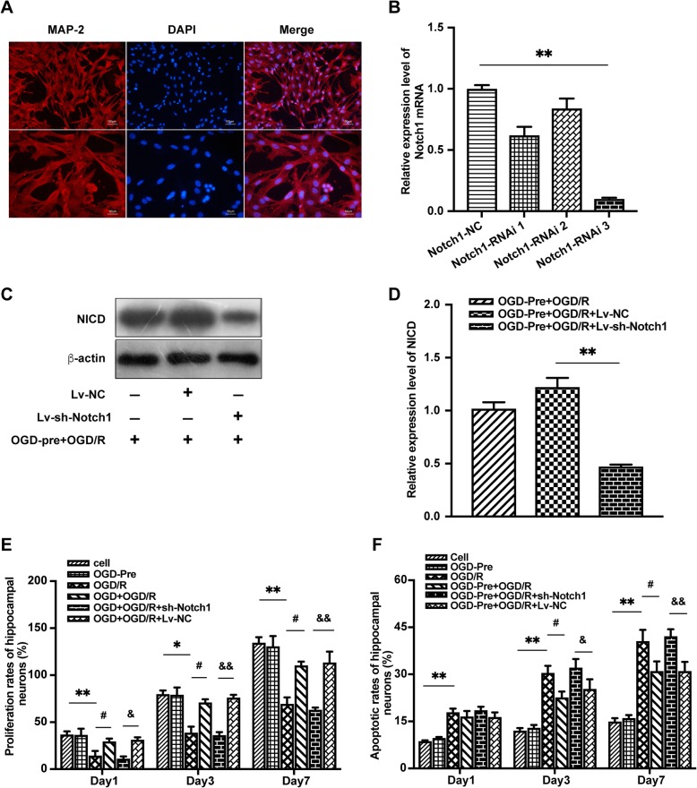
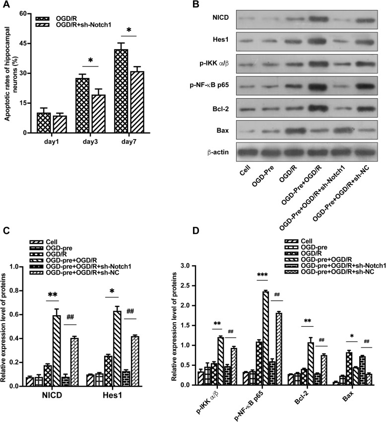
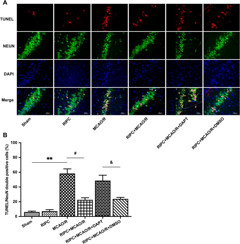
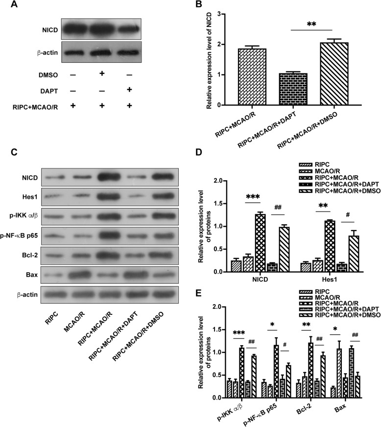
Methods: Middle cerebral artery occlusion and reperfusion (MCAO/R) in adult male rats and oxygen-glucose deprivation and reoxygenation (OGD/R) in primary hippocampal neurons were used as models of I/R injury in vivo and in vitro, respectively. RIPC was induced by a 3-day procedure with 4 cycles of 5 min of left hind limb ischemia followed by 5 min of reperfusion each day before MCAO/R. Intracerebroventricular DAPT injection and sh-Notch1 lentivirus interference were used to inhibit the Notch1 signaling pathway in vivo and in vitro, respectively. After 24 h of reperfusion, neurological deficit scores, infarct volume, neuronal apoptosis, and cell viability were assessed. The protein expression levels of NICD, Hes1, Phospho-IKKα/β (p-IKK α/β), Phospho-NF-κB p65 (p-NF-κB p65), Bcl-2, and Bax were assessed by Western blotting.
Results: RIPC significantly improved neurological scores and reduced infarct volume and neuronal apoptosis in rats subjected to I/R injury. OGD preconditioning significantly reduced neuronal apoptosis and improved cell viability after I/R injury on days 3 and 7 after OGD/R. However, the neuroprotective effect was reversed by DAPT in vivo and attenuated by Notch1-RNAi in vitro. RIPC significantly upregulated the expression of proteins related to the Notch1 and NF-κB pathways. NF-κB signaling pathway activity was suppressed by a Notch1 signaling pathway inhibitor and Notch1-RNAi.
Conclusions: The neuroprotective effect of RIPC against cerebral I/R injury was associated with preactivation of the Notch1 and NF-κB pathways in neurons. The NF-κB pathway is a downstream target of the Notch1 pathway in RIPC and helps protect focal cerebral I/R injury.
Keywords: Cross reaction; Ischemia-reperfusion injury; NF-kappa B; Neuroprotection; Notch1 pathway; Remote ischemic preconditioning.
Conflict of interest statement
The authors declare that they have no competing interests.
Figures


Similar articles
-
Isoquercetin attenuates oxidative stress and neuronal apoptosis after ischemia/reperfusion injury via Nrf2-mediated inhibition of the NOX4/ROS/NF-κB pathway.Chem Biol Interact. 2018 Mar 25;284:32-40. doi: 10.1016/j.cbi.2018.02.017. Epub 2018 Feb 16. Chem Biol Interact. 2018. PMID: 29454613
-
Neuroprotective effects of pioglitazone in a rat model of permanent focal cerebral ischemia are associated with peroxisome proliferator-activated receptor gamma-mediated suppression of nuclear factor-κB signaling pathway.Neuroscience. 2011 Mar 10;176:381-95. doi: 10.1016/j.neuroscience.2010.12.029. Epub 2010 Dec 24. Neuroscience. 2011. PMID: 21185913
-
Neuroprotective Effects of Cerebral Ischemic Preconditioning in a Rat Middle Cerebral Artery Occlusion Model: The Role of the Notch Signaling Pathway.Biomed Res Int. 2018 Aug 6;2018:8168720. doi: 10.1155/2018/8168720. eCollection 2018. Biomed Res Int. 2018. PMID: 30175143 Free PMC article.
-
Loureirin B protects against cerebral ischemia/reperfusion injury through modulating M1/M2 microglial polarization via STAT6 / NF-kappaB signaling pathway.Eur J Pharmacol. 2023 Aug 15;953:175860. doi: 10.1016/j.ejphar.2023.175860. Epub 2023 Jun 16. Eur J Pharmacol. 2023. PMID: 37331681 Review.
-
Remote ischemic preconditioning: a novel protective method from ischemia reperfusion injury--a review.J Surg Res. 2008 Dec;150(2):304-30. doi: 10.1016/j.jss.2007.12.747. Epub 2008 Jan 22. J Surg Res. 2008. PMID: 19040966 Review.
Cited by
-
Anti-cerebral ischemic neuronal injury mechanism of Zhenlong Xingnao capsules: role of the Notch/NF-κB signaling pathway.Am J Transl Res. 2023 Jul 15;15(7):4587-4599. eCollection 2023. Am J Transl Res. 2023. PMID: 37560215 Free PMC article.
-
A Machine Learning-Based Classification of Immunogenic Cell Death Regulators and Characterisation of Immune Microenvironment in Acute Ischemic Stroke.Int J Clin Pract. 2023 Nov 14;2023:9930172. doi: 10.1155/2023/9930172. eCollection 2023. Int J Clin Pract. 2023. PMID: 38020537 Free PMC article.
-
Preclinical evidence of remote ischemic conditioning in ischemic stroke, a metanalysis update.Sci Rep. 2021 Dec 9;11(1):23706. doi: 10.1038/s41598-021-03003-6. Sci Rep. 2021. PMID: 34887465 Free PMC article.
-
Protective role of remote ischemic conditioning in renal transplantation and partial nephrectomy: A systematic review and meta-analysis of randomized controlled trials.Front Surg. 2023 Apr 5;10:1024650. doi: 10.3389/fsurg.2023.1024650. eCollection 2023. Front Surg. 2023. PMID: 37091267 Free PMC article. Review.
-
Neuroadaptive Biochemical Mechanisms of Remote Ischemic Conditioning.Int J Mol Sci. 2023 Dec 1;24(23):17032. doi: 10.3390/ijms242317032. Int J Mol Sci. 2023. PMID: 38069355 Free PMC article. Review.
References
-
- Stenzel-Poore MP, Stevens SL, Xiong Z, Lessov NS, Harrington CA, Mori M, Meller R, Rosenzweig HL, Tobar E, Shaw TE, et al. Effect of ischaemic preconditioning on genomic response to cerebral ischaemia: similarity to neuroprotective strategies in hibernation and hypoxia-tolerant states. Lancet. 2003;362:1028–1037. doi: 10.1016/S0140-6736(03)14412-1. - DOI - PubMed
MeSH terms
Substances
Grants and funding
LinkOut - more resources
Full Text Sources
Other Literature Sources
Research Materials
