. 2019 Sep 16;14(1):106.
doi: 10.1186/s13000-019-0877-2.

LncRNA DNM3OS promotes proliferation and inhibits apoptosis through modulating IGF1 expression by sponging MiR-126 in CHON-001 cells

Affiliations

LncRNA DNM3OS promotes proliferation and inhibits apoptosis through modulating IGF1 expression by sponging MiR-126 in CHON-001 cells

Di Ai et al. Diagn Pathol. .

Abstract

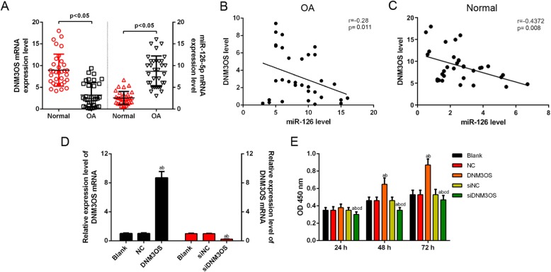
Background: As a degenerative disease, osteoarthritis (OA) greatly affects aged population. The human chondrocyte cell line CHON-001, derived from normal human articular cartilage, has been widely used in vitro in osteoarthritis models. In order to better understand the underlying mechanism of OA pathogenesis, this study was conducted to explore the effects of LncRNA dynamin 3 opposite strand (DNM3OS) on CHON-001 cells.

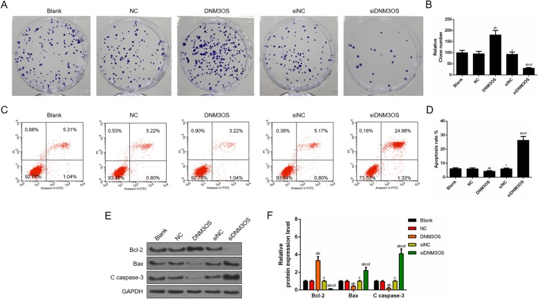
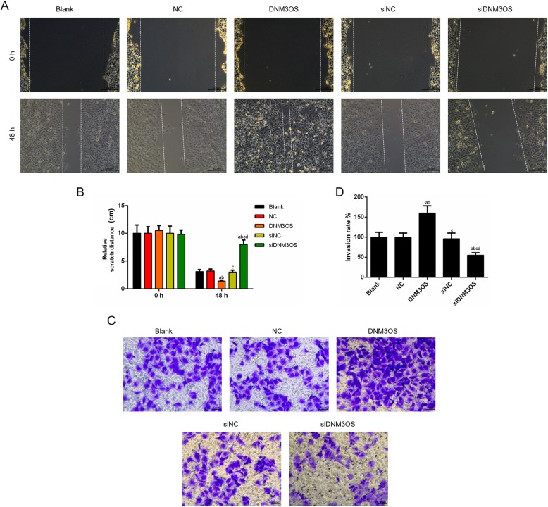
Methods: The expression levels of and correlation between DNM3OS and miR-126 that derived from OA and non-OA tissues were determined by quantitative real time (qRT)-PCR and Spearman's correlation analysis. Cell viability, clone, migration, invasion and apoptosis were respectively determined by cell counting kit-8, colony formation, wound healing assay, transwell and flow cytometry. The target genes were predicted by starbase V2 and targetscan 7.2 and confirmed by luciferase reporter assay. The expressions of apoptosis-related factors were detected by Western blot.

Results: The expression of DNM3OS was down-regulated in OA patients. Functional assays demonstrated that ectopic expression of DNM3OS promoted the proliferation and inhibited apoptosis of CHON-001 cells, and that knocking down DNM3OS suppressed cell proliferation and induced apoptosis. Mechanistic investigation revealed that DNM3OS physically bound to the promoter of miR-126 and suppressed miR-126 expression. Decreased expression of DNM3OS was negatively correlated with miR-126 in OA patients. Furthermore, the effects of siDNM3OS on inhibiting cell proliferation and promoting apoptosis were partially reversed by miR-126 inhibitor. Meanwhile, type insulin-like growth factor-1 (IGF1) was identified as a target gene for miR-126 and was negatively associated with the miR-126 expression. Overexpressed IGF1 restored the effects of miR-126 mimic in suppressing cell proliferation and promoting apoptosis.

Conclusion: Our results showed that DNM3OS could affect the CHON-001 cell proliferation and apoptosis by regulating IGF1 by sponging miR-126.

Keywords: LncRNA dynamin 3 opposite strand; MiR-126; Osteoarthritis; Proliferation; Type insulin-like growth factor-1.


Conflict of interest statement

The authors declare no conflicts of interest.

Figures

Fig. 1
Fig. 1
DNM3OS was low-expressed in OA tissues, had negative correlation with miR-126 and promoted CHON-001 cells viability. (a) The expression of lncRNA DNM3OS in human OA tissues was significantly lower, while miR-126 was obviously higher than in normal tissues using RT-qPCR. (b) The expression of DNM3OS was negatively correlated with the miR-126 level in OA samples by Spearman’s correlation analysis. (c) The expression of DNM3OS was negatively correlated with the miR-126 level in normal samples by Spearman’s correlation analysis. (d) The transfection efficiency of DNM3OS was confirmed by RT-qPCR. (e) CHON-001 cells viability was improved or inhibited by DNM3OS or silencing DNM3OS by CCK-8 assay. aP < 0.05 vs. Blank, bP < 0.05 vs. NC, cP < 0.05 vs. DNM3OS, dP < 0.05 vs. siNC. siDNM3OS: small interfering dynamin 3 opposite strand; NC: negative control; OD: optical density
Fig. 2
Fig. 2
Over-expression of DNM3OS enhanced proliferation and suppressed apoptosis in CHON-001 cells, while DNM3OS silencing had opposite effects. (a) CHON-001 cells proliferation was improved by DNM3OS, while silencing DNM3OS inhibited CHON-001 cells proliferation as indicated by cloning formation experiment. (b) The number of proliferated cells was quantified. (c) CHON-001 cells apoptosis was inhibited by DNM3OS, while DNM3OS silencing induced apoptosis as indicated by flow cytometry. (d) The proportion of apoptosis was quantified. (e) The apoptosis-related proteins (Bcl-2, Bax and c caspase 3) were detected by Western blot. (f) The relative levels of proteins described in (e) were counted by GAPDH as normalization. aP < 0.05 vs. Blank, bP < 0.05 vs. NC, cP < 0.05 vs. DNM3OS, dP < 0.05 vs. siNC. siDNM3OS: small interfering dynamin 3 opposite strand; NC: negative control; Bcl-2: B-cell lymphoma-2; Bax: Bcl2-associated X protein
Fig. 3
Fig. 3
CHON-001 cell migration and invasion were facilitated by the up-regulation DNM3OS, while the knockdown of DNM3OS showed opposite results. (a) CHON-001 cells migration was promoted by DNM3OS, while silencing DNM3OS inhibited cells migration as indicated by wound healing assay. (b) The distance of scratch was quantified. (c) CHON-001 cells invasion was enhanced by DNM3OS, while silencing DNM3OS suppressed cells invasion as indicated by transwell assay. (d) The number of invasion cells was quantified. aP < 0.05 vs. Blank, bP < 0.05 vs. NC, cP < 0.05 vs. DNM3OS, dP < 0.05 vs. siNC. siDNM3OS: small interfering dynamin 3 opposite strand; NC: negative control
Fig. 4
Fig. 4
LncRNA DNM3OS acted as a molecular sponge of miR-126. (a) The predicted binding sequences of miR-126 in DNM3OS using starBase v2.0. (b) The luciferase intensity of cells transfected with DNM3OS wild-type and miR-126 was significantly reduced. (c) The expression of miR-126-5p was negatively associated to DNM3OS in CHON-001 cells by RT-qPCR. (d) MiR-126-5p inhibitor reversed the promotion effects of siDNM3OS on miR-126-5p level as indicated by RT-qPCR. (e) The inhibitory effects of siDNM3OS on cells viability were weakened by miR-126 inhibitor. aP < 0.05 vs. DNM3OS-WT or Blank or MC, bP < 0.05 vs. NC or M, cP < 0.05 vs. DNM3OS or IC, dP < 0.05 vs. I, eP < 0.05 vs. siDNM3OS. WT: wild-type; MUT: mutated-type; NC: negative control; mimic: miR-126 mimic; M: miR-126 mimic; I: miR-126 inhibitor; MC: miR-126 mimic control; IC: miR-126 inhibitor control; siDNM3OS: small interfering dynamin 3 opposite strand
Fig. 5
Fig. 5
The down-regulation of miR-126 abolished the DNM3OS knockdown-mediated inhibition of cells proliferation and promotion of cell apoptosis. (a) Co-transfecting si-DNM3OS and miR-126 inhibitor promoted cell proliferation as shown by cloning formation experiment. (b) The number of proliferated cells was quantified. (c) Cells apoptosis was triggered by silencing DNM3OS, which partially reversed by miR-126 inhibitor. (d) The proportion of apoptosis was quantified. (e) The apoptosis-related proteins (Bcl-2, Bax and c caspase 3) were detected by Western blot. (f) The relative levels of proteins described in (e) were counted by GAPDH as normalization. aP < 0.05 vs. MC, bP < 0.05 vs. M, cP < 0.05 vs. IC, dP < 0.05 vs. I, eP < 0.05 vs. siDNM3OS. M: miR-126 mimic; I: miR-126 inhibitor; MC: miR-126 mimic control; IC: miR-126 inhibitor control; siDNM3OS: small interfering dynamin 3 opposite strand; Bcl-2: B-cell lymphoma-2; Bax: Bcl2-associated X protein
Fig. 6
Fig. 6
DNM3OS down-regulation repressed migration and invasion by acting as a miR-126 sponge. (a) CHON-001 cells migration was inhibited by silencing DNM3OS, which reversed by miR-126 inhibitor using wound healing assay. (b) The distance of scratch was quantified. (c) CHON-001 cells invasion was inhibited by silencing DNM3OS, which was reversed by miR-126 inhibitor using transwell assay. (d) The number of invasion cells was quantified. aP < 0.05 vs. MC, bP < 0.05 vs. M, cP < 0.05 vs. IC, dP < 0.05 vs. I, eP < 0.05 vs. siDNM3OS. M: miR-126 mimic; I: miR-126 inhibitor; MC: miR-126 mimic control; IC: miR-126 inhibitor control; siDNM3OS: small interfering dynamin 3 opposite strand
Fig. 7
Fig. 7
MiR-126 directly targeted IGF1 in CHON-001 cells. (a) The predicted binding sequences of IGF1 in miR-126 using targetscan 7.2. (b) The luciferase intensity of cells transfected with miR-126 wild-type and IGF1 was significantly reduced. (c) The protein level of IGF1 was decreased in miR-126 mimic-treated cells. (d) The mRNA and protein level of IGF1 were quantified. (e) CHON-001 cells viability was higher in mimic+IGF1 than that in mimic group. aP < 0.05 vs. IGF1-WT or MC + NC, bP < 0.05 vs. MC + IGF1, cP < 0.05 vs. mimic+NC. Mimic: miR-126 mimic; MC: miR-126 mimic control; WT: wild-type; MUT: mutated-type
Fig. 8
Fig. 8
Restored IGF1 expression improved the suppressive effects of miR-126 overexpression in cell proliferation and attenuated the promotion effects of miR-126 overexpression in cell apoptosis. (a) Co-transfecting miR-126 mimic and IGF1 promoted cell proliferation as shown by cloning formation experiment. (b) The number of proliferated cells was quantified. (c) Cells apoptosis was triggered by miR-126 mimic, which partially reversed by IGF1. (d) The proportion of apoptosis was quantified. (e) The apoptosis-related proteins (Bcl-2, Bax and c caspase 3) were detected by Western blot. (f) The relative levels of proteins described in (e) were counted by GAPDH as normalization. aP < 0.05 vs. MC + NC, bP < 0.05 vs. MC + IGF1, cP < 0.05 vs. mimic+NC
Fig. 9
Fig. 9
The up-regulation of miR-126 suppressed migration and invasion by regulating IGF1. (a) CHON-001 cells migration was inhibited by miR-126 mimic, which was reversed by IGF1 using wound healing assay. (b) The distance of scratch was quantified. (c) CHON-001 cells invasion was inhibited by miR-126 mimic, which reversed by IGF1 using transwell assay. (d) The numbers of invasion cells was quantified. aP < 0.05 vs. MC + NC, bP < 0.05 vs. MC + IGF1, cP < 0.05 vs. mimic+NC

References

    1. Liu C, Wang B, Xiao L, Li Y, Xu L, Zhao Z, Zhang L. Protective effects of the pericellular matrix of chondrocyte on articular cartilage against the development of osteoarthritis. Histol Histopathol. 2018;33:757–764. - PubMed
    1. Queen R. Directing clinical care using lower extremity biomechanics in patients with ankle osteoarthritis and ankle arthroplasty. J Orthop Res. 2017;35:2345–2355. doi: 10.1002/jor.23609. - DOI - PubMed
    1. Zhang W, Wang S, Zhang R, Zhang Y, Li X, Lin Y, Wei X. Evidence of Chinese herbal medicine Duhuo Jisheng decoction for knee osteoarthritis: a systematic review of randomised clinical trials. BMJ Open. 2016;6:e008973. doi: 10.1136/bmjopen-2015-008973. - DOI - PMC - PubMed
    1. Ni GX. Development and prevention of running-related osteoarthritis. Curr Sports Med Rep. 2016;15:342–349. doi: 10.1249/JSR.0000000000000294. - DOI - PubMed
    1. Mobasheri A. The future of osteoarthritis therapeutics: targeted pharmacological therapy. Curr Rheumatol Rep. 2013;15:364. doi: 10.1007/s11926-013-0364-9. - DOI - PMC - PubMed