. 2019 Oct:48:425-441.
doi: 10.1016/j.ebiom.2019.09.014. Epub 2019 Sep 13.

High levels of serum hyaluronan is an early predictor of dengue warning signs and perturbs vascular integrity

Affiliations

High levels of serum hyaluronan is an early predictor of dengue warning signs and perturbs vascular integrity

Chun-Yu Lin et al. EBioMedicine. 2019 Oct.

Abstract

Background: A main pathological feature of severe dengue virus infection is endothelial hyper-permeability. The dengue virus nonstructural protein 1 (NS1) has been implicated in the vascular leakage that characterizes severe dengue virus infection, however, the molecular mechanisms involved are not known.

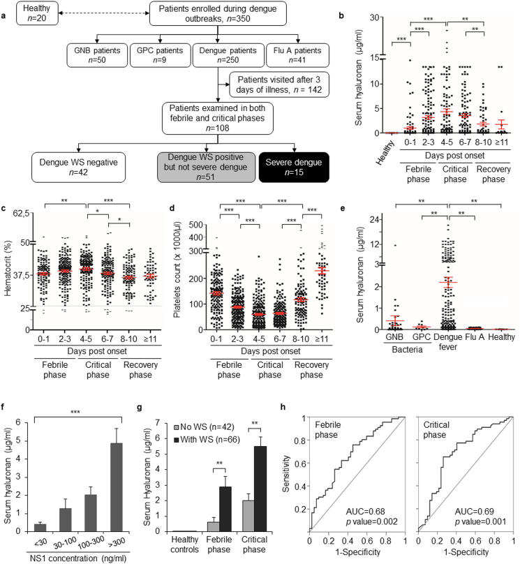
Methods: A cohort of 250 dengue patients has been followed from the onset of symptoms to the recovery phase. Serum hyaluronan levels and several other clinical parameters were recorded. The effect of NS1 treatment of cultured fibroblasts and endothelial cells on the expressions of hyaluronan synthetic and catabolic enzymes and the hyaluronan receptor CD44, were determined, as have the effects on the formation of hyaluronan-rich matrices and endothelial permeability.

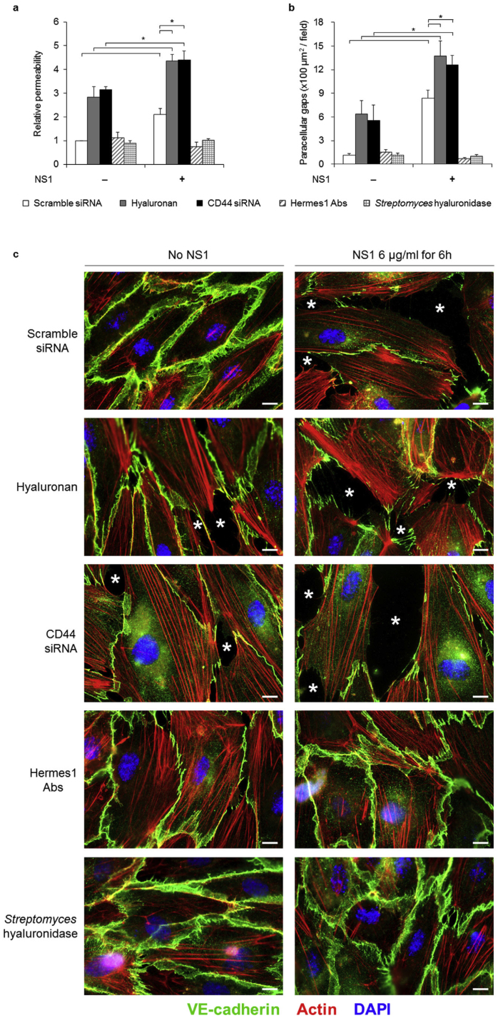
Findings: Elevated serum hyaluronan levels (≥70 ng/ml) during early infection was found to be an independent predictor for occurrence of warning signs, and thus severe dengue fever. High circulating levels of the viral protein NS1, indicative of disease severity, correlated with high concentrations of serum hyaluronan. NS1 exposure decreased the expression of CD44 in differentiating endothelial cells impairing the integrity of vessel-like structures, and promoted the synthesis of hyaluronan in dermal fibroblasts and endothelial cells in synergy with dengue-induced pro-inflammatory mediators. Deposited hyaluronan-rich matrices around cells cultured in vitro recruited CD44-expressing macrophage-like cells, suggesting a mechanism for enhancement of inflammation. In cultured endothelial cells, perturbed hyaluronan-CD44 interactions enhanced endothelial permeability through modulation of VE-cadherin and cytoskeleton re-organization, and exacerbated the NS1-induced disruption of endothelial integrity.

Interpretation: Pharmacological targeting of hyaluronan biosynthesis and/or its CD44-mediated signaling may limit the life-threatening vascular leakiness during moderate-to-severe dengue virus infection. FUND: This work was supported in part by grants from the Swedish Cancer Society (2018/337; 2016/445), the Swedish Research Council (2015-02757), the Ludwig Institute for Cancer Research, Uppsala University, the Ministry of Science and Technology, Taiwan (106-2314-B-037-088- and 106-2915-I-037-501-), Kaohsiung Medical University Hospital (KMUH103-3 T05) and Academy of Finland. The funders played no role in the design, interpretation or writing of the manuscript.

Keywords: CD44; Cytokines; Dengue; HAS2; HYAL2; Hyaluronan; TGFbeta; VE-cadherin; Vascular leakage.


Conflict of interest statement

CYL, YHC and PH have submitted a patent application based on the findings in this manuscript in United states (US 15/881,350) and EU (EP 18155997.2). CYL, YHC and PH are the inventors but not owner of a patent proved in Taiwan (TW I651535). The other authors have declared that no conflict of interest exists.

Figures

Fig. 1
Fig. 1
Serum hyaluronan levels increase robustly in patients with dengue fever and constitute a biomarker for disease severity. (a) Flow chart of the enrollment of patients during dengue outbreaks. Twenty healthy adults were enrolled as control group for comparison. Laboratory-confirmed dengue patients, and gram-negative bacilli (GNB), gram-positive cocci (GPC) bacteremia and influenza A virus (Flu A) infected patients were enrolled and analyzed. For further comparison between dengue patients without and with warning signs (WS), patients who did not visit hospital or were not evaluated day by day within 3 days after symptoms onset (n = 142) were excluded from the analyses described in panels g and h below. (b-d) Serum hyaluronan levels (b), hematocrit (c) and platelet counts (d) in 250 dengue virus infected patients the days after symptoms onset. (e) Serum hyaluronan levels of patients infected with bacteria, dengue virus or influenza virus, as well as of healthy controls. (f) Correlation between circulating NS1 concentrations and serum hyaluronan levels in patients with dengue fever (n = 182) are depicted. Statistical analysis was carried out by One-way ANOVA (analysis of variance). (g) Serum hyaluronan levels from 108 patients who provided paired serum samples during their febrile and critical phases, respectively, exhibiting WS or not. (h) ROC (receiver operating characteristic) curve of the serum hyaluronan concentration in febrile and critical phases show that it is a predictor of the occurrence of WS throughout the entire period of illness. Area under curve (AUC) was 0.68 and 0.69 respectively (See also Fig. S1 e). In (b-g), * p < .05, ** p < .01, *** p < .001.
Fig. 2
Fig. 2
NS1-treatment of dermal fibroblasts and microvascular endothelial cells enhances synthesis and alters organization of hyaluronan, promoting adhesion of macrophages. (a-b) RNA was prepared from human dermal fibroblast (a) and proliferative microvascular endothelial (b) cultures treated or not with NS1 (3 μg/ml) for 24 h, and reversed transcribed to cDNA; real-time PCR was run using primers for HAS1,2,3, HYAL1,2 and CD44 (n = 3). (c) Analysis of hyaluronan content in conditioned media of fibroblasts (upper panel) and endothelial cells (lower panel) treated or not with NS1 (n = 3). (d-g) Immunofluorescence of untreated and NS1-treated fibroblasts (d and f) and endothelial cells (e and g), exposed to PMA-activated THP-1 human monocyte-derived macrophage-like cells (f and g) or not (d and e), were fixed with 3.7% formaldehyde and stained for hyaluronan (green) or CD44 (red); hyaluronan-rich cable-like structures (arrows) and dot-like structures (arrowheads) are depicted. Representative THP-1 cells among several THP-1 cells are marked by black triangles (f and g). See also Fig. S3. (h and i) Fibroblasts (h) and endothelial cells (i) were treated with 3 μg/ml NS1 protein for 24 h, or not, in the absence or presence of Hermes1 (50 μg/ml) antibodies. Then 200,000 PMA-activated THP-1 human monocyte-derived macrophage-like cells (round cells) were co-cultured with fibroblasts and endothelial cells for 20 min, where after non-adherent cells were removed and adherent cells were stained. Ten randomly taken fields (20×) were obtained and the bound THP-1 cells were counted. Quantification of four independent experiments is depicted. In (a-c) and (h-i), * p < .05. In (d-g), scale bars, 10 μm.
Fig. 3
Fig. 3
NS1-treatment of differentiating microvascular endothelial cells results in impaired tubulogenesis. (a) Microvascular endothelial TIME cells grown in Matrigel for 16 h, after which cells had differentiated and formed tubular structures, were treated, or not, with 6 μg NS1/ml for an additional 9 h. After harvesting the cells, RNA was prepared and qRT-PCR analyses performed for the expression of mRNAs of HAS1, 2, 3, HYAL1, 2 and CD44. The data shown is from three independent experiments performed in triplicates ± SEM. (b) Cell lysates of the differentiated tubular endothelial cells were subjected to SDS-PAGE and immunoblotting for HAS2, HYAL2 and CD44, as well as GAPDH as loading control; the densities of the bands were quantified from three independent experiments by ImageJ (depicted by numbers). (c) The hyaluronan in 25 h conditioned media from untreated or NS1-treated differentiating endothelial cells, were analyzed for size by agarose electrophoresis, as described in Materials and Methods. (d) Microvascular endothelial cells were subjected to NS1 treatment (lower panel) or not (upper panel) during their growth in Matrigel for an additional 9 h after formation of tubular structures. The effect of NS1 on the vessel-like structures are shown. Numbers 1, 2, 3 indicate the order of asynchronous disassembly of vessel-like structures. Scale bars, 100 μm. In (a-b), * p < .05.
Fig. 4
Fig. 4
Dengue virus-induced pro-inflammatory cytokines synergize with NS1 to promote hyaluronan synthesis involving several signaling pathways. (a) The levels of TGFβ, MCP-1, IL-6, TNFα, IL-8 and IL-10 were analyzed in 108 dengue patients both in febrile and critical phases, and compared to twenty healthy individuals. (b-c) Dermal fibroblasts (b) and microvascular endothelial cells grown under proliferative conditions (c) were treated (filled bars), or not (open bars), with NS1 (3 μg/ml) alone or in combination with TGFβ (1 ng/ml) or TNFα (10 ng/ml) for 24 h. The expression levels of mRNAs for HAS2, CD44 and TLR4 were determined by RT-PCR. The levels of hyaluronan in 24 h conditioned media were also determined (right panels) (n = 3, triplicate determinations of each independent experiment). (d-e) Dermal fibroblasts (d) and endothelial cells (e) were treated with NS1 (3 μg/ml) for the indicated time periods. Cell lysates were prepared and proteins were separated by SDS-PAGE and immunoblotted for the phosphorylated and total amount of p65 NF-B, Akt, Erk1/2 and p38 MAP-kinase. (f) Quantification of three independent experiments of dermal fibroblasts and endothelial cells are depicted in gray and black bars respectively. * p < .05 when compared to the non-treated controls. In (a-c) and (f), results are means ± SEM. * p < .05, ** p < .01, *** p < .001.
Fig. 5
Fig. 5
Hyaluronan-CD44 interactions are important for the stability of intercellular adhesion junctions. (a) Confluent monolayers of microvascular endothelial cells expressing or depleted of CD44, were pre-treated or not with exogenous hyaluronan (25 μg/ml) or Hermes1 antibodies (50 μg/ml) overnight. Then, cell monolayers were co-treated or not with 6 μg/ml NS1 protein and some cultures also with Streptomyces hyaluronidase (5 units/ml) for 6 h. The permeability was determined by using an in vitro transwell permeability assay (n = 3). (b-c) Confluent microvascular endothelial cells, expressing or depleted of CD44, grown on Matrigel-coated coverslips, were treated as described in (a), fixed and stained for VE-cadherin (green), phalloidin (red), and DAPI (blue) (c; quantified in b). Asterisks indicate representative para-cellular gaps between endothelial cells. See also Fig. 6. In (a–b), data are presented as mean ± SEM. * p < .05. In (c), scale bars, 10 μm.
Fig. 6
Fig. 6
Effect of NS1, CD44 depletion and hyaluronan on VE-cadherin localization. Human microvascular endothelial TIME cells (2 × 105 cells/well in 12-well plates), transfected with CD44 siRNA or control, scrambled siRNA, were stimulated with 25 g/ml hyaluronan overnight, fixed in 4% paraformaldehyde and stained for VE-cadherin (green) and actin filaments (TRITC-phalloidin; red). Photographs were taken with a Zeiss Axioplan 2 immunofluorescence microscope. Arrows indicate the co-localization of VE-cadherin with actin fibers. Arrowheads indicate the discontinuity and fragility of VE-cadherin. Scale bars, 10 μm.
Fig. 7
Fig. 7
Schematic illustration of a possible mechanism for NS1-induced increased endothelium permeability. (a) Under physiological conditions, balanced expression of CD44 and hyaluronan levels contribute to cell-to-cell adhesion by cross-bridging between cells and maintaince of an intact glycocalyx. (b) During severe dengue virus infection, the excessive levels of partly fragmented hyaluronan occupy the reduced number of CD44 receptors, thus preventing efficient cross-bridging. In addition, disruption of the glycocalyx layer may contribute to increased permeability. It should be noted that in cells in which CD44 had been knocked down, NS1 still enhanced the disruption of junctional proteins, such as VE-cadherin (Fig. 5, Fig. 6), suggesting that perturbed hyaluronan-CD44 interactions is not the only mechanism involved in NS1-induced vascular leakage.

References

    1. Lennon F.E., Singleton P.A. Hyaluronan regulation of vascular integrity. Am J Cardiovasc Dis. 2011;1(3):200–213. - PMC - PubMed
    1. Takahashi Y., Li L., Kamiryo M., Asteriou T., Moustakas A., Yamashita H. Hyaluronan fragments induce endothelial cell differentiation in a CD44- and CXCL1/GRO1-dependent manner. J Biol Chem. 2005;280(25):24195–24204. - PubMed
    1. West D.C., Hampson I.N., Arnold F., Kumar S. Angiogenesis induced by degradation products of hyaluronic acid. Science. 1985;228:1324–1326. - PubMed
    1. Laurent T.C., Hyaluronan Fraser J.R.E. FASEB J. 1992;6:2397–2404. - PubMed
    1. Heldin P., Lin C.Y., Kolliopoulos C., Chen Y.H., Skandalis S.S. Regulation of hyaluronan biosynthesis and clinical impact of excessive hyaluronan production. Matrix Biol. 2019;78-79:100–117. - PubMed

MeSH terms