. 2020 Jan 1;457(1):43-56.
doi: 10.1016/j.ydbio.2019.09.003. Epub 2019 Sep 14.

Early ear neuronal development, but not olfactory or lens development, can proceed without SOX2

Affiliations

Early ear neuronal development, but not olfactory or lens development, can proceed without SOX2

Martina Dvorakova et al. Dev Biol. .

Abstract

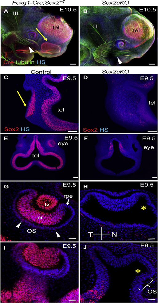
SOX2 is essential for maintaining neurosensory stem cell properties, although its involvement in the early neurosensory development of cranial placodes remains unclear. To address this, we used Foxg1-Cre to conditionally delete Sox2 during eye, ear, and olfactory placode development. Foxg1-Cre mediated early deletion of Sox2 eradicates all olfactory placode development, and disrupts retinal development and invagination of the lens placode. In contrast to the lens and olfactory placodes, the ear placode invaginates and delaminates NEUROD1 positive neurons. Furthermore, we show that SOX2 is not necessary for early ear neurogenesis, since the early inner ear ganglion is formed with near normal central projections to the hindbrain and peripheral projections to the undifferentiated sensory epithelia of E11.5-12.5 ears. However, later stages of ear neurosensory development, in particular, the late forming auditory system, critically depend on the presence of SOX2. Our data establish distinct differences for SOX2 requirements among placodal sensory organs with similarities between olfactory and lens but not ear placode development, consistent with the unique neurosensory development and molecular properties of the ear.

Keywords: Eye; Inner ear; Neuronal projections; Olfactory system; Placode development.


Conflict of interest statement

Competing interests

The authors have no competing interests to declare.

Figures

Fig. 1.
Fig. 1.. SOX2 is eliminated in the ear and pre-lens placodes in early Sox2cKO embryos.
Whole mount analysis of SOX2 expression pattern in E8.5 control, heterozygous Foxg1-Cre;Sox2+/f, and Sox2cKO embryos (11 somites), displayed in a dorsal view (control n = 9, Sox2cKO n = 5, Foxg1-Cre;Sox2+/f n = 5). Foxg1-Cre expression is visualized by using anti-Cre antibody in heterozygous Foxg1-Cre;Sox2+/f, and Sox2cKO embryos (C-F). Split channel images show SOX2 (C’-F’) and Cre immunolabeling (C”-F”). The dotted oval indicates the area of the otic pit (A, C-C”, D-D”). SOX2 expression is lost in the Sox2cKO ear invaginating placode (D, D’) compared to control (A), and heterozygous Foxg1-Cre;Sox2+/f embryos (C, C’). In the developing eye, SOX2 is eliminated in the presumptive lens ectoderm (PLE) and decreased SOX2 levels are evident in many cells of the optic vesicle (OV) in Sox2cKO (F, F’) compared to control embryos (B) and Foxg1-Cre;Sox2+/f (E, E’). Note Foxg1-Cre expression in the surface ectoderm in the area of the PLE (E, F, E”, F”). HS, Hoechst nuclear staining. Scale bars: 50 μm.
Fig. 2.
Fig. 2.. Complete SOX2 deletion in the otocyst, pre-lens and olfactory placodes, and in the nasal part of the eye vesicle mirrors the tdTomato reporter pattern for Foxg1-cre at E9.0 embryos (18 somites).
(A, D, G) In the control embryo, SOX2 is expressed in the neural tube and olfactory placode (op, arrowhead in A, D), eye vesicle prior to eye cup formation as well as the pre-lens placode (p-lp, arrowhead in D), and ear vesicle (G). (B-I) In Sox2cKO, SOX2 protein is detectable in the neural tube (B), and in the temporal domain of the eye vesicle (dotted area indicates the nasal domain of the eye vesicle in E, F), and it is completely missing in the epithelium of the olfactory placode (B), pre-lens placode (E) and the ear epithelium (H). A reporter of Foxg1-cre expression, tdTomato, indicates that Foxg1-cre expression matches the SOX2 deletion pattern in Sox2cKO (C, F, I). Note the few tdTomato positive cells anteroventral to the ear (I). HS, Hoechst nuclear staining. Scale bars: A-C 200 μm; D-I 50 μm.
Fig. 3.
Fig. 3.. The olfactory and lens placodes fail to invaginate in Sox2cKO.
(A, B) Whole-mount immunostaining shows the Cre expression pattern and innervation (tubulin) at E10.5. The Foxg1-Cre+ domain of the olfactory epithelium in Sox2cKO is missing (yellow arrow indicates the olfactory epithelium in Foxg1-Cre;Sox2+/f). The snout has an abnormal shape and a smaller eye with missing lens vesicle is apparent in Sox2cKO. Note, neuronal fibers from the trigeminal ganglion (arrowhead) innervate the presumptive olfactory area in Sox2cKO similar to controls, and the oculomotor nerve (III) extends from the midbrain in both control and mutant mice. (C, D) Formation of the olfactory placode with SOX2 expression is indicated in cross-sections by arrow in E9.5 controls (n = 2; arrow indicates the olfactory epithelium). In Sox2cKO, no SOX2 expression is detected in the area of presumptive olfactory placode (n = 2). Note the reduced SOX2 expression in the telencephalon (tel) of Sox2cKO (D), consistent with Foxg1-Cre expression in the telencephalon in (A, B). (E, F) Cross-sections show that SOX2 is undetectable in the developing Sox2cKO eye compared to the control at E9.5. (G-J) Higher magnification images show SOX2 expression in the neural retina (nr), lens vesicle (lv), and in the ventral area of the eye cup in the optic stalk (OS, arrowheads) in control embryos, whereas in Sox2cKO, no SOX2 expression is detectable in the eye. Note the failure of invagination of the lens placode to form the lens vesicle in Sox2cKO, the decreased number of neural retina cells, and lack of the U-shape formation of the optic cup with a large gap remaining between the neural retina and the retinal pigment epithelium (yellow asterisk in H, J). HS, Hoechst nuclear staining; N, nasal; T, temporal; tel, telencephalon vesicle; rpe, retinal pigment epithelium. Scale bars: A, B 200 μm; C, D, E, F 100 μm; G-J 50 μm
Fig. 4.
Fig. 4.. Deletion of Sox2 results in a complete loss of lens formation and abnormal eye development.
(A, B) In E11.5 controls and heterozygous Sox2 mutants (Foxg1-Cre;Sox2+/f), the lens vesicle (lv), optic stalk (OS) and the two-layered optic cup with an outer layer of retinal pigment epithelium (rpe) and the inner neural retina (asterisk) are formed. (C) The Sox2cKO developing eye appears to be smaller without any detectable lens vesicle, with a reduced neural retina domain, and abnormal formation of the optic stalk at E11.5. (D-I) At E14.5 and E17.5, the size of the optic cup is significantly reduced without the optic lens in Sox2cKO compared to controls and heterozygous Sox2 mutants. The retinal pigment epithelium and neural retina are present, although significantly diminished in Sox2cKO (n = 3 per genotype and time point). Note the presence of extraocular muscles in the Sox2cKO mutant at E14.5 (arrows in F). tel, telencephalon. Scale bars: A-C 200 μm; D-I 100 μm.
Fig. 5.
Fig. 5.. Delamination of NEUROD1+ neuroblasts from the SOX2-deficient epithelium and formation of the vestibular ganglion in Sox2cKO is comparable to control embryos.
(A, B) In E9.5 controls, SOX2 is expressed in the otocyst (ot) and in developing neurons of the vestibular ganglion (VG), whereas SOX2 is not detectable in the ear epithelium or neurons in Sox2cKO (n = 5 embryos per genotype). The boundaries of the Sox2cKO ear vesicle (otocyst) are delineated by yellow lines (B). Note, in Sox2cKO, SOX2 is not expressed in ISL1+ VG neurons and all SOX2+ cells co-express SOX10, consistent with glial cell molecular characteristics. Split channel images show SOX2 (A’, B’) and SOX10 immunolabeling (A”, B”). (C, D) NEUROD1+ neuroblasts (arrowheads) delaminate from the SOX2+ otic epithelium in controls and SOX2 negative epithelium in Sox2cKO. (E-G) The quantification of neurons in the VG area show no significant differences between control and Sox2cKO embryos at E10.5 (ISL1+ neurons per 1000 μm2; mean ± s.d.; n = 4 embryos per genotype and 2 ganglia per embryo, multiple t test with the Holm-Sidak method, P = NS). Dotted lines indicate the boundaries of the VG in A-A”, B-B”, E, F. (H) Plane of sections A-F is displayed. (I, J) Whole-mount immunostaining with anti-acetylated α-tubulin (nerve fibers) and anti-CRE shows apparently diminished innervation of the ear epithelium in Sox2cKO compared to littermate heterozygous Foxg1-Cre;Sox2+/f (arrowheads), indicating abnormalities in inner ear development at E10.5 (n = 3 embryos per genotype). (K, L) Immunostaining for the proliferating nuclear antigen Ki67 shows proliferating ISL1+ neurons in the VG of Sox2cKO and control embryos at E9.5 (n = 2 embryos per genotype). (M, N) The proliferation pattern evaluated by double-immunolabeling of EdU (injected at E9.5) and BrdU (injected at E10.5) shows an overall similar proliferation pattern in the VG between control and Sox2cKO littermates at E11.5, although decreased BrdU-labeling (green) in the VG of Sox2cKO is apparent (n = 2 embryos per genotype). GG, geniculate ganglion; HS, Hoechst nuclear staining; ot, otocyst. Scale bars: 50 μm; M, N 100 μm.
Fig. 6.
Fig. 6.. Neuronal development is arrested by apoptosis in Sox2cKO vestibular ganglion neurons at E11.5.
(A, B) Cleaved caspase-3 whole-mount immunostaining shows significantly increased apoptosis in the mutant vestibular ganglion (VG, demarcated by dotted line) compared to controls. Note the drastically reduced innervation of the spiral ganglion (SG, arrowhead) in Sox2cKO compared to control. A’, B’ display higher magnification images of the VG. (C) Cleaved caspase-3 immunostaining was quantified using ImageJ in the VG of E11.5 control (n = 6) and Sox2cKO embryos (n = 5). **P = 0.0043, Mann-Whitney U test. The data are presented as mean ± s.d. (D, E) Whole-mount immunostaining with anti-NeuN, a neuronal soma marker, shows a diminished VG domain at E12.5. GG, geniculate ganglion; HS, Hoechst nuclear staining; PC, posterior crista. Scale bars: 100 μm, 50 μm for A’ and B‘.
Fig. 7.
Fig. 7.. Abnormalities in the specification of the sensory epithelium of the cochlea are associated with a few residual spiral ganglion neurons in the Sox2cKO inner ear.
(A-D) Immunohistochemistry for the transcription factors GATA3 and PAX2 in cross sections reveals abnormalities in the formation of sensory epithelium with a broader expression of GATA3 and a lack of a clearly defined prosensory region in the epithelium of the cochlear duct (CD) in Sox2cKO (n = 3 embryos per genotype). The prosensory domain in the control cochlea is marked by co-expression of Pax2 and GATA3 and is indicated by the arrow in (A). Spiral ganglion (SG) neurons express GATA3 (arrowheads indicate the SG in the control and the residual SG in Sox2cKO). The inserts show GATA3 expression. (E, F) Representative vibratome sections show the expression of TrkC, tyrosine kinase receptor for the nerve growth factor NTF3, in SG neurons. HS, Hoechst nuclear staining. Scale bars: 100 μm.
Fig. 8.
Fig. 8.. Inner ear structural malformations in Sox2cKO are revealed by computer-assisted 3D-reconstruction.
The inner ear of Sox2cKO shows cochlear agenesis and a rudimental saccule but all other structures are missing or undiscernible compared to the control inner ear at E14.5 (n = 2 embryos per genotype). aa, anterior ampulla; asc, anterior semicircular canal; cd, cochlear duct; ed, endolymphatic duct; la, lateral ampulla; lsc, lateral semicircular canal; pa, posterior ampulla; psc, posterior semicircular canal; sac, saccule; ut, utricle. Note the enlargement of the apex.
Fig. 9.
Fig. 9.. The central and peripheral projections of the inner ear are comparable between controls and Sox2cKO at E11.5.
(A-B’”) Lateral views of whole-mounted hindbrains show analogous triple-lipophilic dye labeling (see Materials and Methods) from the ear (green), from the trigeminal ganglion (red), and from the jugular foramen of the IX/X nerve (magenta) in the control (A) and Sox2cKO mutant (B; n = 3 embryos per genotype). Vestibular afferents of the VIIIth cranial nerve (green), projecting to reach the cerebellum (CB) and the caudal part of the hindbrain, are comparable between the control (A) and mutant (B). Dye applications into the ear at this stage often in addition to labeling vestibular afferents also label the facial nerve (FN) extending around the developing ear and FN components (FBM, facial branchial motor neurons; IN, intermediate nerve; SSN, superior salivatory nucleus), as shown in control embryo (A). Trigeminal afferents (red) projecting in the descending tract of the trigeminal nerve (dV), glossopharyngeal (IX) and vagal (X) afferents (magenta) of the control are comparable to Sox2cKO afferent projections. (A’-A”’, B’-B”’) Details of vestibular afferent central projections (green) show fewer fibers in Sox2cKO (B’-B’”) compared to the control (A’-A”‘) but at the same time these fibers extend as far rostral (A’, B’) and caudal (A’“, B’”) as afferents in littermate controls. Inner ear projections are somewhat reduced near the VIIIth nerve root in the mutant (arrowheads in A” and B”). (C, D) Insertion of green dye into rhombomere 4 labels the FN and inner ear efferents extending to the posterior canal crista region (PC). Insertion of red colored dye into the vestibular nuclei labels vestibular ganglion neurons and vestibular afferents extending to the region of the PC in control and mutant mice as well as the saccule (S) in control. The same red dye application labeled the PC but not the saccula in Sox2cKO (D). Note that at this stage both inner ear afferents (red) and efferents (green) extend to the region of the PC despite the fact that no PC sensory epithelium ever forms in the Sox2cKO mice. Cranial nerves: V, trigeminal nerve; VII, facial nerve (FN); VIII, vestibulocochlear nerve; IX, glossopharyngeal nerve; X, vagus nerve; XI, accessory nerve; FBM, facial branchial motor neurons; IN, intermediate nerve (component of the facial nerve); SSN, superior salivatory nucleus (the visceromotor component of the facial nerve); VG, vestibular ganglion. Scale bars: 100 μm.
Fig. 10.
Fig. 10.. Vestibular projection remains near normal at E12.5 but no cochlear projection can be labeled in Sox2cKO.
(A, B) Representative images of the lateral views of whole-mounted hindbrains show analogous triple-dye labeling from the ear (green), from the trigeminal ganglion (magenta), and from the jugular foramen of the IX/X nerve (red) in control and mutant littermates at E12.5 (n = 3 embryos per genotype). Inner ear vestibular afferents (green) nearly identically project centrally in the control and Sox2cKO, although the number of afferents is reduced in Sox2cKO. (C, D) To evaluate the earliest central projections from spiral ganglion neurons to the cochlear nucleus at E12.5, lipophilic dye was inserted into the cochlear duct (red). Vestibular afferent fibers of the vestibular nerve (VN) are labeled by green dye inserted into the vestibular organs. Control animals show a profound cochlear projection (red) to the cochlear nucleus (CN), segregated from vestibular afferents (green, C). No cochlear projection to the CN is detectable in Sox2cKO only overlapping labeling of vestibular afferents (D). (E, F) Fillings from rhombomere 4 to label vestibular afferents (green), and from rhombomere 5 to label vestibular and cochlear afferents (red, see Materials and Methods) reveal basal turn spiral ganglia (SG) in the control (detail of red dye-labeled SG in insert). No cochlear innervation or neurons are labeled in mutant littermates (F). Note the larger vestibular ganglion (VG) in the control compared to Sox2cKO mice. VIII, vestibulocochlear nerve; IX, glossopharyngeal nerve; X, vagus nerve; CB, cerebellum; dV, the descending tract of trigeminal nerve; FN, facial nerve; PC, posterior crista. Scale bars: 100 μm.
Fig. 11.
Fig. 11.. Only efferent fibers project to the Sox2cKO ear at E18.5.
(A, B) Lipophilic dye applied to rhombomere 4 (red) labels the facial and intermediate nerve, and dye injected into the cerebellum (green) labels vestibular neurons, vestibular afferents, and contralateral inner ear efferents (IEE) in controls. In Sox2cKO, facial and intermediate nerve fibers are labeled but no vestibular or spiral ganglion neurons are labeled/found (n = 3 embryos per genotype). Some IEE remain at least until this stage and form a meshwork of fibers where the vestibular ganglion used to be and occasionally extend toward but never reach the remaining rudimentary ear of Sox2cKO. (C) Dye applications show the distribution of afferents and efferents in the cochlea and spiral ganglion of controls with inner ear efferent fibers forming the intraganglionic bundle (IGSB, green). (D) The transmitted light image (blue background) shows a reduced residual ear vesicle in Sox2cKO. All vestibular afferents and the facial nerve are labeled by green dye injected into the ipsilateral rhombomere 4 and intermediate nerve afferents are labeled by red dye from rhombomere 5/6. Note that no vestibular neurons or any innervation in the cochlea are labeled in Sox2cKO. VIII, vestibulocochlear nerve; GG, geniculate ganglion; FN, facial nerve; IN, intermediate nerve; RF, radial fibers; OC, the organ of Corti; SG, spiral ganglion; VG, vestibular ganglion. Scale bars: 100 μm.
Fig. 12.
Fig. 12.. Schematic of the morphological ear defects and central projection changes in Sox2cKO.
In control mice (right), the inner ear is formed by a complex labyrinth of ducts and recesses with six sensory epithelia, five vestibular: the maculae of the utricle (U) and saccule (S), and the cristae of the three semicircular canal ampullae (PC, AC, HC), and the auditory sensory organ of the cochlea, the organ of Corti (OC). Neurons of two distinct vestibular ganglia (VG, green) and spiral ganglia (SG, red) form afferent projections from the ear to the brain. The ear is also innervated by the inner ear efferents (IEE, lilac) that end on the sensory cells. In Sox2cKO, the deletion of Sox2 results in inner ear dysmorphology with cochlear agenesis, a rudimental saccule, and with all other structures being unrecognizable (left) at E14.5. The absence of SOX2 does not affect the initial formation of the vestibular ganglion and neurite navigation to their central targets, the vestibular nuclei and cerebellum, (green line). Similarly, dye tracing from the trigeminal ganglion (magenta), and from the jugular foramen of the IX/X nerve (blue) show comparable central projections in control and Sox2cKO mutant littermates at E11.5 and E12.5. In contrast to control embryos, no afferent innervation (red) could be traced from the cochlea to the cochlear nucleus in Sox2cKO. The red dot in the mutant shows the possible transient formation of some SG neurons. Black arrowheads indicate applications of lipophilic fluorescent dyes. Trigeminal (V), vestibulocochlear (VIII), glossopharyngeal (IX), vagal (X) cranial nerves; AC, anterior canal crista; CC, semicircular canal; CB, cerebellum; CD, cochlear duct; ED, endolymphatic duct; HC, horizontal canal crista; PC, posterior canal crista; r2, r4, r6, rhombomere 2, 4, 6; S, saccule; SG, spiral ganglion; U, utricle.

References

    1. Abello G, Khatri S, Radosevic M, Scotting PJ, Giraldez F, Alsina B, 2010. Independent regulation of Sox3 and Lmx1b by FGF and BMP signaling influences the neurogenic and non-neurogenic domains in the chick otic placode. Dev Biol 339, 166–178. - PubMed
    1. Ahmed M, Xu J, Xu PX, 2012. EYA1 and SIX1 drive the neuronal developmental program in cooperation with the SWI/SNF chromatin-remodeling complex and SOX2 in the mammalian inner ear. Development 139, 1965–1977. - PMC - PubMed
    1. Ashery-Padan R, Marquardt T, Zhou X, Gruss P, 2000. Pax6 activity in the lens primordium is required for lens formation and for correct placement of a single retina in the eye. Genes Dev 14, 2701–2711. - PMC - PubMed
    1. Barrionuevo F, Naumann A, Bagheri-Fam S, Speth V, Taketo MM, Scherer G, Neubuser A, 2008. Sox9 is required for invagination of the otic placode in mice. Dev Biol 317, 213–224. - PubMed
    1. Bhattacharyya S, Bailey AP, Bronner-Fraser M, Streit A, 2004. Segregation of lens and olfactory precursors from a common territory: cell sorting and reciprocity of Dlx5 and Pax6 expression. Dev Biol 271, 403–414. - PubMed

Publication types

Substances

LinkOut - more resources