. 2019 Oct 1;116(40):19930-19938.
doi: 10.1073/pnas.1911119116. Epub 2019 Sep 16.

Tubulin lattice in cilia is in a stressed form regulated by microtubule inner proteins

Affiliations

Tubulin lattice in cilia is in a stressed form regulated by microtubule inner proteins

Muneyoshi Ichikawa et al. Proc Natl Acad Sci U S A. .

Abstract

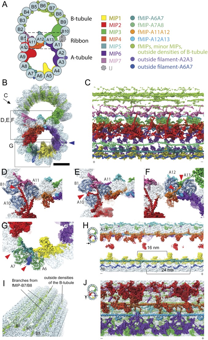
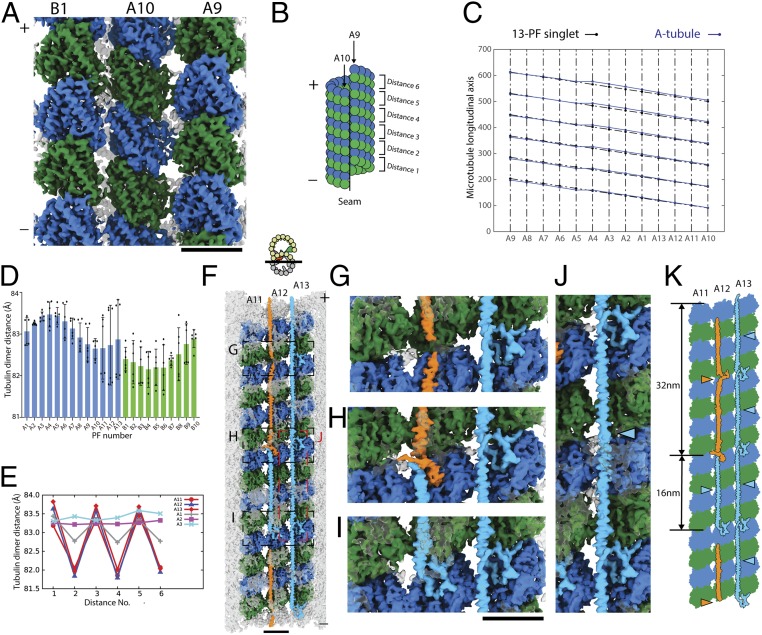
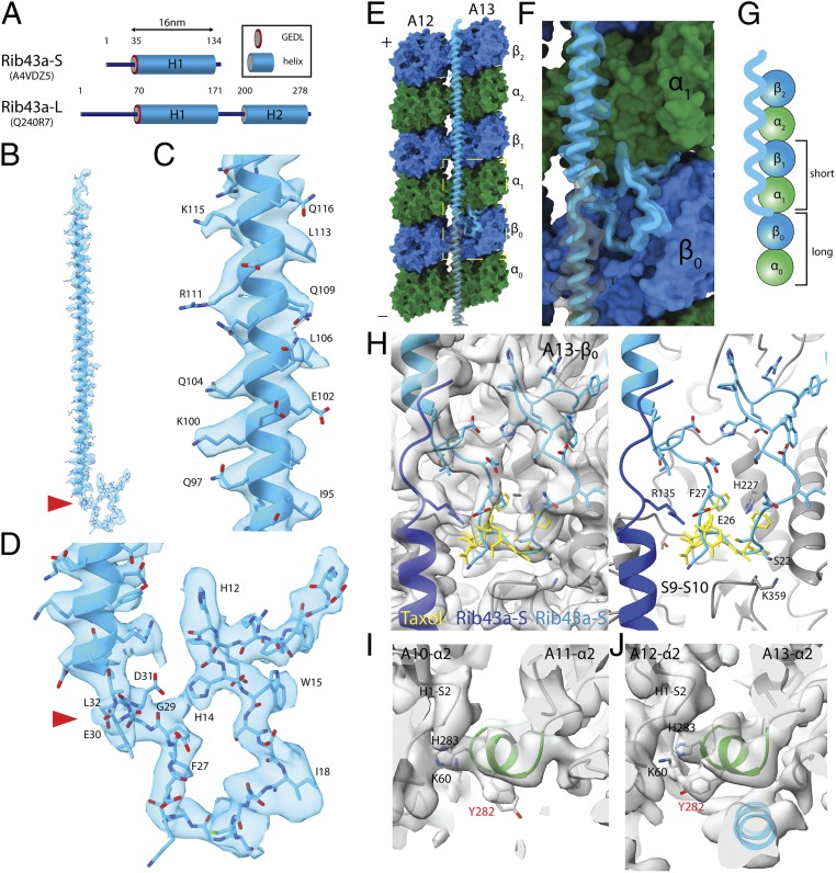
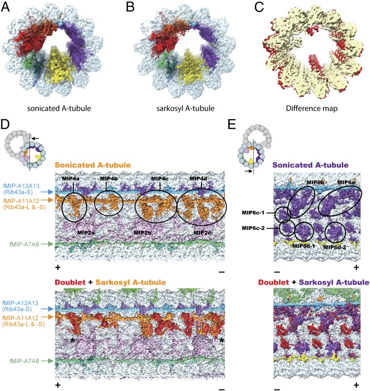
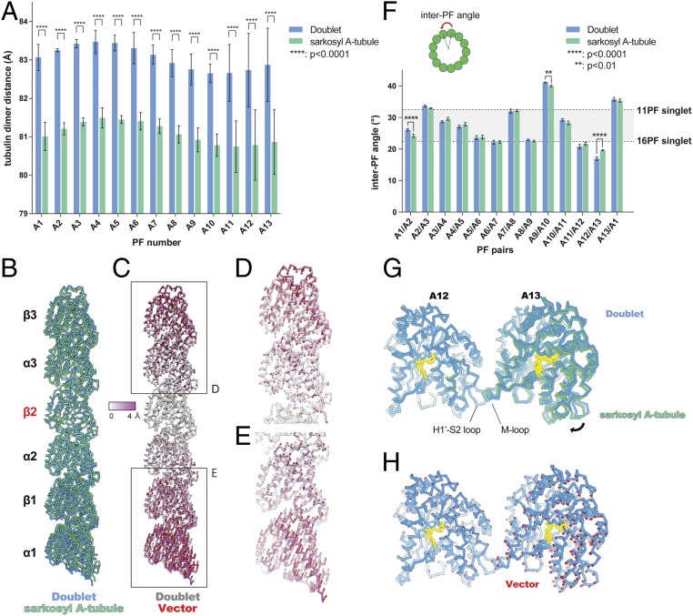
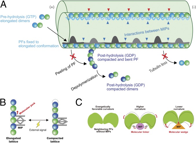
Cilia, the hair-like protrusions that beat at high frequencies to propel a cell or move fluid around are composed of radially bundled doublet microtubules. In this study, we present a near-atomic resolution map of the Tetrahymena doublet microtubule by cryoelectron microscopy. The map demonstrates that the network of microtubule inner proteins weaves into the tubulin lattice and forms an inner sheath. From mass spectrometry data and de novo modeling, we identified Rib43a proteins as the filamentous microtubule inner proteins in the protofilament ribbon region. The Rib43a-tubulin interaction leads to an elongated tubulin dimer distance every 2 dimers. In addition, the tubulin lattice structure with missing microtubule inner proteins (MIPs) by sarkosyl treatment shows significant longitudinal compaction and lateral angle change between protofilaments. These results are evidence that the MIPs directly affect and stabilize the tubulin lattice. It suggests that the doublet microtubule is an intrinsically stressed filament and that this stress could be manipulated in the regulation of ciliary waveforms.

Keywords: axoneme; cilia; ciliopathies; cryoelectron microscopy; microtubule.


Conflict of interest statement

The authors declare no conflict of interest.

Figures

Fig. 1.
Fig. 1.
Networks of the MIPs are woven into the tubulin lattice. (A) Schematic cartoon of the doublet from Tetrahymena viewed from the tip of the cilia. PF numbers are shown, and MIPs are colored as on the Right. The PF ribbon region is indicated as the dashed box. The inner junction (IJ), not presented in our structure, is known to bridge the B- and A-tubules (60). (B) Surface rendering of the 48-nm unit of the doublet is colored according to A. Blue arrowhead indicates the outside filament-A2A3/the outside molecular ruler (19). (Scale bar, 10 nm.) Views of CG are indicated in B. (C) The weaving network of MIPs inside the tubulin lattice with tubulin densities removed. Plus and minus ends are indicated by (+) and (−) signs, which are consistent throughout. (DG) Insertions of the MIPs into the tubulin lattice. Red arrowheads indicate the insertion densities. (D) α-Helical branches from the MIP2 go in between PFs A10 and A11. (E) A branch of MIP2 (red arrowhead) goes in between PF pairs A10/A11 and A10/B1, reaching outside the tubulin lattice. (D and E) Cross-sections with different depths along the longitudinal axis of the doublet. (F) Branches from fMIPs-A11A12 and -A12A13 (red arrowheads) go in between the tubulin lattice. (G) Branches from fMIP-A6A7 reach the outside surface and contact densities outside (red arrowheads). (H) Sectional view showing the outside filament-A6A7 (blue). Outside filament-A6A7 appears as a 24-nm repeating unit. (I) Outside densities of the B-tubule. (J) fMIPs appear as single α-helical structures running in between the inner ridges of the PF pairs A11/A12 and A12/A13. The globular MIPs and fMIPs are connected by branches.
Fig. 2.
Fig. 2.
The complex tubulin lattice within the 48-nm repeating unit of the doublet. (A) Outside view of surface rendering of the doublet at PFs A9, A10, and B1. α- and β-tubulins are colored green and blue. (B) Schematic diagram of the A-tubule and the tubulin dimer distance measurement. (C) Two-dimensional plot of the tubulin lattice of the A-tubule of the doublet (blue) and the 13-PF singlet (black) (29). The tubulin lattice is cut and unfurled at the seam as in B. Despite having the same 13-3 B-lattice as the 13-PF singlet, the A-tubule of the doublet shows a nonuniform tubulin dimer distance and Z-shift. (D) Plot of the dimer distance measurements among PFs in the doublet. For each PF, 6 dimer distances within the 48-nm repeating unit (illustrated in B) were measured and plotted as dots. Mean value (bar) with SD (error bar) for each PF are shown. (E) Bimodal pattern of the tubulin dimer distances. The dimer distances from PFs A11–A13 and A1–A3 of the doublet were plotted in the same longitudinal order as in the 48-nm unit. The dimer distances of PF A11 to A1 oscillate with every 2 tubulin units (∼16 nm). (F) Luminal view of the PF ribbon region A11–A13. fMIP densities in the A11–A13 region are now colored based on its tracing and morphology. The short 16-nm filament density is colored in light blue while the longer filament density is colored in orange. Tubulins in PFs A11–A13 are colored while all other densities are transparent. Magnified views of GJ are indicated by a dashed box in F. (K) Schematic diagram of the filament density identified in the PF ribbon region. Per 48 nm, there are 3 short filaments between PFs A12 and A13 (light blue arrowheads) and there is 1 short filament (light blue arrowheads) and 1 longer filament (orange arrowhead) between PFs A11 and A12.
Fig. 3.
Fig. 3.
Rib43a leads to the bimodal distance in the PF regions. (A) Secondary structure prediction of Rib43a-S and Rib43a-L. Only the large stretch of α-helices more than 20 residues from the structure prediction is shown. The GEDL consensus sequence is a conserved region of Rib43a (PFAM PF05914). (B) Model of the Rib43a-S inside its segmented density. (C and D) Magnified views of the helical region (C) and the N-terminal region (D) of Rib43a-S. The location of the GEDL motif is shown by the red arrowheads in B and D. (E) Model of Rib43a binds to the PF pair A12/A13. Yellow dashed box shows the magnified view in F. (F) The N terminus of Rib43a-S inserts into the interdimer interface in PF A13. (G) Schematic model of how Rib43a-S binds to the PF leading to the bimodal dimer distance pattern. (H) Superimposed views of taxol (PDB: 5SYF, yellow) and Rib43a-S with map (Left) and without map (Right) show similar topology. R135 in the C terminus of the lower Rib43a-S (dark blue) might interact with E26 of the N terminus of the upper Rib43a-S (light blue) in a head-to-tail dimerization mechanism. (I and J) M-loop conformations in the lateral interaction with Rib43a (PFs A12 and A13) and without (PFs A10 and A11). The side chain of Y282 adopts a different conformation in the presence of Rib43a, potentially due to steric clash. In this conformation, Y282 might interact with K60 of the neighboring α-tubulin.
Fig. 4.
Fig. 4.
Sarkosyl treatment removes some MIPs from the doublet. (A and B) Surface renderings of the sonicated A-tubule (A) and sarkosyl A-tubule (B) maps. (C) Difference map between the sonicated and sarkosyl A-tubule maps. Superimposition of the 2 maps reveals the missing MIP densities in the sarkosyl A-tubule map (red regions). Parts of the MIP2 and MIP6 are missing in the sarkosyl A-tubule map. (D and E) Sonicated A-tubule map (Top) and the overlap of doublet and sarkosyl A-tubule maps (Bottom). The MIP4 and MIP6 regions of the doublet (red) are mapped onto corresponding regions from the sarkosyl A-tubule map (MIP4 in orange and MIP6 in purple). The views are indicated in the illustrations on the Top Left. Remaining fMIPs are indicated on the side. The coloring of MIP2 and MIP4 is different from other figures to avoid confusion (see the illustration for the coloring). Some densities at the MIP4 and MIP6 regions are missing after the sarkosyl treatment while the fMIPs appear intact. The slight shifts in MIP4 (indicated by asterisks) at both + and − end are due to lateral compaction of the tubulin lattice.
Fig. 5.
Fig. 5.
Longitudinal tubulin lattice length and curvature are regulated by the MIPs. (A) Plot of tubulin dimer distances from doublet and the sarkosyl A-tubule. Mean values with SD for each PF are shown. The average value of each PF from the sarkosyl A-tubule shows a lateral compression of ∼2 Å. Statistical analysis was performed by 2-way ANOVA, Bonferroni post hoc test (see also SI Appendix, Table S2). (B) Comparison of tubulin models refined in PF-A12 from doublet (blue) and sarkosyl A-tubule (green) showing a longitudinal compaction after missing some MIPs. Models were aligned by β2-tubulin. (C) Tubulin models of PF-A12 from the doublet are colored according to the degree of displacement. Vectors of the Cα displacement toward the sarkosyl A-tubule model are shown in red. (D and E) Close-up views of the tubulins from the periphery with vectors. (F) Plot of inter-PF angles in the doublet and sarkosyl A-tubule. Inter-PF angles were measured as shown in the schematic diagram on Top and mean values were plotted (see also SI Appendix, Fig. S5C and Table S3). Error bars represent SD. Two-way ANOVA, Bonferroni post hoc test was performed to compare the mean values. PF pairs with P values smaller than 0.01 are highlighted by asterisks (see also SI Appendix, Table S4). The gray area in the plot represents the PF pair angles commonly seen for in vitro reconstituted singlets (29). (G) Alignment of the models of PF pair A12/A13 from the doublet (blue) and sarkosyl A-tubule (green) based on the tubulin unit of PF-A12 reveals ∼3° difference in rotation (black arrow). (H) The model of PFs A12/A13 from the doublet with the vectors (red) of the displacement of Cα compared to the sarkosyl A-tubule model. Nucleotides, yellow.
Fig. 6.
Fig. 6.
Model of stabilization mechanisms of the doublet tubulin lattice by MIPs. (A) Model of the impacts of the MIPs on the doublet. First, elongated tubulin dimers in GTP prehydrolysis state are incorporated into the tubulin lattice. This elongated and stable conformation is fixed after assembly into the lattice through the interactions with the MIPs. The network of MIPs (blue arrowheads) also holds the tubulin lattice from the inside to prevent the loss of tubulin or breakage. At the plus end, MIPs prevent the peeling of PFs and depolymerization by keeping PFs in a stable and elongated conformation. Hence, the doublet is stabilized by the MIPs at several different levels to ensure that it can withstand the mechanical stress and prevent catastrophic events for the cilia. Some MIPs, such as Rib43a, have insertions into the tubulin lattice (red arrowheads), causing the larger interdimer gap and bimodal dimer distance. (B) Schematic diagram of the function of the MIPs in regulating tubulin lattice length. Some MIPs work as a molecular jack to keep the tubulin lattice elongated. External signals could change the MIP property and thereby the tubulin lattice. (C) MIPs regulate the angles between PFs. Without MIPs, tubulin lattice takes an energetically favorable curvature. Some MIPs work as molecular linkers, which hold adjacent tubulin pairs together so that it will take a higher curvature such as in the PFs A9/A10. Other MIPs, in particular, Rib43a work as molecular wedges and open the PF pairs and induce a lower curvature.

References

    1. Bui K. H., Yagi T., Yamamoto R., Kamiya R., Ishikawa T., Polarity and asymmetry in the arrangement of dynein and related structures in the Chlamydomonas axoneme. J. Cell Biol. 198, 913–925 (2012). - PMC - PubMed
    1. Stepanek L., Pigino G., Microtubule doublets are double-track railways for intraflagellar transport trains. Science 352, 721–724 (2016). - PubMed
    1. Fliegauf M., Benzing T., Omran H., When cilia go bad: Cilia defects and ciliopathies. Nat. Rev. Mol. Cell Biol. 8, 880–893 (2007). - PubMed
    1. Akhmanova A., Steinmetz M. O., Tracking the ends: A dynamic protein network controls the fate of microtubule tips. Nat. Rev. Mol. Cell Biol. 9, 309–322 (2008). - PubMed
    1. Ichikawa M., et al. , Subnanometre-resolution structure of the doublet microtubule reveals new classes of microtubule-associated proteins. Nat. Commun. 8, 15035 (2017). - PMC - PubMed

Publication types

LinkOut - more resources