. 2019 Oct 15;93(16):e1561-e1571.
doi: 10.1212/WNL.0000000000008326. Epub 2019 Sep 16.

Pediatric postoperative cerebellar cognitive affective syndrome follows outflow pathway lesions

Affiliations

Pediatric postoperative cerebellar cognitive affective syndrome follows outflow pathway lesions

Fatimah M Albazron et al. Neurology. .

Abstract

Objective: To evaluate lesion location after pediatric cerebellar tumor resection in relation to the development of severe cognitive and affective disturbances, or cerebellar cognitive affective syndrome (CCAS).

Methods: The postsurgical lesion location of 195 pediatric patients with cerebellar tumors was mapped onto a template brain. Individuals with CCAS were matched to 2 participants without CCAS by sex, age, and lesion volume. Lesion analyses included both a hypothesis-driven evaluation of the cerebellar outflow pathway (deep nuclei and superior cerebellar peduncles) and data-driven multivariate lesion symptom mapping. Lesion-associated networks were evaluated by comparing connectivity patterns between the lesion location of cases with and those without CCAS with resting-state functional connectivity MRI data from large normative adult and pediatric cohorts.

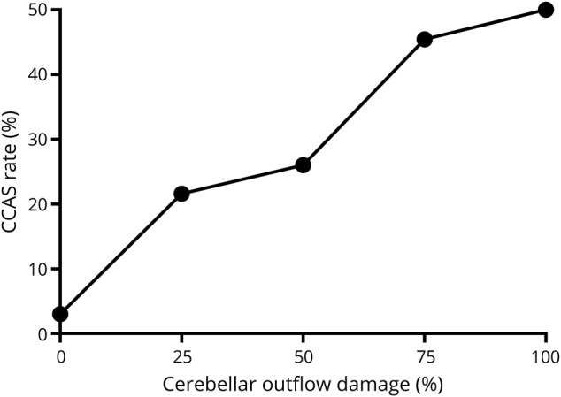
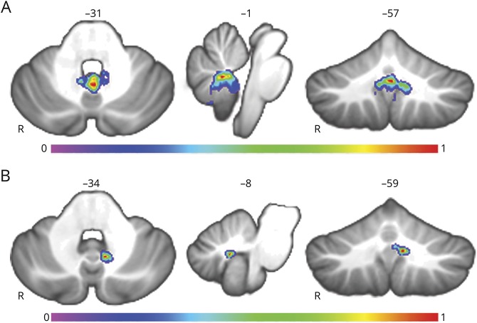
Results: CCAS was present in 48 of 195 participants (24.6%) and was strongly associated with cerebellar outflow tract lesions (p < 0.0001). Lesion symptom mapping also highlighted the cerebellar outflow pathway, with peak findings in the fastigial nuclei extending into the inferior vermis. Lesion network mapping revealed that the cerebellar region most associated with CCAS was functionally connected to the thalamic mediodorsal nucleus, among other sites, and that higher connectivity between lesion location and the mediodorsal nucleus predicts CCAS occurrence (p < 0.01). A secondary analysis of 27 participants with mutism revealed similar localization of lesions and lesion-associated networks.

Conclusion: Lesions of the cerebellar outflow pathway and inferior vermis are associated with major cognitive and affective disturbances after pediatric cerebellar tumor resection, and disrupted communication between the cerebellum and the thalamic mediodorsal nucleus may be important.


Figures

Figure 1
Figure 1. Cerebellar outflow pathway lesion load
The cerebellar outflow pathway is shown in blue on the left extending from the deep cerebellar nuclei along the superior cerebellar peduncles. The entire region of interest was separated into 1-mm oblique coronal slices perpendicular to the angle of the superior cerebellar peduncles (17°), of which 3 examples are shown. A sample lesion is shown in red that overlaps with the cerebellar outflow pathway. The extent the lesion overlap with the outflow pathway is quantified as a proportion of total voxels for each slice. Slice 83 (1) has a damaged proportion value of 0.15; slice 77 (2) has a damaged proportion of 0.73; and slice 71 (3) has a proportion of damage of 0.30. Across all slices, the maximum proportional value served as the cerebellar outflow pathway lesion load, which was used for subsequent analyses. This value would come from slice 77 (2) in the right panel among the slices shown, where the lesion overlaps with 73% of voxels in the outflow pathway for that slice.
Figure 2
Figure 2. Lesion overlap maps
(A) Lesion overlap of all participants had a peak of 110 of 195 at the vermal lobule IX, Montreal Neurological Institute (MNI) coordinate 0, −52, −38. (B) Patients with cerebellar cognitive affective syndrome (CCAS) had a peak lesion overlap of 39 of 48 at vermal lobule IX (MNI coordinate −1, −51, −39), and (C) participants without CCAS had a peak overlap of 71 or 147 also at vermal lobule IX (MNI coordinate 0, −52, −38). (D) A proportional subtraction, or difference map, of non-CCAS lesions subtracted from CCAS-associated lesions showed a regional peak involving vermis lobule IX (MNI coordinate −4, −53, −31) and involving the adjacent fastigial, interposed, and medial dentate nuclei (coordinates −3, −52, −28; −5, −59, −32; and −12, −53, −33, respectively).
Figure 3
Figure 3. Risk of CCAS stratified by cerebellar outflow pathway lesion load
This figure demonstrates that cerebellar cognitive affective syndrome(CCAS) rates are higher as the proportion of cerebellar outflow damage increases. The number of participants with CCAS compared to those without CCAS was as follows: no cerebellar outflow damage CCAS = 1, no CCAS = 32; 0% to 25% damage CCAS = 16, no CCAS = 58; 25% to 50% damage CCAS = 13, no CCAS = 37; 50% to 75% damage CCAS = 10, no CCAS = 12; and 75% to 100% damage CCAS = 8, no CCAS = 8.
Figure 4
Figure 4. Lesion symptom mapping results
(A) Results of the lesion symptom mapping analysis of all 195 participants. Warmer voxels have a higher association with the development of cerebellar cognitive affective syndrome with the overall map being statistically significant (p < 0.0001). (B) The same map with a higher statistical threshold highlights the strongest findings in the fastigial and interposed nuclei of the cerebellum and vermal lobule IX with peak Montreal Neurological Institute coordinates: 3, −55, −30; −7, −59, −34; and −1, −57, −31, respectively.
Figure 5
Figure 5. Lesion-associated networks derived from adult data
(A) Network of regions functionally connected to the cerebellar region associated with cerebellar cognitive affective syndrome (CCAS) is shown. This network was derived from seed-based functional connectivity from a large normative dataset of healthy adults using the result shown in figure 4B as the seed region. Regions with positively correlated oscillatory patterns are shown in warm colors, and negatively correlated networks did not reach significance at this threshold. Peak coordinates for the parvocellular division of the mediodorsal nucleus of the thalamus, left temporal stem, right temporal stem, and right red nucleus are as follows: (−1, −10, 10), (−25, −20, −6), (29, −21,-4), and (5, −24, −9), respectively. (B) Fisher r-to-z transformed scores show significantly higher functional connectivity between lesion sites causing CCAS compared to those not associated with CCAS at the mediodorsal (MD) nucleus of the thalamus (parvocellular), right temporal stem, and right red nucleus with values of p = 0.0029, 0.0012, and 0.0080, respectively. Other sites of the network had higher correlation with CCAS lesions but did not reach statistical significance after correction for multiple compressions (left temporal stem and anterior cingulate cortex with p = 0.0180 and 0.1790, respectively). Error bars represent SEM.
Figure 6
Figure 6. Lesion-associated network derived from pediatric data
Spatial distribution of the network derived from the cerebellum region most associated with developing cerebellar cognitive affective syndrome is shown. Results are similar to those from the adult dataset (figure 5A), including regional peaks in the mediodorsal nucleus of the thalamus (parvocellular) and right red nucleus at Montreal Neurological Institute coordinates (−3, −16, 8) and (5, −19, −12), respectively.
Figure 7
Figure 7. Cerebellar mutism lesion symptom mapping
(A) Multivariate lesion symptom mapping results of cerebellar mutism syndrome (CMS) are shown. The overall map is similar to that of figure 4A with a regional peak in vermal lobule IX (−1, −57, −31) extending to the fastigial nuclei and vermal lobule X. (B) The same analysis as in panel (A) in the cohort with cerebellar cognitive affective syndrome without mutism has a peak finding in the region of the interposed nucleus and extending to the medial dentate nucleus (−8, −59, −34).

Comment in

References

    1. Lanier JC, Abrams AN. Posterior fossa syndrome: review of the behavioral and emotional aspects in pediatric cancer patients. Cancer 2017;123:551–559. - PubMed
    1. Wisoff JH, Epstein FJ. Pseudobulbar palsy after posterior fossa operation in children. Neurosurgery 1984;15:707–709. - PubMed
    1. Rekate HL, Grubb RL, Aram DM, Hahn JF, Ratcheson RA. Muteness of cerebellar origin. Arch Neurol Am Med Assoc 1985;42:697–698. - PubMed
    1. Hoche F, Guell X, Vangel MG, Sherman JC, Schmahmann JD. The cerebellar cognitive affective/Schmahmann syndrome scale. Brain 2018;141:248–270. - PMC - PubMed
    1. Levisohn L, Cronin-Golomb A, Schmahmann JD. Neuropsychological consequences of cerebellar tumour resection in children: cerebellar cognitive affective syndrome in a paediatric population. Brain 2000;123:1041–1050. - PubMed

Publication types