. 2019 Sep 17;11(18):7639-7662.
doi: 10.18632/aging.102276. Epub 2019 Sep 17.

miR-125a-5p inhibits tumorigenesis in hepatocellular carcinoma

Affiliations

miR-125a-5p inhibits tumorigenesis in hepatocellular carcinoma

Xin Xu et al. Aging (Albany NY). .

Abstract

Hepatocellular carcinoma (HCC) is one of the most prevalent and deadly cancers world-wide. miR-125a-5p is a tumor suppressor in HCC and other cancers, but its mechanisms of action during HCC tumorigenesis remain largely unknown. In this study, we found that miR-125a-5p expression was significantly lower in HCC tissues and cell lines than matched normal tissues and liver cells. miR-125a-5p overexpression inhibited HCC cell proliferation and induced apoptosis in vitro and in vivo, while miR-125a-5p knockdown had the opposite effects. In addition, PTPN1 and MAP3K11 were identified as targets of miR-125a-5p. Knocking down PTPN1 and MAP3K11 activated the JNK MAPK signaling pathway to suppress HCC cell proliferation and induce apoptosis. Our findings suggest that miR-125a-5p may be a useful therapeutic target for treatment of HCC patients.

Keywords: HCC; MAP3K11; MAPK signaling pathway; PTPN1; miR-125a-5p.


Conflict of interest statement

CONFLICTS OF INTEREST: All authors declare that they have no competing interests.

Figures

Figure 1
Figure 1
miR-125a-5p is down-regulated in HCC tissues and cells. (A) Heat map of differentially expressed miRNAs in eight matched pairs of HCC tissues and adjacent normal tissues. (B) qRT-PCR indicated that miR-125a-5p expression was down-regulated in HCC tissues compared to the adjacent normal tissues. Student’s t-test, mean ± SD, *P<0.05, **P<0.01. (C) miR-125a-5p expression was down-regulated in HCC tumor tissues compared to adjacent normal tissues in TCGA in Starbase. (D) qRT-PCR indicated that miR-125a-5p expression was lower in HCC cell lines (Bel-7404, SK-Hep1, Bel-7404, and MHCC-97H) than in a normal liver cell line (L02). Student’s t-test, mean ± SD, **P<0.01, ***P<0.001. (E) Kaplan-Meier curves indicated that overall survival of HCC patients was not associated with miR-125a-5p expression level in Kaplan Meier Plotter.
Figure 2
Figure 2
miR-125a-5p suppresses proliferation and induces apoptosis in HCC cells in vitro. (A) qRT-PCR showed that miR-125a-5p was up-regulated after transfection with mimics and down-regulated after transfection with inhibitors in Bel-7404 and SK-Hep1 cells. Student’s t-test, mean ± SD, ***P<0.001. (B and C) Cell proliferation ability was assessed using CCK8 and colony formation assays in transfected Bel-7404 and SK-Hep1 cells. Two-way ANOVA, mean ± SD, *P<0.05, **P<0.01. (D) Western blots were used to analyze the expression of p53, Bax, Bcl-2, and active caspase-3 after transfection in Bel-7404 and SK-Hep1 cells. (E) Immunofluorescence staining of cleaved caspase substrate was detected after transfection in Bel-7404 and SK-Hep1 cells. Scale bars: 100μm.
Figure 3
Figure 3
PTPN1 and MAP3K11 are direct targets of miR-125a-5p in HCC. (A) Three target prediction databases were used to predict the targets of miR-125a-5p; 131 common elements were identified. (B and C) These 131 target genes were analyzed with ClueGO and CluePedia in Cytoscape. (D and F) miR-125a-5p and its putative binding sequences in the 3′-UTRs of PTPN1 and MAP3K11. (E and G) miR-125a-5p overexpression inhibited, while knockdown increased, luciferase activity of the wild-type (WT), but not mutant (MT), PTPN1 or MAP3K11 3′-UTRs. Student’s t-test, mean ± SD, **P<0.01, ns: not statistically significant. (H) PTPN1 and MAP3K11 protein expression was assessed via Western blot in transfected Bel-7404 and SK-Hep1 cells.
Figure 4
Figure 4
PTPN1 and MAP3K11 are up-regulated and negatively correlated with the expression of miR-125a-5p in HCC. (A and B) PTPN1 and MAP3K11 were up-regulated in TCGA in Starbase. (C and D) High expression of PTPN1 and MAP3K11 was associated with poorer overall survival in the GEPIA database. (E and F) qRT-PCR indicated that PTPN1 and MAP3K11 were up-regulated in the eight matched pairs of HCC tissues compared to the adjacent normal tissues. Student’s t-test, mean ± SD, *P<0.05, **P<0.01, ***P<0.001, ****P<0.0001. (GI) qRT-PCR and Western blots indicated that PTPN1 and MAP3K11 were up-regulated in HCC cell lines. One-way ANOVA, mean ± SD, *P<0.05, **P<0.01. (J and K) Spearman correlation analysis showed that miR-125a-5p expression was negatively correlated with PTPN1 and MAP3K11 expression.
Figure 5
Figure 5
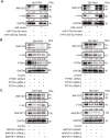
miR-125a-5p suppresses PTPN1 and MAP3K11 expression via the MAPK signaling pathway in HCC. (A) Western blots indicated that miR-125a-5p overexpression reduced, while miR-125a-5p knockdown increased, the expression of JNK1/2/3 and c-Jun in Bel-7404 and SK-Hep1 cells. (B and C) Western blots showed that PTPN1 and MAP3K11 knockdown decreased the expression of JNK1/2/3 and c-Jun in Bel-7404 and SK-Hep1 cells.
Figure 6
Figure 6
miR-125a-5p suppresses cell proliferation and induces apoptosis in HCC by targeting PTPN1 and MAP3K11 via the MAPK signaling pathway. (AD) Cell proliferation ability was assessed using CCK8 and colony formation assays after Bel-7404 and SK-Hep1 cells were transfected with miR-control, miR-125a-5p inhibitors, PTPN1 siRNA 3, or MAP3K11 siRNA 1. Two-way ANOVA, mean ± SD, **P<0.01, ***P<0.001. (E and F) Immunofluorescence staining of cleaved caspase substrate was detected after transfection in Bel-7404 and SK-Hep1 cells. Scale bars: 100μm. (G and H) Expression of PTPN1, MAP3K11, JNK1/2/3, and c-Jun was examined in transfected Bel-7404 and SK-Hep1 cells using Western blots.
Figure 7
Figure 7
miR-125a-5p suppresses cell proliferation and induces apoptosis by targeting PTPN1 and MAP3K11 via the MAPK signaling pathway in vivo. (A and B) Representative images of mice and xenografted tissues. (C and D) Xenograft tissue volumes and weights were analyzed (n=5). Two- and One-way ANOVAs, mean ± SD. **P<0.01, ***P<0.001. (E) PTPN1 and MAP3K11 expression was higher in HCC tumor tissues than in normal liver tissues in the Human Protein Atlas database. (F) Brdu, Ki67, PTPN1, MAP3K11, JNK1/2/3, and c-Jun expression were detected via immunofluorescence in xenografted tissuesat 400× magnification.
Figure 8
Figure 8
Proposed mechanism of miR-125a-5p function in HCC. miR-125a-5p inhibits HCC cell proliferation and induces cell apoptosis by directly targeting PTPN1 and MAP3K11 via the MAPK signaling pathway.

Similar articles

Cited by

References

    1. Bray F, Ferlay J, Soerjomataram I, Siegel RL, Torre LA, Jemal A. Global cancer statistics 2018: GLOBOCAN estimates of incidence and mortality worldwide for 36 cancers in 185 countries. CA Cancer J Clin. 2018; 68:394–424. 10.3322/caac.21492 - DOI - PubMed
    1. Gomaa AI, Khan SA, Toledano MB, Waked I, Taylor-Robinson SD. Hepatocellular carcinoma: epidemiology, risk factors and pathogenesis. World J Gastroenterol. 2008; 14:4300–8. 10.3748/wjg.14.4300 - DOI - PMC - PubMed
    1. Welzel TM, Graubard BI, Quraishi S, Zeuzem S, Davila JA, El-Serag HB, McGlynn KA. Population-attributable fractions of risk factors for hepatocellular carcinoma in the United States. Am J Gastroenterol. 2013; 108:1314–21. 10.1038/ajg.2013.160 - DOI - PMC - PubMed
    1. El-Serag HB, Rudolph KL. Hepatocellular carcinoma: epidemiology and molecular carcinogenesis. Gastroenterology. 2007; 132:2557–76. 10.1053/j.gastro.2007.04.061 - DOI - PubMed
    1. Coleman MP. Cancer survival:global surveillance will stimulate health policy and improve equity. Lancet. 2014; 383:564–73. 10.1016/S0140-6736(13)62225-4 - DOI - PubMed

Publication types

MeSH terms