. 2019 Sep 16;10(1):4216.
doi: 10.1038/s41467-019-12110-y.

A phyB-PIF1-SPA1 kinase regulatory complex promotes photomorphogenesis in Arabidopsis

Affiliations

A phyB-PIF1-SPA1 kinase regulatory complex promotes photomorphogenesis in Arabidopsis

Inyup Paik et al. Nat Commun. .

Abstract

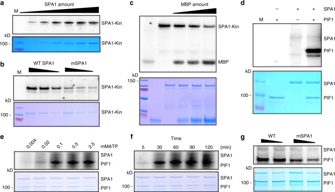
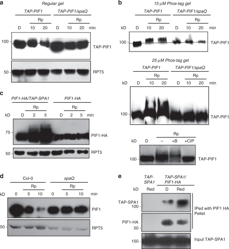
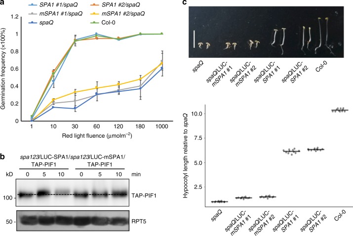
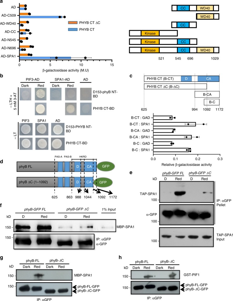
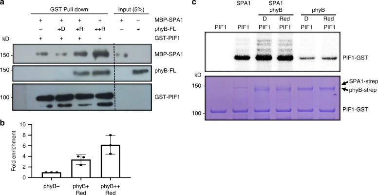
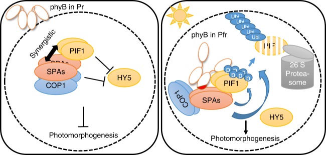
CONSTITUTIVELY PHOTOMORPHOGENIC1 (COP1) is a highly conserved E3 ubiquitin ligase from plants to animals and acts as a central repressor of photomorphogenesis in plants. SUPPRESSOR OF PHYA-105 1 family members (SPA1-SPA4) directly interact with COP1 and enhance COP1 activity. Despite the presence of a kinase domain at the N-terminus, no COP1-independent role of SPA proteins has been reported. Here we show that SPA1 acts as a serine/threonine kinase and directly phosphorylates PIF1 in vitro and in vivo. SPAs are necessary for the light-induced phosphorylation, ubiquitination and subsequent degradation of PIF1. Moreover, the red/far-red light photoreceptor phyB interacts with SPA1 through its C-terminus and enhances the recruitment of PIF1 for phosphorylation. These data provide a mechanistic view on how the COP1-SPA complexes serve as an example of a cognate kinase-E3 ligase complex that selectively triggers rapid phosphorylation and removal of its substrates, and how phyB modulates this process to promote photomorphogenesis.


Conflict of interest statement

The authors declare no competing interests.

Figures

Fig. 1
Fig. 1
SPA1 acts as a ser/thr protein kinase. a SPA1 kinase domain (SPA1-Kin) purified from E. coli showed an auto-phosphorylation activity in a concentration-dependent manner (autoradiogram on top panel). Bottom panel shows the protein level in a Coomassie-stained gel. M, indicates protein marker. b A conserved amino acid mutation on the SPA1 kinase domain reduces the auto-phosphorylation activity of SPA1 (autoradiogram on top panel). Bottom panel shows the protein level in a Coomassie-stained gel. c The N-terminal kinase domain of SPA1 exhibits kinase activity in the presence of myelin basic protein (MBP); a general kinase substrate (autoradiogram on top panel). Bottom panel shows the protein levels in a Coomassie-stained gel. d Full-length SPA1 purified from Pichia pastoris phosphorylates PIF1 in vitro (autoradiogram on top panel). Bottom panel shows the protein levels in a Coomassie-stained gel. e ATP-dependent kinase assays of full-length SPA1 on PIF1 (autoradiogram on top panel). ATP concentrations used were 0.004, 0.02, 0.1, 0.5, and 2.5 mM. Bottom panel shows the protein levels in a Coomassie-stained gel. f Kinetic analysis of the kinase activity of the full-length SPA1 on PIF1 (autoradiogram on top panel). Bottom panel shows the protein levels in a Coomassie-stained gel. g A conserved amino acid mutation on the full-length SPA1 kinase domain reduces the SPA1 kinase activity toward PIF1 (autoradiogram on top panel). Bottom panel shows the protein levels in a Coomassie-stained gel
Fig. 2
Fig. 2
SPAs are necessary for the light-induced phosphorylation and degradation of PIF1 in vivo. a Immunoblots showing the light-induced phosphorylation of TAP-PIF1 is defective in the spaQ mutant compared to wild type. Four-day-old dark-grown seedlings were either kept in darkness or exposed to a pulse of red light (300 μmolm−2) and then incubated in the dark for the duration indicated before being sampled for protein extraction. All dash lines show the dark position of the PIF1 band. b Immunoblots show a defect in the light-induced phosphorylation of TAP-PIF1 in spaQ background compared to wild type in gels containing 15 (top) and 25 (middle) µM phostag. (Bottom panel) The red light-induced slow-migrating band is a phosphorylated form of TAP-PIF1 as indicated by the phosphatase treatment. +CIP, native Calf Intestinal Phosphatase; +B, heat-inactivated boiled CIP. c Overexpression of SPA1 induces faster phosphorylation of PIF1-HA than wild type under weak red-light pulse (1 μmolm−2). Seedlings were pretreated with the proteasome inhibitor (40 μM bortezomib) for 5 h before being exposed to red light. Blots were probed with anti-HA and anti-RPT5 antibodies. d Immunoblots showing native PIF1 level in spaQ mutant compared to wild type. e Immunoblots show that TAP-SPA1 can interact with PIF1-HA in a red light-inducible manner in vivo
Fig. 3
Fig. 3
The kinase activity of SPA1 is necessary for its biological function. a Seed germination phenotypes of wild type and mutant SPA1 expressed in the spaQ background in response to an increasing fluence of red light. Col-0 and spaQ were used as controls. Homozygous transgenic plants expressing similar levels of wild type and mutant SPA1 were selected. Error bars indicate s.e.m. (n = 3). b Immunoblots showing the phosphorylation and degradation of TAP-PIF1 in spa123 seedlings expressing either the wild type or the mutant form of LUC-SPA1. c (top) Photograph showing the seedling phenotypes of two independent lines of wild type LUC-SPA1 and LUC-mSPA1 expressed in spaQ background grown in darkness for 4 days. The mutant LUC-mSPA1 failed to complement the short hypocotyl phenotype whereas the wild type LUC-SPA1 largely complements the phenotype. (Bottom) Dot plot shows the hypocotyl lengths of seedlings shown in the top panel. Scale bar = 5 mm. Line = median, (n = 11)
Fig. 4
Fig. 4
PhyB interacts with SPA1 in a light-dependent manner through its C-terminal domain. a Mapping of the interaction domains for phyB and SPA1 using the Yeast-two-hybrid assays. Right panel shows the full-length and various domains of SPA1. Left, β-galactosidase assays show that the Coiled-Coil (CC) domain and WD40 repeat at the C-terminus of SPA1 is necessary for interaction with the C-terminal domain of phyB (PHYB CT, aa 625-1172). On the other hand, the extreme C-terminal 80 amino acids of PHYB-CT is necessary for SPA1 interaction. β-galactosidase activity was normalized by activation domain (AD) empty vector control. LexA-PHYB CT (aa 625-1172), LexA-PHYB CT ΔC (aa 625-1092). Error bar = SD, (n = 2). b (Top panel) Yeast-two-hybrid assays in the presence of chromophore phycocyanobillin (PCB). Yeast colonies were grown under red light for two days on –LTH (5 mM 3-AT) drop-out media containing 20 µM of PCB. PIF3 was used as a positive control. The full-length phyB (D153-PHYB) can interact with PIF3 in both dark and red light, while the N-terminal half of phyB (D153-PHYB NT) can only interact with PIF3 under red light. The C-terminal half of phyB also showed interaction with PIF3 independent of light. However, under the same condition both full-length and N-terminal half of phyB failed to show interaction with SPA1, whereas the C-terminal half of phyB showed interaction with SPA1. (Bottom panel) Growth on control plate is normal for all combinations of constructs. c The C-terminal 80 amino acids of phyB are sufficient for interaction with SPA1. (Top panel) Schematic diagram of various truncated constructs of phyB fused to BD vector. Full-length SPA1 was fused to AD vector. Error bar = SD, (n = 2). d Schematic diagram showing the structures of phyB-GFP FL and phyB-GFP ΔC as a fusion protein with green fluorescent protein (GFP). e In vivo co immunoprecipitation assay shows that phyB interacts with SPA1 in response to red light using the last 80 amino acids. phyB-SPA1 interaction is drastically reduced when the putative SPA1-interaction domain in phyB C-terminus was deleted. f Semi-in vitro pull-down assay shows that the C-terminal domain of phyB is necessary for SPA1 interaction. g In vitro pull-down assay with phyB-GFP purified from yeast and MBP-SPA1 expressed from E. coli shows light- and C-terminal-dependent interaction between the two proteins. h Integrity of the purified phyB-GFP and phyB ΔC-GFP has been shown by in vitro light-dependent interaction with GST-PIF1
Fig. 5
Fig. 5
PhyB interaction with SPA1 promotes phyB-SPA1-PIF1 trimolecular complex formation and enhanced phosphorylation of PIF1 in vitro. a In vitro pull-down assay shows that phyB enhances PIF1-SPA1 interaction in a light- and concentration-dependent manner. b Bar graph shows the enhanced interaction between SPA1 and PIF1 in the presence of phyB. Error bar = SD, (n = 3). c (top panel) Autoradiogram shows that phyB enhances SPA1-mediated phosphorylation of PIF1 in vitro. (Bottom panel) Coomassie-stained gel shows the amount of various proteins used in the assay
Fig. 6
Fig. 6
The C-terminal SPA1-interaction domain of phyB is essential for photobody formation and PIF1 phosphorylation. a Immunoblots showing the level of native PIF1 in two independent transgenic lines of phyB-GFP FL (#31 and #24) and phyB-GFP ΔC (#44 and #27) along with wild type and phyAB as controls. Total protein was extracted from 4-day-old dark-grown seedlings either kept in darkness or exposed to continuous red light (3.5 μmolm−2 s−1) over time. b A graph showing the amount of PIF1 levels in response to red light exposure over time. c, d The light-induced phosphorylation of PIF1 is defective in phyB-GFP ΔC compared to phyB-GFP FL. e The phyB-SPA1 interaction is necessary for phyB nuclear transport and photobody formation. The phyB-GFP FL produces nuclear photobodies after 5 h of red light (10 µmolm−2 s−1) treatment (left), whereas phyB-GFP ΔC showed dispersed nuclear signals (right). Bar = 15 μm. f The cytoplasmic GFP signals were quantified from phyB-GFP FL and phyB-GFP ΔC images by imageJ (n = 12), Error bar = SD. ***p-value < 0.00001, student’s t-test
Fig. 7
Fig. 7
Direct interaction between phyB and SPA1 is necessary for phyB-mediated seed germination. a Schematic diagram of phyB-28, phyB-GFP ΔC and phyB-GFP FL proteins. b, c The C-terminal SPA1-interaction domain of phyB is necessary for phyB-mediated seed germination. phyB-GFP ΔC displays reduced germination compared to phyB-GFP FL lines, while phyB-28 displays slower germination compared to wild type (phyA-211 that contains wild type PHYB). The error bars indicate s.e.m. (n = 3). d The light-induced phosphorylation and degradation of TAP-PIF1 is defective in phyB-28 compared to wild type (phyA-211 that contains wild type PHYB). Total protein was extracted from four-day-old dark-grown seedlings either kept in darkness or exposed to continuous red light (3.5 μmolm−2 s−1) over time
Fig. 8
Fig. 8
RNA-sequencing revealed unique roles of SPAs in PIF-regulated gene expression. a Venn diagram of differentially expressed genes (DEGs) in a three-way comparison as indicated (Col-0 R/D, spaQ R/D and cop1-4 R/D). Genes showing differential expression in red light vs dark in three genotypes are presented. b Venn diagram of DEGs in phyB-GFP FL vs phyB-GFP ΔC in response to red light. A total of 561 genes was identified as DEGs in phyB-GFP FL that did not show differential expression in phyB-GFP ΔC. c Venn diagram showing DEGs in a pairwise comparison between SPA-dependent red light responsive genes and phyB C-terminal-dependent red light responsive genes. Sixty percent of phyB C terminal-dependent red light responsive genes are overlapped with SPA-dependent red responsive genes, supporting a role of the phyB C-terminal domain in SPA-regulated gene expression in response to red light. d Venn diagram showing DEGs in a pairwise comparison between pifQ-dependent genes in darkness and spaQ-dependent red light responsive genes. Approximately, 54% of the PIF target genes are mis-regulated in spaQ mutant. e Hierarchical clustering of 1086 DEGs in two different pairwise comparisons as indicated (pifQ D/Col-0 D and spaQ R/Col-0 R). f Bar graph showing expression level of one light-inducible and one light-repressive gene in three genotypes as indicated. Reads Per Kilobase of transcript per Million (RPKM) values were obtained from RNAseq data. Error bar = SD, (n = 3)
Fig. 9
Fig. 9
Model showing phyB-SPA1-COP1-PIF1 relationships in dark and light conditions. In darkness, inactive Pr form of phyB is present in the cytoplasm, while COP1-SPA complex together with PIF1 as a co-factor induces degradation of positively acting transcription factors (HY5/HFR1 and possibly others). However, upon light exposure, activated Pfr form of phyB translocates into the nucleus and interacts with PIF1 as well as SPA1 to trigger rapid phosphorylation of PIF1. phyB interacts with PIFs mostly through its N-terminal domain, while its extreme C-terminal 80 amino acids (shown as a red patch) are necessary for SPA1 interaction. By stabilizing the phyB-SPA1-PIF1 tripartite complex, phyB can initiate the light-induced phosphorylation of PIF1 by SPA1 kinase. Phosphorylated PIF1 is then recognized by the CUL4COP1-SPA E3 ubiquitin ligase for rapid poly-ubiquitination and subsequent degradation through the 26S proteasome. Degradation of PIFs and stabilization of HY5 result in promotion of photomorphogenesis

References

    1. Deshaies RJ. SCF and Cullin/RING H2-based ubiquitin ligases. Annu. Rev. Cell Dev. Biol. 1999;15:435–467. doi: 10.1146/annurev.cellbio.15.1.435. - DOI - PubMed
    1. Vierstra RD. The ubiquitin–26S proteasome system at the nexus of plant biology. Nat. Rev. Mol. Cell Biol. 2009;10:385–397. doi: 10.1038/nrm2688. - DOI - PubMed
    1. Castillon A, Shen H, Huq E. Phytochrome Interacting Factors: central players in phytochrome-mediated light signaling networks. Trends Plant Sci. 2007;12:514–521. doi: 10.1016/j.tplants.2007.10.001. - DOI - PubMed
    1. Yi C, Deng XW. COP1 - from plant photomorphogenesis to mammalian tumorigenesis. Trends Cell Biol. 2005;15:618–625. doi: 10.1016/j.tcb.2005.09.007. - DOI - PubMed
    1. Marine J-C. Spotlight on the role of COP1 in tumorigenesis. Nat. Rev. Cancer. 2012;12:455–464. doi: 10.1038/nrc3271. - DOI - PubMed

Publication types

MeSH terms