. 2019 Sep 16;9(1):13393.
doi: 10.1038/s41598-019-49706-9.

The reduction of miR146b-5p in monocytes and T cells could contribute to the immunopathogenesis of hepatitis C virus infection

Affiliations

The reduction of miR146b-5p in monocytes and T cells could contribute to the immunopathogenesis of hepatitis C virus infection

Yasuteru Kondo et al. Sci Rep. .

Abstract

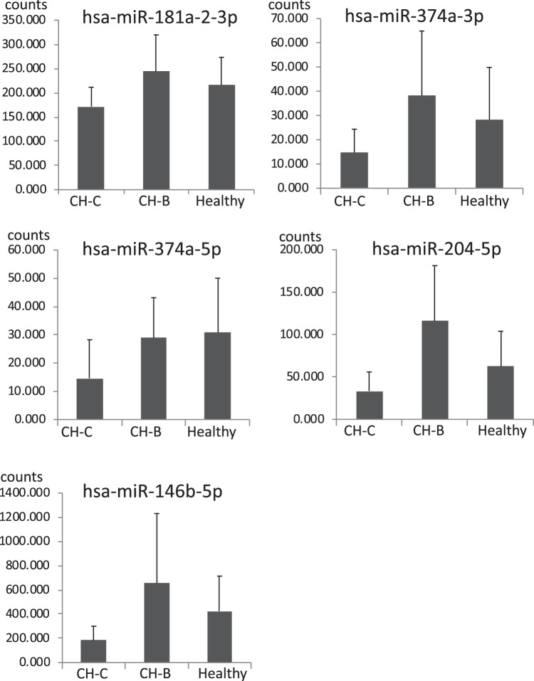
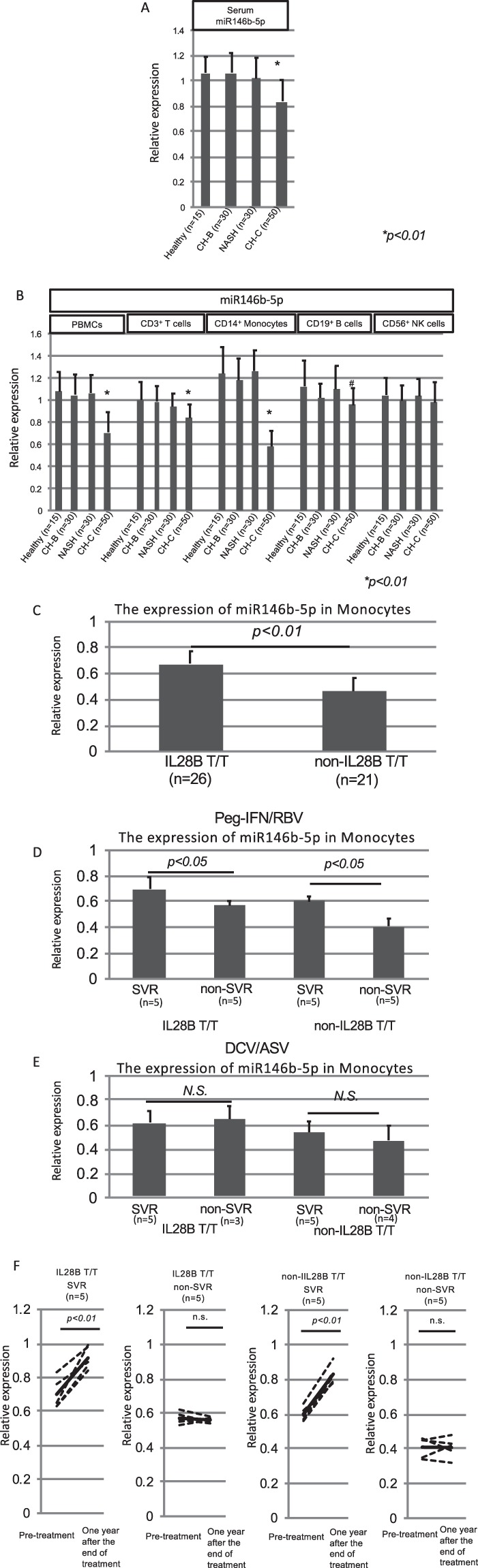
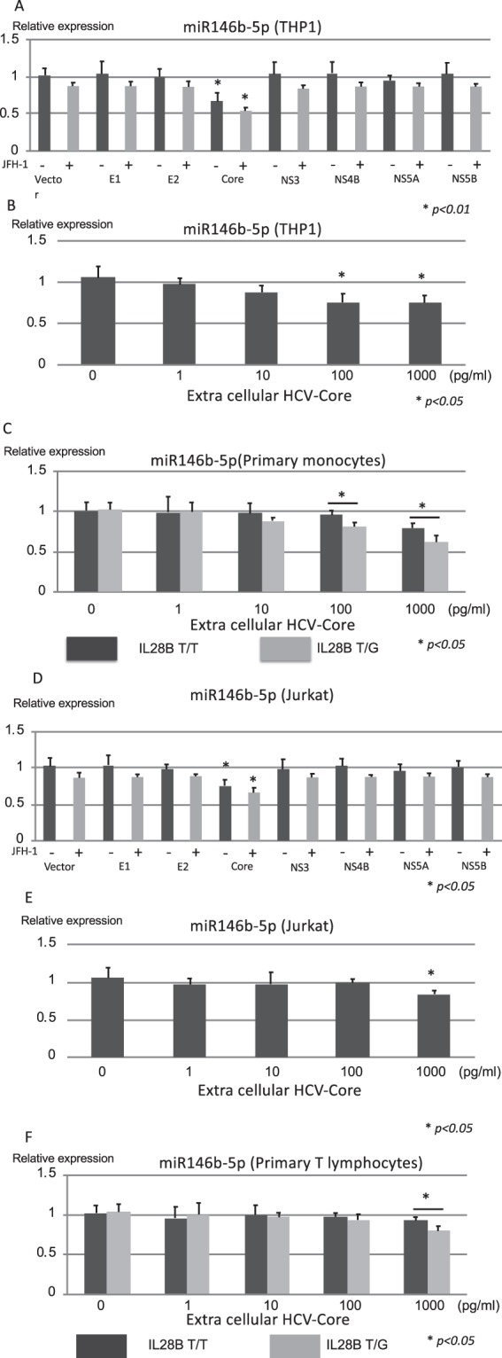
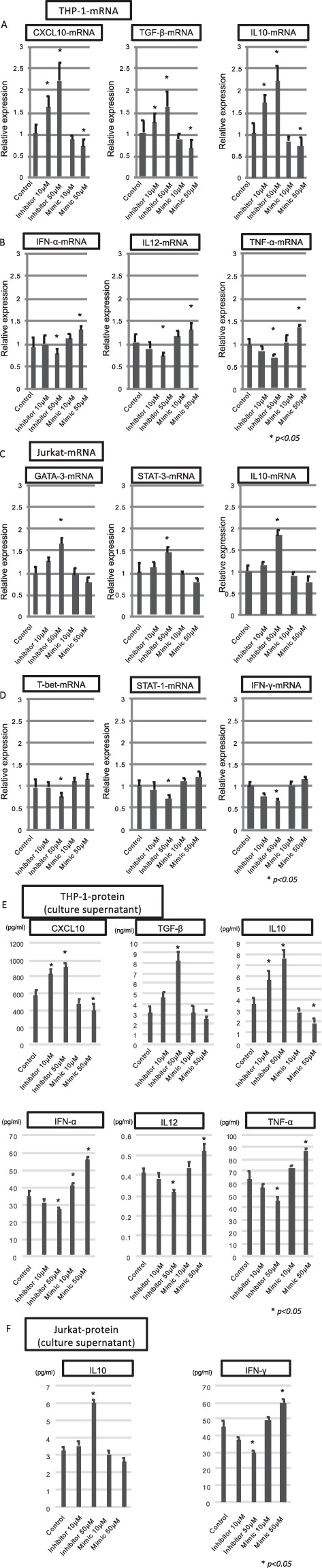
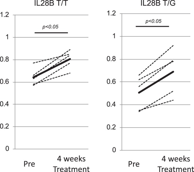
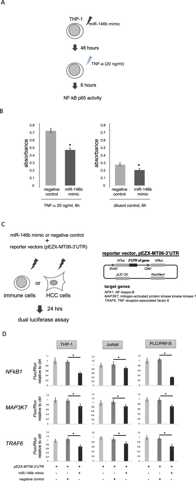
It has been reported that various kinds of miRNAs could affect the pathogenesis of hepatitis C virus infection. Recently, our group reported that deep-sequencing analysis was useful to detect disease-specific miRNAs. The aim of this study is to identify the HCV-specific miRNAs that could contribute to the immunopathogenesis of HCV by using clinical samples and in vitro analysis. Five miRNAs (hsa-miR181a-2-3p, hsa-miR-374a-3p, hsa-miR374a-5p, hsa-miR-204-5p and hsa-miR146b-5p) were shown to be significantly downregulated in CH-C by deep sequence analysis. The average ratio (PBMCs miRNAs/serum miRNAs) of hsa-miR146b-5p was highest among all the miRNAs. Moreover, serum hsa-miR146b-5p was significantly down-regulated in CH-C patients in comparison to CH-B patients and healthy subjects. The expression of hsa-miR146b-5p in CD3+ T cells and CD14+ monocytes of CH-C patients was significantly lower than that of the other groups. The hsa-miR146b-5p expression in CD14+ monocytes of SVR patients treated with Peg-IFN/RBV was significantly higher than in those of non-SVR patients treated with Peg IFN/RBV. However, the hsa-miR146b-5p expression in CD14+ monocytes of SVR patients treated with DCV and ASV was comparable to that in monocytes of non-SVR patients treated with DCV and ASV. Moreover, the expression levels of hsa-miR146b-5p in CD14+ monocytes were significantly increased after achieving SVR and 1(OH)Vitamin D3 treatment. Further, the expression of HCV-Core could suppress miR146b-5p expression in immune cells and affect the expression of various kinds of cytokines by affecting the NF-κB signaling. In conclusion, the reduction of miR146b-5p in monocytes and T cells could contribute to the immunopathogenesis of hepatitis C virus infection.


Conflict of interest statement

The authors declare no competing interests.

Figures

Figure 1
Figure 1
The scheme of this study is shown in this figure. A comprehensive, validation and functional analysis was carried out in this study.
Figure 2
Figure 2
The CH-C specific suppression of miRNAs in the serum. This figure shows the five miRNAs that had significantly lower expression in CH-C patients in comparison to those in the other groups. Y-axis indicates the counts of miRNAs. Error-bars indicate standard deviation.
Figure 3
Figure 3
The validation analysis of miR146b-5p expression and the identification of responsive immune cells producing hsa-miR146b-5p. The quantification of miR146b-5p in the serum was carried out to validate the comprehensive analysis using real-time PCR. The Cel-miR-39-3p was spiked in the serum for the control miRNA. The relative expressions of miR146b-5p are shown in the Y-axis. One healthy subject indicated one relative expression. We normalized the other subjects using the relative expression (A). Then, we analyzed the expression of hsa-miR146b-5p in various kinds of isolated immune cells (PBMCs, CD3+ T cells, CD14+ monocytes, CD19+ B cells and CD56+ NK cells). The quantification of miR146b-5p in the various kinds of cells was carried out using real-time PCR. One PBMC sample in a healthy subject indicated one relative expression. Then, we normalized the other samples using the relative expression. The relative expressions of miR146b-5p are shown in the Y-axis (B). A comparison of hsa-miR146b-5p expression in monocytes between IL28B T/T (n = 26) and non-IL-28B T/T (n = 21) patients was carried out (C). A comparison of hsa-miR146b-5p expression between the SVR patients (n = 10) and non-SVR patients (n = 10) after receiving PEG-IFN/RBV treatment was carried out (D). A comparison of hsa-miR146b-5p expression between the SVR patients (n = 10) and non-SVR patients (n = 7) after receiving DCV/ASV treatment was carried out (E). Error-bars indicate standard deviation. The expression levels of hsa-miR146b-5p in CD14+ monocytes were compared between before and after achieving SVR. (F) The relative expressions of miR146b-5p are shown in the Y-axis.
Figure 4
Figure 4
The HCV-antigen responsible for suppressing the expression of hsa-miR146b-5p in monocytes and T cells. The relative expression of miR146b-5p in THP-1 (A) and Jurkat (D) cells is shown after the transfection of various kinds of HCV antigen expressing plasmids (HCV-core, E1, E2, NS3, NS4B, NS5A and NS5B) with or without JFH-1 full length strain. The relative expressions of miR146b-5p in THP-1 (B) and Jurkat (E) cells are shown after adding the extra-cellular HCV-core protein. The relative expressions of hsa-miR146b-5p in CD14+ monocytes (C) and CD3+ T cells (F) from IL28B T/T subjects and IL28B T/G subjects were analyzed after adding the extra-cellular HCV-core protein. Error-bars indicate standard deviation.
Figure 5
Figure 5
The biological effects of hsa-miR146b-5p in monocytes and T cells. CXCL10, TGF-β and IL10 produced from CD14+ monocytes were representative cytokines that could induce favorable effects for eradicating HCV. The expressions of CXCL10-mRNA, TGF-β-mRNA and IL10-mRNA in THP-1 cells are shown after the transfection of the inhibitor or mimic of miR146b-5p (A). IFN-α, IL12, and TNF-α produced from CD14+ monocytes were representative cytokines that could induce a favorable effect to eradicate HCV. The expressions of IFN-α, IL12, and TNF-α in THP-1 cells are shown after the transfection of the inhibitor or mimic of miR146b-5p (B). GATA-3-mRNA, STAT-3-mRNA and IL10-mRNA expressed in T cells were representative factors that could induce unfavorable effects for eradication of HCV. The expressions of GATA-3-mRNA, STAT-3-mRNA and IL10-mRNA in Jurkat cells are shown after the transfection of inhibitor or mimic of miR146b-5p (C). T-bet-mRNA, STAT-1-mRNA and IFN-γ-mRNA expressed in T cells were representative factors that could induce favorable effects for eradicating HCV. The expressions of T-bet-mRNA, STAT-1-mRNA and IFN-γ in Jurkat cells are shown after the transfection of inhibitor or mimic of miR146b-5p (D). The amounts of CXCL10, TGF-β, IL10, IFN-α, IL12, and TNF-α in the culture supernatant of THP-1 cells are shown after the transfection of the inhibitor or mimic of miR146b-5p (E). The amounts of IL10 and IFN-γ in the culture supernatant of Jurkat cells were shown after the transfection of the inhibitor or mimic of miR146b-5p (F).Error-bars indicate standard deviation.
Figure 6
Figure 6
The effect of 1(OH)ViaminD3 treatment on the expression of hsa-miR146b-5p in CH-C patients. The expression of hsa-miR146b-5p in monocytes before and after treatment with 1(OH)Vitamin D3 is shown. The relative expression of hsa-miR146b-5p in monocytes is shown on the Y-axis. One healthy subject indicated one relative expression. Then, we normalized the other subjects using the relative expression.
Figure 7
Figure 7
NF-kB transcription assay and reporter assay. THP-1 cells were electroporated with 300 nM of miR-146b mimic or negative control (Ambion) and treated with TNF-α (20 ng/ml) or diluent control for 6 hours (A). Nuclear extracts from the cells were prepared using a TransAM Nuclear Extract Kit (Active Motif). The transcription activity of NF-kB was measured using an NF-kB p65 transcription factor assay kit (Active Motif). Bars represent mean absorbance of three determinants ± SE. *p < 0.05 (B). THP-1, Jurkat and PLC/PRF/5 cells were electroporated with pEZX-MT06 target reporter vectors containing 3′ UTR of NFKB1, MAP3K7 and TRAF6 (GeneCopoeia) together with miR-146b mimic or negative control (Ambion) (C). After 24 hours, luciferase assay was performed using Luc-Pair Luciferase Assay Kit 2.0 (GeneCopoeia). Luminescence of the firefly luciferase was normalized using the Renilla luciferase. Relative luminescence of a reporter vector co-transfected with miR-146b mimic was calculated as a ratio for negative control. Bars represent mean relative luminescence of five determinants ± SE. *p < 0.05 (D).

References

    1. Alter MJ, et al. The prevalence of hepatitis C virus infection in the United States, 1988 through 1994. The New England journal of medicine. 1999;341:556–562. doi: 10.1056/nejm199908193410802. - DOI - PubMed
    1. Park SH, Rehermann B. Immune responses to HCV and other hepatitis viruses. Immunity. 2014;40:13–24. doi: 10.1016/j.immuni.2013.12.010. - DOI - PMC - PubMed
    1. Aleman S, et al. A risk for hepatocellular carcinoma persists long-term after sustained virologic response in patients with hepatitis C-associated liver cirrhosis. Clinical infectious diseases: an official publication of the Infectious Diseases Society of America. 2013;57:230–236. doi: 10.1093/cid/cit234. - DOI - PubMed
    1. Asahina Y, et al. alpha-fetoprotein levels after interferon therapy and risk of hepatocarcinogenesis in chronic hepatitis C. Hepatology (Baltimore, Md.) 2013;58:1253–1262. doi: 10.1002/hep.26442. - DOI - PubMed
    1. Chayama Kazuaki, Notsumata Kazuo, Kurosaki Masayuki, Sato Ken, Rodrigues Lino, Setze Carolyn, Badri Prajakta, Pilot-Matias Tami, Vilchez Regis A., Kumada Hiromitsu. Randomized trial of interferon- and ribavirin-free ombitasvir/paritaprevir/ritonavir in treatment-experienced hepatitis C virus-infected patients. Hepatology. 2015;61(5):1523–1532. doi: 10.1002/hep.27705. - DOI - PMC - PubMed

Publication types