Review
. 2019 Sep 12:10:22.
doi: 10.1186/s13227-019-0133-6. eCollection 2019.

Peripheral sensory neurons govern development of the nervous system in bivalve larvae

Affiliations
Review

Peripheral sensory neurons govern development of the nervous system in bivalve larvae

Olga V Yurchenko et al. Evodevo. .

Abstract

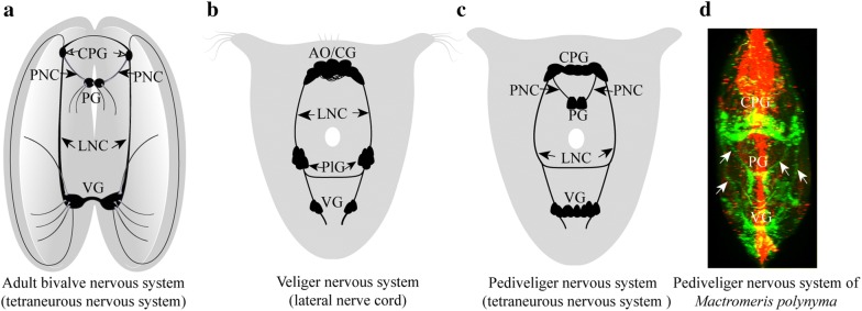
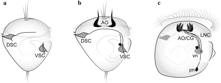
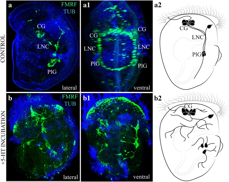
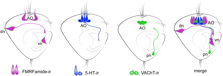
Recent findings regarding early lophotrochozoan development have altered the conventional model of neurogenesis and revealed that peripheral sensory elements play a key role in the initial organization of the larval nervous system. Here, we describe the main neurogenetic events in bivalve mollusks in comparison with other Lophotrochozoa, emphasizing a novel role for early neurons in establishing larval nervous systems and speculating about the morphogenetic function of the apical organ. We demonstrate that during bivalve development, peripheral sensory neurons utilizing various transmitters differentiate before the apical organ emerges. The first neurons and their neurites serve as a scaffold for the development of the nervous system. During veliger stage, cerebral, pleural, and visceral ganglia form along the lateral (visceral) nerve cords in anterior-to-posterior axis. The pedal ganglia and corresponding ventral (pedal) nerve cords develop much later, after larval settlement and metamorphosis. Pharmacological abolishment of the serotonin gradient within the larval body disrupts the navigation of "pioneer" axons resulting in malformation of the whole nervous system architecture. Comparative morphological data on neurogenetic events in bivalve mollusks shed new light on the origin of the nervous system, mechanisms of early axon navigation, and sequence of the tetraneurous nervous system formation. Furthermore, this information improves our understanding of the basic nervous system architecture in larval Bivalvia and Mollusca.

Keywords: Ganglia; Mollusk; Nerve cords; Neurotransmitters; Tetraneuralia.


Conflict of interest statement

Competing interestsThe authors declare that they have no competing interests.

Figures

Fig. 1
Fig. 1
Architecture of the nervous system of adult bivalves (a) and larvae before and after metamorphosis (bd). a Tetraneurous nervous system (ventral + pedal cords) of the adult bivalve. b Nervous system of bivalve larvae. c The tetraneurous bivalve nervous system after settlement/metamorphosis. d Nervous system of a mactrid juvenile Mactromeris polynyma from plankton as visualized by whole-mount immunostaining for FMRFamide (green) and acetylated alpha-tubulin (red) (the arrows indicate the lateral nerve cord and ventral (pedal) nervous cords). CPG cerebropleural ganglion, PNC pedal nervous cord, PG pedal ganglion, LNC lateral nerve cord, VG visceral ganglion, AO/CG apical organ/cerebral ganglion, PlG pleural ganglion
Fig. 2
Fig. 2
Neurogenesis of the oyster Crassostrea gigas: FMRF-immunoreactive nervous system. a Early neurons that appear in the dorsal (DSC) and ventral (VSC) peripheral sensory centers, before appearance of transmitters in the apical organ (AO). b, c They provide morphogenetic scaffolding for the anlagen of the lateral nerve cords (LNC) during oyster development. vn ventral neurons, pn posterior neurons (Schematic drawings adapted by Olga Kharchenko from Yurchenko et al. [14])
Fig. 3
Fig. 3
Schematic depiction of the location of early sensory elements in representative members of different lophotrochozoan groups. The long axons of early peripheral neurons (gray color) are arranged similarly in two parallel nerve bundles along the ventral side of the larvae, where the anlagen of the lateral nerve cords will later form. a Larvae of the mussel Mytilus trossulus and the chiton Ischnochiton hakodadensis; b Larva of the oyster Crassostrea gigas; c Larva of the gastropods Tritonia diomedea, Aplysia californica, and Lymnaea stagnalis; d Larva of the annelids, Phyllodoce maculata and Platynereis dumerilii
Fig. 4
Fig. 4
Effect of serotonin on the growth of FMRFamide-immunoreactive processes during development of the mollusk Mytilus trossulus. Mussel trochophores (32 hpf) were cultured in filtered seawater in the presence of 5-HT (10−6 M) or without any additives (the control). Larvae were collected at the veliger stage (60 hpf) and subjected to whole-mount immunostaining for FMRFamide (green) and acetylated alpha-tubulin (blue) as described earlier [14]. ab2 right lateral view of untreated (a, a2) and 5-HT-treated (b, b2) larvae. a1, b1 Ventral view of untreated and 5-HT-treated larvae. Note the enhanced sprouting and absence of compact ventral neurite bundles in the 5-HT-treated larvae. CG cerebral ganglion, LNC lateral nerve cord, PlG pleural ganglion
Fig. 5
Fig. 5
Arrangement of neurons expressing different transmitters at the veliger stage of the bivalve mollusk Crassostrea gigas. Only the right side of the body is shown. FMRFamide-immunoreactive fibers first grow dorso-ventrally and then turn posteriorly. 5-HT-ir fibers grow in anterior-to-posterior direction from the apical organ (AO), while VAChT-ir grows in the posterior-to-anterior direction towards the AO. Altogether, these processes form a compact neurite bundle along the ventral side of the larval body. The right and left bundles run in parallel on the respective sides of the larval body, organizing rudiments of the paired lateral nerve cords. AO apical organ, dn dorsal neurons, vn ventral neurons, pn posterior neurons (Schematic drawings adapted by Olga Kharchenko from Yurchenko et al. [14])

References

    1. Erdmann W. Nr. 5. Ober die Entwicklung und die Anatomie der ansatzreifen Larve von Ostrea edulis mit Bemerkungen iiber die Lebensgeschichte der Auster. Untersuchungen iiber die Lebensgeschichte der Auster; 1934. p. 1–25.
    1. Nielsen C. Trochophora larvae: cell lineages, ciliary bands, and body regions. 1. Annelida and Mollusca. J Exp Zool B Mol Dev Evol. 2004;302:35–68. doi: 10.1002/jez.b.20001. - DOI - PubMed
    1. Voronezhskaya EE, Nezlin LP, Odintsova NA, Plummer JT, Croll RP. Neuronal development in larval mussel Mytilus trossulus (Mollusca: Bivalvia) Zoomorphology. 2008;127:97–110. doi: 10.1007/s00435-007-0055-z. - DOI
    1. Wanninger A. Evolutionary developmental biology of invertebrates 2: lophotrochozoa spiralia. Wien: Springer; 2015.
    1. Martín-Durán JM, Pang K, Børve A, Lê HS, Furu A, Cannon JT, Jondelius U, Hejnol A. Convergent evolution of bilaterian nerve cords. Nature. 2018;553:45–50. doi: 10.1038/nature25030. - DOI - PMC - PubMed

LinkOut - more resources