

Assessment of the changes in corneal biomechanical properties after collagen cross-linking in patients with keratoconus
- PMID: 31528759
- PMCID: PMC6742757
- DOI: 10.1016/j.joco.2019.02.002
Assessment of the changes in corneal biomechanical properties after collagen cross-linking in patients with keratoconus
Abstract
Purpose: To assess the changes in biomechanical properties of the cornea after treatment of keratoconus patients with UV-A/riboflavin corneal collagen cross-linking (CXL) using Corvis ST (Oculus, Wetzlar, Germany) and Ocular Response Analyzer (ORA; Reichert Ophthalmic Instruments, Inc., Buffalo, NY, USA) devices.
Methods: In this prospective, observational case series, 48 eyes from 48 consecutive patients with progressive keratoconus were enrolled. Patients with history or signs of ocular disorders other than keratoconus, previous eye surgery, systemic diseases, or inability to cooperate with any measurement device were excluded. Corvis ST and ORA images were obtained at baseline and 4 months after CXL. The primary outcome measures comprised Corvis ST corneal biomechanical factors [time of highest concavity (T), time of applanation 1 (T1), time of applanation 2 (T2), length of applanation 1 (L1), length of applanation 2 (L2), velocity of applanation 1 (V1), velocity of applanation 2 (V2), deformation amplitude (DA), peak distance (PD), and radius (R)] and the ORA parameters [corneal hysteresis (CH), corneal resistance factor (CRF), Goldmann-related IOP (IOPg), cornea-compensated IOP (IOPcc), and waveform score (WS)].
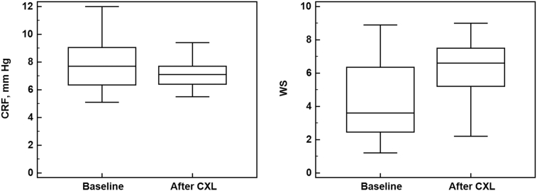
Results: The mean [± standard deviation (SD)] age of patients was 20 ± 5 years, and 27 (56%) were male. At baseline, the averages of the refraction, mean keratometry, and keratometric astigmatism were -3.0 ± 1.8 diopter (D), 47.0 ± 1.8 D, and 3.5 ± 1.5 D, respectively. According to Corvis ST, L2 increased from 0.83 ± 0.25 mm at baseline to 1.15 ± 0.57 mm after CXL; and V2 decreased from -0.81 ± 0.08 to -0.94 ± 0.26 m/s (P = 0.001 and P = 0.032, respectively). ORA parameters showed significant decrease in the CRF (from 7.82 ± 1.72 to 7.21 ± 1.05 mmHg; P = 0.036) and increase in the WS (from 4.58 ± 2.55 to 6.12 ± 1.92; P = 0.002).
Conclusions: According to in vivo observation with Corvis ST and ORA, CXL induces significant changes in corneal biomechanical properties in cases with keratoconus. The parameters with significant changes (L2 and V2) may reflect increased stiffness of the treated cornea. The importance of such observations should be elucidated in future studies.
Keywords: Corneal biomechanics; Corneal collagen cross-linking; Corvis ST; Ocular response analyzer.
Figures

References
-
- Krachmer J.H., Feder R.S., Belin W.M. Keratoconus and related noninflammatory corneal disorders. Surv Ophthalmol. 1984;28(4):293–322. - PubMed
-
- Kenney C., Nesburn A., Burgeson E.R., Butkowski J.R., Ljubimov A. Abnormalities of the extracellular matrix in keratoconus corneas. Cornea. 1997;16(3):345–351. - PubMed
-
- Sawaguchi S., Yue B.Y., Sugar J., Gilboy J.E. Lysosomal enzyme abnormalities in keratoconus. Arch Ophthalmol. 1989;107(10):1507–1510. - PubMed
-
- Rehany U., Lahav M., Shoshan S. Collagenolytic activity in keratoconus. Ann Ophthalmol. 1982;14(8):751–754. - PubMed
-
- Kao W.W., Vergnes J.P., Ebert J., Sundar-Raj C.V., Brown S.I. Increased collagenase and gelatinase activities in keratoconus. Biochem Biophys Res Commun. 1982;107(3):929–936. - PubMed
LinkOut - more resources
Full Text Sources
