

In vivo identification of small molecules mediating Gpr126/Adgrg6 signaling during Schwann cell development
- PMID: 31529518
- PMCID: PMC7189964
- DOI: 10.1111/nyas.14233
In vivo identification of small molecules mediating Gpr126/Adgrg6 signaling during Schwann cell development
Abstract
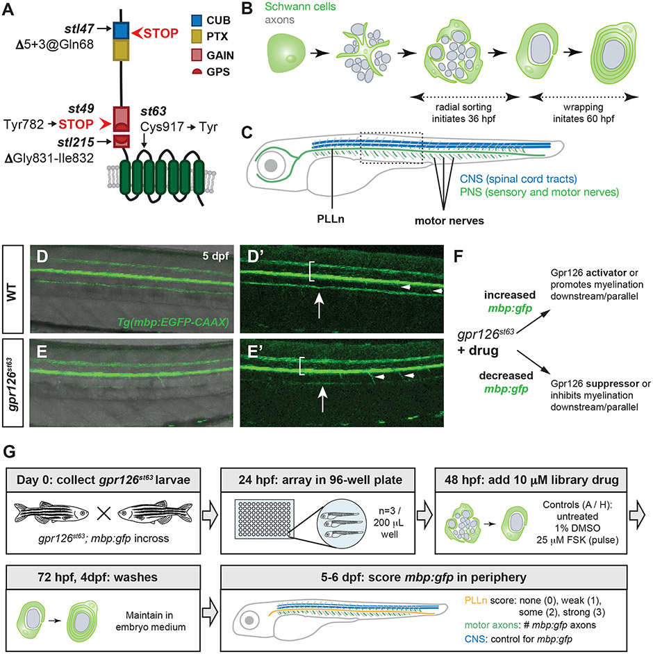
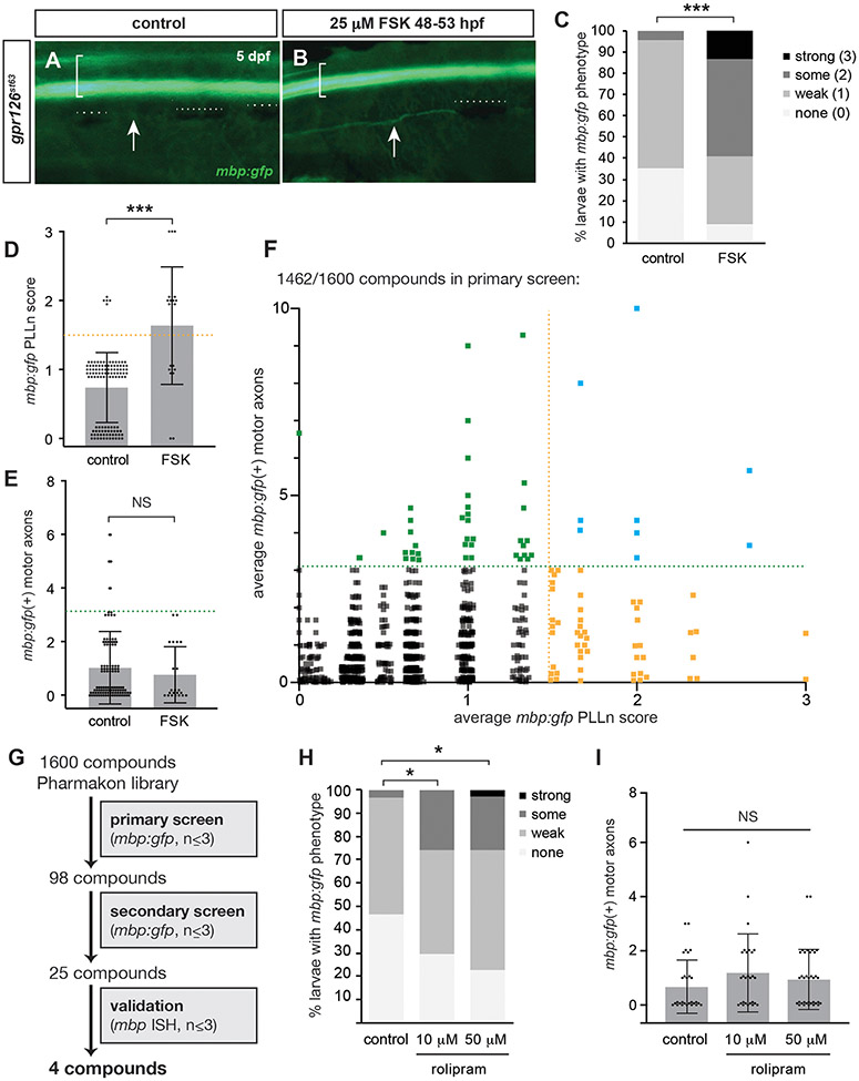
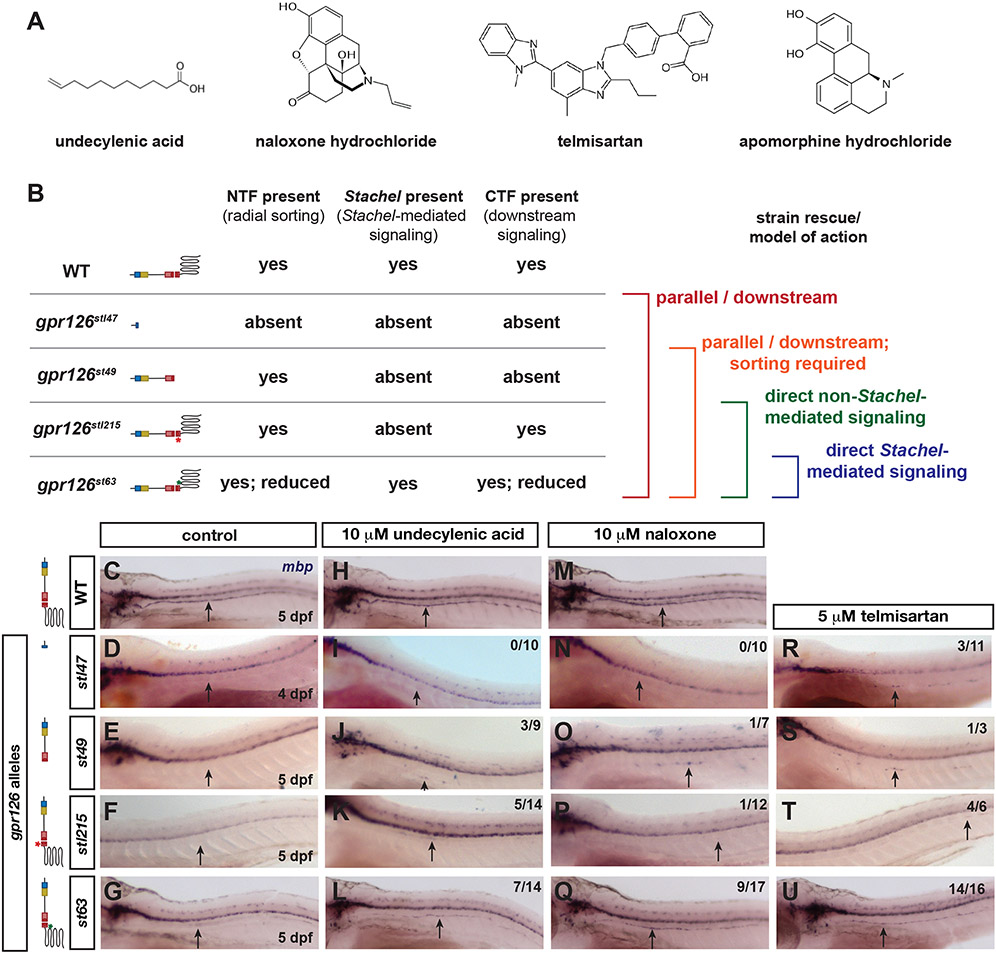
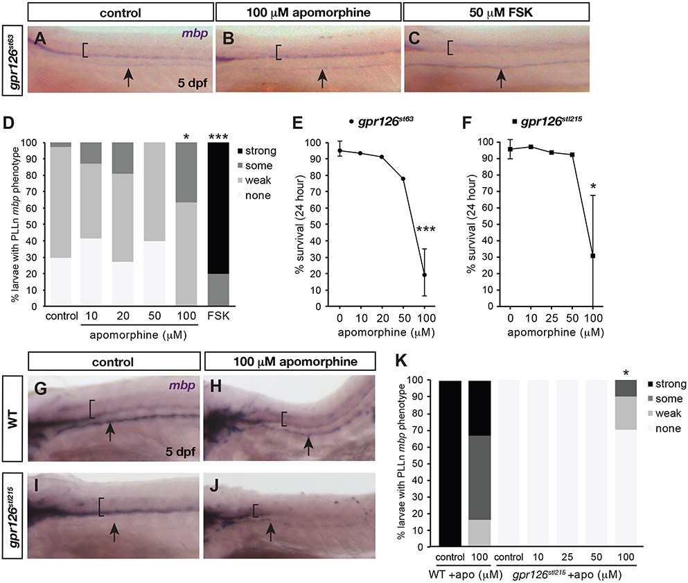
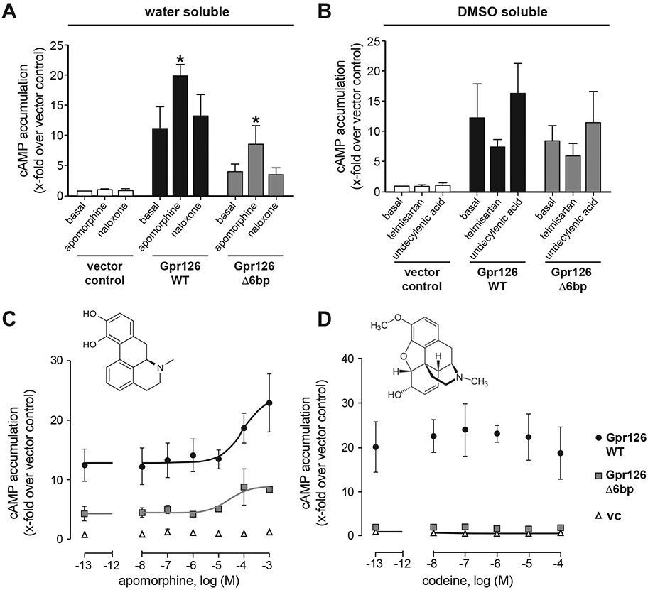
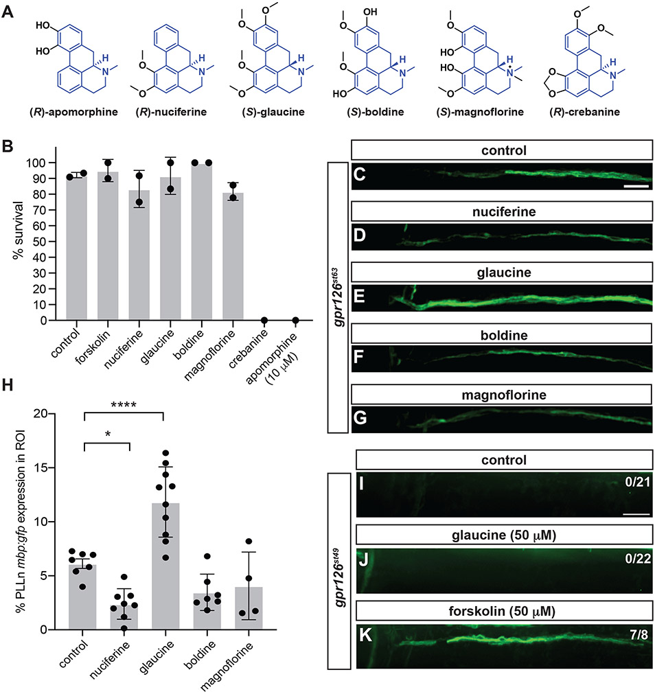
Gpr126/Adgrg6, an adhesion family G protein-coupled receptor (aGPCR), is required for the development of myelinating Schwann cells in the peripheral nervous system. Myelin supports and insulates vertebrate axons to permit rapid signal propagation throughout the nervous system. In mammals and zebrafish, mutations in Gpr126 arrest Schwann cells at early developmental stages. We exploited the optical and pharmacological tractability of larval zebrafish to uncover drugs that mediate myelination by activating Gpr126 or functioning in parallel. Using a fluorescent marker of mature myelinating glia (Tg[mbp:EGFP-CAAX]), we screened hypomorphic gpr126 mutant larvae for restoration of myelin basic protein (mbp) expression along peripheral nerves following small molecule treatment. Our screens identified five compounds sufficient to promote mbp expression in gpr126 hypomorphs. Using an allelic series of gpr126 mutants, we parsed the ability of small molecules to restore mbp, suggesting differences in drug efficacy dependent on Schwann cell developmental state. Finally, we identify apomorphine hydrochloride as a direct small molecule activator of Gpr126 using combined in vivo/in vitro assays and show that aporphine class compounds promote Schwann cell development in vivo. Our results demonstrate the utility of in vivo screening for aGPCR modulators and identify small molecules that interact with the gpr126-mediated myelination program.
Keywords: Gpr126/Adgrg6; Schwann cells; adhesion GPCR; myelin; zebrafish.
© 2019 New York Academy of Sciences.
Conflict of interest statement
COMPETING INTERESTS
The authors declare no conflicts of interest.
Figures
Similar articles
-
The adhesion GPCR Adgrg6 (Gpr126): Insights from the zebrafish model.Genesis. 2021 Apr;59(4):e23417. doi: 10.1002/dvg.23417. Epub 2021 Mar 18. Genesis. 2021. PMID: 33735533 Free PMC article. Review.
-
A screen of pharmacologically active compounds to identify modulators of the Adgrg6/Gpr126 signalling pathway in zebrafish embryos.Basic Clin Pharmacol Toxicol. 2023 Oct;133(4):364-377. doi: 10.1111/bcpt.13923. Epub 2023 Jul 16. Basic Clin Pharmacol Toxicol. 2023. PMID: 37394692 Free PMC article.
-
Identification of compounds that rescue otic and myelination defects in the zebrafish adgrg6 (gpr126) mutant.Elife. 2019 Jun 10;8:e44889. doi: 10.7554/eLife.44889. Elife. 2019. PMID: 31180326 Free PMC article.
-
Analysis of Gpr126 function defines distinct mechanisms controlling the initiation and maturation of myelin.Development. 2013 Aug;140(15):3167-75. doi: 10.1242/dev.093401. Epub 2013 Jun 26. Development. 2013. PMID: 23804499 Free PMC article.
-
New insights into signaling during myelination in zebrafish.Curr Top Dev Biol. 2011;97:1-19. doi: 10.1016/B978-0-12-385975-4.00007-3. Curr Top Dev Biol. 2011. PMID: 22074600 Free PMC article. Review.
Cited by
-
The adhesion GPCR Adgrg6 (Gpr126): Insights from the zebrafish model.Genesis. 2021 Apr;59(4):e23417. doi: 10.1002/dvg.23417. Epub 2021 Mar 18. Genesis. 2021. PMID: 33735533 Free PMC article. Review.
-
Adhesion GPCRs as a paradigm for understanding polycystin-1 G protein regulation.Cell Signal. 2020 Aug;72:109637. doi: 10.1016/j.cellsig.2020.109637. Epub 2020 Apr 16. Cell Signal. 2020. PMID: 32305667 Free PMC article. Review.
-
A screen of pharmacologically active compounds to identify modulators of the Adgrg6/Gpr126 signalling pathway in zebrafish embryos.Basic Clin Pharmacol Toxicol. 2023 Oct;133(4):364-377. doi: 10.1111/bcpt.13923. Epub 2023 Jul 16. Basic Clin Pharmacol Toxicol. 2023. PMID: 37394692 Free PMC article.
-
Mechanisms of adhesion G protein-coupled receptor activation.J Biol Chem. 2020 Oct 9;295(41):14065-14083. doi: 10.1074/jbc.REV120.007423. Epub 2020 Aug 6. J Biol Chem. 2020. PMID: 32763969 Free PMC article. Review.
-
Structural basis for the tethered peptide activation of adhesion GPCRs.Nature. 2022 Apr;604(7907):763-770. doi: 10.1038/s41586-022-04619-y. Epub 2022 Apr 13. Nature. 2022. PMID: 35418678
References
-
- Arac D, Aust G, Calebiro D, et al. 2012. Dissecting signaling and functions of adhesion G protein-coupled receptors. Annals of the New York Academy of Sciences. 1276: 1–25. - PubMed
-
- Langenhan T, Aust G & Hamann J. 2013. Sticky signaling--adhesion class G protein-coupled receptors take the stage. Science signaling. 6: re3. - PubMed
-
- Jessen KR & Mirsky R. 2005. The origin and development of glial cells in peripheral nerves. Nat Rev Neurosci. 6: 671–682. - PubMed
-
- Woodhoo A & Sommer L. 2008. Development of the Schwann cell lineage: from the neural crest to the myelinated nerve. Glia. 56: 1481–1490. - PubMed
Publication types
MeSH terms
Substances
Grants and funding
- DGE‐1745038/National Science Foundation/International
- Medical Faculty of the University of Leipzig/International
- R01 NS079445/NH/NIH HHS/United States
- R01 HD080601/NH/NIH HHS/United States
- BMBF/International
- CRC 1052 B06/German Research Foundation/International
- Research Unit FOR 2149/German Research Foundation/International
- European Social Fund and the Free State of Saxony/International
- 266061011/German Research Foundation/International
- R01 HD080601/HD/NICHD NIH HHS/United States
- R01 NS079445/NS/NINDS NIH HHS/United States
- Collins Medical Trust/International
- 266022790/German Research Foundation/International
- 209933838/German Research Foundation/International
LinkOut - more resources
Full Text Sources
Molecular Biology Databases
Miscellaneous
