. 2019 Nov;18(21):3010-3029.
doi: 10.1080/15384101.2019.1667189. Epub 2019 Sep 18.

Blocking Notch signal pathway suppresses the activation of neurotoxic A1 astrocytes after spinal cord injury

Affiliations

Blocking Notch signal pathway suppresses the activation of neurotoxic A1 astrocytes after spinal cord injury

Dingfei Qian et al. Cell Cycle. 2019 Nov.

Abstract

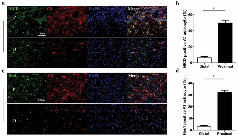
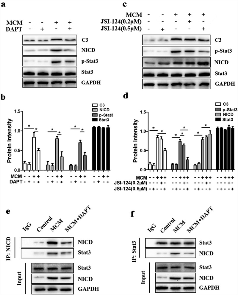
Spinal cord injury (SCI) is a catastrophic disease which has complicated pathogenesis including inflammation, oxidative stress and glial scar formation. Astrocytes are the most abundant cells in central nervous system and fulfill homeostatic functions. Recent studies have described a new reactive phenotype of astrocytes, A1, induced by inflammation, which may have negative effects in SCI. As the Notch signaling pathway has been linked to cell differentiation and inflammation, we aimed to investigate its potential role in the differentiation of astrocytes in SCI. Contusive SCI rat model showed elevated A1 astrocyte numbers at the damage site 28 days after SCI and the expression levels of Notch signaling and its downstream genes were upregulated parallelly. Western blotting, RT-qPCR and immunofluorescence revealed that blocking of Notch pathway using γ-secretase blocker (DAPT) suppressed the differentiation of A1 astrocytes. Flow cytometry, and TUNEL staining indicated that DAPT alleviated neuronal apoptosis and axonal damage caused by A1 astrocytes likely through the Notch-dependent release of pro-inflammatory factors. CO-IP and western blotting revealed an interaction between Notch pathway and signal transducer and activator of transcription 3 (Stat3), which played a vital role in differentiation of A1 astrocytes. We conclude that phenotypic transition of A1 astrocytes and their neurotoxity were controlled by the Notch-Stat3 axis and that Notch pathway in astrocytes may serve as a promising therapeutic target for SCI.

Keywords: A1 astrocytes; DAPT; Notch signaling; Spinal cord injury; Stat3.


Figures

Figure 1.
Figure 1.
Increased A1 astrocyte numbers at the lesion site 28 days following experimental spinal cord injury (SCI) in rats. (a) Representative images of rat spinal cord sections immunohistochemically stained for the A1 astrocyte marker complement component 3 (C3, red) and the general reactive astrocyte marker glial fibrillary acid protein (GFAP, green) 3 and 28 days post-SCI. Cell nuclei were counterstained with DAPI (blue). The upper row shows the proximal (P) end of the lesion and the lower row shows the distal (d) uninjured area for each treatment group. C3-positive astrocytes were more numerous in the lesion area on day 28 post-injury (Virtual coil) compared to day 3 (Arrow). Scale bar, 200 µm. (b) Quantification of C3 immunofluorescence intensity. Immunoexpression of C3 was significantly elevated at the lesion site on day 28 post-injury compared to uninjured spinal cord. *p < 0.05. (c) Representative western blots of C3 expression levels at the lesion area on days 3 and 28 post-injury compared to uninjured spinal cord. (d) Semi-quantification of relative C3 protein expression (normalized to GAPDH expression). *p < 0.05.
Figure 2.
Figure 2.
Activation of Notch signaling in A1 astrocytes at the lesion site following SCI. (a, c) Representative images of spinal cord sections immunohistochemically stained for C3 (red), the Notch transcriptional regulator notch intracellular domain (NICD, green), and the NICD target gene Hes1 (green) in A1 astrocytes at the lesion area. Cell nuclei were counterstained with DAPI (blue). P in images represents the proximal lesion area and D the distal and uninjured area. Scale bar, 100 µm. (b) The proportions of NICD-positive A1 astrocytes at the distal and proximal ends of the lesion, *p < 0.05. (d) The proportion of Hes1-positive A1 astrocytes at the distal and proximal ends of the lesion, *p < 0.05.
Figure 3.
Figure 3.
Notch signaling induction and reactive transformation of astrocytes by microglial conditioned medium (MCM) in vitro. (a) Representative images of astrocyte cultures immunostained for GFAP (green) and Notch-1 (red) (with nuclear counterstaining by DAPI, blue) after 24 h in MCM. Notch-1 protein expression in A1 astrocytes was markedly enhanced by MCM treatment compared to untreated astrocytes. Scale bar, 50 µm. (b) Representative images of astrocyte cultures immunostained for GFAP (green) and NICD (red) (with nuclear counterstaining by DAPI, blue) after 24 h in MCM. The expression of NICD was intensely augmented both in the cytoplasm and nucleus (arrows) compared to untreated control cultures. Scale bar, 50 µm. (c-e) RT-qPCR analysis of Notch-1, Hey1, and Hes-1 mRNA expression levels in primary astrocytes treated as indicated. GAPDH was used as the internal control. There were significant increases in Notch-1, Hey1, and Hes-1 mRNA expression after MCM treatment for 12, 24, and 48 h. *p < 0.05. (f) Western blot analysis of Notch-related proteins (Notch-1, NICD, Hey1, and Hes1) after MCM treatment for 24 and 48 h. (g, h) Semi-quantification (normalized to GAPDH) of Notch-related proteins showing that Notch signaling was activated in A1 astrocytes by MCM treatment for 24 and 48 h. In contrast, Notch activation was markedly lower in response to MCM plus the Notch inhibitor DAPT. *p < 0.05.
Figure 4.
Figure 4.
The Notch signaling inhibitor DAPT blocks MCM-induced activation of A1 astrocytes. (a) Representative images of astrocyte cultures immunostained for GFAP (green) and C3 (red) (with nuclear counterstaining by DAPI, blue) after 24 h in MCM or MCM+DAPT (10 μM). Expression of the A1 marker C3 was significantly enhanced by MCM but not MCM+DAPT. Scale bar, 100 µm. (b) Quantification of C3 fluorescence intensity in each treatment group, *p < 0.05. (c-e) RT-qPCR analysis of TNF-α, IL-1β, and IL-6 mRNA expression in primary astrocytes following the indicated treatment. GAPDH was used as the internal control. Pro-inflammatory mRNAs were significantly upregulated in A1 astrocytes by MCM but not MCM+DAPT (10 μM). *p < 0.05. (f,g) RT-qPCR measurements of A1 and A2 markers in astrocytes treated with MCM with or without DAPT (10 μM) for 24 h compared to untreated controls. DAPT reduced expression of the A1 markers Serping1, Ligp1, C3 and Amigo2. Alternatively, expression of the A2 markers, Slc10a6, Tm4sf1 and S100a10 was increased, but the difference did not reach statistical significance., *p < 0.05. (h) Western blot analysis of C3 protein expression in astrocytes following MCM or MCM+DAPT (10 μM) treatment for 24 h. (i) Semi-quantification (normalized to GAPDH) of C3 expression in astrocytes treated with MCM or MCM+DAPT. *p < 0.05.
Figure 5.
Figure 5.
Blockade of astrocytic Notch signaling reduces the neurotoxicity of astrocyte conditioned medium (ACM). (a) Annexin V-FITC/PI double staining and flow cytometry was used to detect neuronal apoptosis induced by astrocyte condition medium (ACM) for 24 h and by ACM generated in the presence of the Notch inhibitor DAPT (ACM+DAPT). (b) Quantitative results of flow cytometry that double positive neurons were counted demonstrated that DAPT pretreatment of astrocytes reduced ACM-induced apoptosis of primary neurons, *p < 0.05. (c) TUNEL staining (red) for detection of apoptosis in neurons cultured in ACM or ACM+DAPT for 24 h. Cell nuclei were counterstained with DAPI (blue). Scale bar, 100 µm. (d) Quantitative estimation of the proportion of apoptotic cells in each treatment group. Pretreatment of astrocytes with DAPT for ACM generation substantially reduced ACM-induced neuronal apoptosis. *p < 0.05. (e) Western blot analysis of neuronal apoptosis-related proteins in each treatment group. (f) Semi-quantitative analysis (normalized to GAPDH) showing that the increase in pro-apoptotic proteins Bax and cleaved-caspase 3 following ACM treatment for 24 h was substantially lower in the ACM+DAPT treatment group, whereas expression of the anti-apoptotic Bcl-2 was higher in the ACM+DAPT treatment group. *p < 0.05.
Figure 6.
Figure 6.
Blockade of astrocytic Notch signaling reduces the damaging effects of ACM on neuronal axons. (a) Top row: Bright-field images of primary neurons before and after treatment with ACM or ACM+DAPT for 24 h. Scale bar, 50 µm; Lower row: Immunofluorescence staining of MAP2 (green) and DAPI counterstaining (blue) in each treatment group. Scale bar, 20 µm. The conditioned medium of A1 astrocytes disrupts axon extension, an effect lost when ACM was generated in the presence of DAPT. (b,c) The effect of ACM and ACM+DAPT on axon length and axon branching in each group, *p < 0.05. (d) Quantification of MAP2 fluorescence intensity from (a). *p < 0.05. (e) RT-qPCR measures of MAP2 mRNA expression in primary neurons treated with ACM or ACM+DAPT. Note the substantial reduction in expression of this neuronal marker by ACM but not ACM+DAPT compared to untreated neuronal cultures. *p < 0.05.
Figure 7.
Figure 7.
Activation of A1 astrocytes is mediated by the Notch/Stat3 pathway. (a) Western blot analysis of C3, NICD, phosphorylated (p)-Stat3, and Stat3 expression levels in astrocytes treated with MCM or MCM+DAPT for 24 h. (b) Semi-quantitative analysis (normalized to GAPDH) of C3, NICD, p-Stat3, and Stat3 expression in each group. The expression levels of NICD and p-Stat3 were substantially increased by MCM but not by MCM+DAPT. *p < 0.05. (c) Western blot analysis of C3, NICD, p-Stat3, and Stat3 expression in astrocytes treated with MCM or MCM plus the Stat3 inhibitor JSI-124 (0.2 or 0.5 µM) for 24 h. (d) Semi-quantitative analysis (normalized to GAPDH) showing that JSI-124 decreased C3 and p-Stat3 expression but increased NICD expression, *p < 0.05. (e) Association of NICD and Stat3 in A1 astrocytes. Astrocyte lysates obtained from Control (untreated), MCM, and MCM+DAPT treatment groups were immunoprecipitated with NICD antibody and the bound proteins analyzed using a Stat3 antibody. IgG was used as an internal loading control. (f) Astrocyte lysates were immunoprecipitated with Stat3 antibody and the bound proteins were analyzed using a NICD antibody. IgG was used as an internal loading control. The interaction of Stat3 and NICD was significantly increased in MCM-treated A1 astrocytes.
Figure 8.
Figure 8.
Schematic model showing the possible interaction between Notch and Stat3 pathway. Notch pathway in astrocytes is activated by MCM, NICD is cleaved by γ-secretase from Notch1 and released into the cytoplasm, which can be blocked by DAPT. In the cytoplasm, NICD binds to Stat3 and promotes phosphorylation of Stat3. JSI-124 can inhibit the phosphorylation of Stat3. The complex of NICD and phosphorylated Stat3 enters the nucleus and activates the expression of A1 astrocyte related genes, which promotes the transformation of astrocytes to A1 phenotype. Figure was produced using Photoshop.

References

    1. Ahuja CS, Wilson JR, Nori S, et al. Traumatic spinal cord injury. Nat Rev Dis Primers. 2017;3:17018. - PubMed
    1. Singh A, Tetreault L, Kalsi-Ryan S, et al. Global prevalence and incidence of traumatic spinal cord injury. Clin Epidemiol. 2014;6:309–331. - PMC - PubMed
    1. Jazayeri SB, Beygi S, Shokraneh F, et al. Incidence of traumatic spinal cord injury worldwide: a systematic review. Eur Spine J. 2015;24:905–918. - PubMed
    1. Pineau I, Lacroix S.. Proinflammatory cytokine synthesis in the injured mouse spinal cord: multiphasic expression pattern and identification of the cell types involved. J Comp Neurol. 2007;500:267–285. - PubMed
    1. Milich LM, Ryan CB, Lee JK. The origin, fate, and contribution of macrophages to spinal cord injury pathology. Acta Neuropathol. 2019;137:785–797. - PMC - PubMed

MeSH terms

Substances