. 2019 Nov;78(11):1559-1565.
doi: 10.1136/annrheumdis-2019-215210. Epub 2019 Sep 17.

Evidence that tissue resident human enthesis γδT-cells can produce IL-17A independently of IL-23R transcript expression

Affiliations

Evidence that tissue resident human enthesis γδT-cells can produce IL-17A independently of IL-23R transcript expression

Richard James Cuthbert et al. Ann Rheum Dis. 2019 Nov.

Abstract

Objectives: Murine models of interleukin (IL)-23-driven spondyloarthritis (SpA) have demonstrated entheseal accumulation of γδT-cells which were responsible for the majority of local IL-17A production. However, IL-23 blockers are ineffective in axial inflammation in man. This study investigated γδT-cell subsets in the normal human enthesis to explore the biology of the IL-23/17 axis.

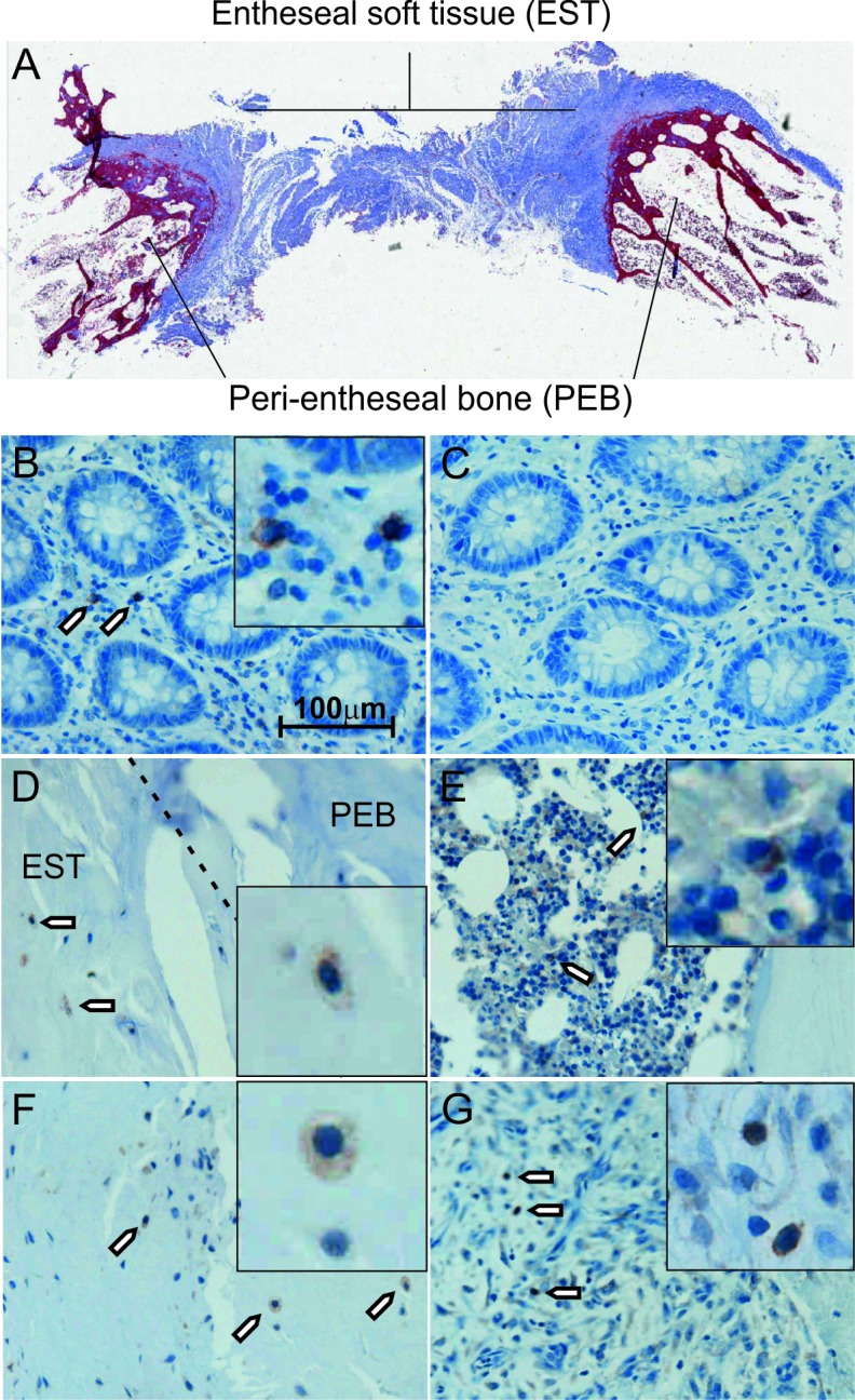
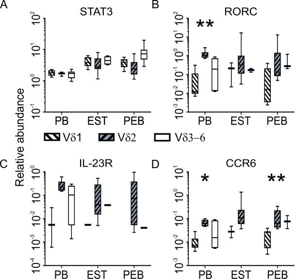
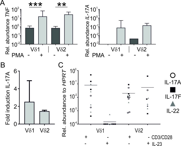
Methods: Human spinous processes entheseal soft tissue (EST) and peri-entheseal bone (PEB) were harvested during elective orthopaedic procedures. Entheseal γδT-cells were evaluated using immunohistochemistry and isolated and characterised using flow cytometry. RNA was isolated from γδT-cell subsets and analysed by qPCR. Entheseal γδT-cells were stimulated with phorbol 12-myristate 13-acetate (PMA) and ionomycin, anti-CD3/28 or IL-23 and IL-17A production was measured by high-sensitivity ELISA and qPCR.

Results: Entheseal γδT-cells were confirmed immunohistochemically with Vδ1 and Vδ2 subsets that are cytometrically defined. Transcript profiles of both cell populations suggested tissue residency and immunomodulatory status. Entheseal Vδ2 cells expressed high relative abundance of IL-23/17-associated transcripts including IL-23R, RORC and CCR6, whereas the Vδ1 subset almost completely lacked detectable IL-23R transcript. Following PMA stimulation IL-17A was detectable in both Vδ1 and Vδ2 subsets, and following CD3/CD28 stimulation both subsets showed IL-17A and IL-17F transcripts with neither transcript being detectable in the Vδ1 subset following IL-23 stimulation.

Conclusion: Spinal entheseal Vδ1 and Vδ2 subsets are tissue resident cells with inducible IL-17A production with evidence that the Vδ1 subset does so independently of IL-23R expression.

Keywords: T cells; ankylosing spondylitis; psoriatic arthritis; spondyloarthritis.


Conflict of interest statement

Competing interests: None declared.

Figures

Figure 1
Figure 1
γδT-cell localisation and phenotyping. Masson’s trichrome stained section showing the area of the spine harvested for analysis. Outer edges of the spinous process are labelled peri-entheseal bone (PEB) and the interspinous ligament labelled entheseal soft tissue (EST) (A). Immunohistochemistry showing γδT-cell receptor expression in human colon tissue, inset shows high power image (B). Staining is absent with omission of the primary antibody (C). Positive staining is observed in the border between the EST and the PEB (D) as well as in the haematopoietic bone marrow (E) and deeper within the EST (F). Inflammatory infiltrate of ruptured Achilles also contains positively stained cells (G). Brown colour and arrows indicate regions of positive staining.
Figure 2
Figure 2
γδT-cell phenotyping in blood and enthesis using flow cytometry. γδT-cells were identified based on positive expression of CD45 and CD3 and positive expression of the γδT-cell receptor, and then subdivided based on the Vδ isoform of the receptor expressed (A). In entheseal tissue (n=11), peri-entheseal bone contained a significantly higher proportion of the Vδ1 and a lower proportion of the Vδ2 expressing cells compared with healthy control blood (n=14) (B). There was no difference in subset proportion in spondyloarthritis patients (n=20) compared with healthy controls (C). Analysis of γδ subsets showing naïve, tissue resident memory (TRM), central memory (CM) and effector memory phenotypes (EM) (D). *P<0.05.
Figure 3
Figure 3
γδT-cells in enthesis and blood are transcriptionally distinct. Unmatched entheseal tissue derived subsets were compared with healthy blood derived cells. They had significantly higher expression of transforming growth factor β1 (TGFβ1), nuclear receptor subfamily 4 group a member 1 (Nr4a1) and lower expression of Krupple-like factor 2 (KLF2) and T-box 21 (TBX21) (A). All γδT-cell subsets expressed high levels of signal transduction molecules and immunomodulatory genes, Expression of IL-23/IL-17 axis cytokines was low or absent. Colour denotes relative expression to HPRT blue-low, black-equal, yellow-high, grey-below detection, Arrows indicate higher expression in γδT-cells (all subsets) from entheseal tissue (EST and PEB) compared with blood. Numbers show difference in median relative abundance. The ‘un-sorted’ category represents gene expression in an unsorted mixture of all cells released from entheseal digests (B) (PEB n=12, EST n=12, PB n=6). *P<0.05, **P<0.01. EST, entheseal soft tissue; PEB, peri-entheseal bone.
Figure 4
Figure 4
The Vδ2 subset expressed higher levels of genes involved or associated with IL-23-driven IL-17 signalling. Entheseal tissue derived subsets had generally higher expression of STAT3 compared with blood (A) and the Vδ2 subset had the highest expression of RORC (B), IL-23R (C) and CCR6 transcript (D) (PEB n=12, EST n=12, PB n=6). *P<0.05, **P<0.01. EST, entheseal soft tissue; PEB, peri-entheseal bone.
Figure 5
Figure 5
TNFα and IL-17A are produced by both γδT-cell subsets quantitative PCR showing TNFα and IL-17A transcript expression in γδT-cell subsets with or without stimulation (n=8) (A). Fold induction of secreted IL-17A protein following PMA/ionomycin stimulation compared with unstimulated fraction of purified Vδ1 and Vδ2 subsets (n=3) bar shows that mean whiskers represent 1 SD (B). Transcript expression of IL-17A, IL-17F and IL-22 following 48-hour stimulation of Vδ1 and Vδ2 subsets with combined anti-CD3/CD28 or IL-23 stimulation (n=10). Line denotes mean expression (all genes combined) (C). Genes for which a housekeeping value was obtained, but for which a target value was not, were assigned a value of 0.0001, as 0 cannot be plotted on a log scale. *P<0.05, **P<0.01, ***P≤0.001.

References

    1. Taurog JD, Chhabra A, Colbert RA. Ankylosing spondylitis and axial spondyloarthritis. New England Journal of Medicine 2016;374:2563–74. 10.1056/NEJMra1406182 - DOI - PubMed
    1. Jacques P, Lambrecht S, Verheugen E, et al. . Proof of concept: enthesitis and new bone formation in spondyloarthritis are driven by mechanical strain and stromal cells. Ann Rheum Dis 2014;73:437–45. 10.1136/annrheumdis-2013-203643 - DOI - PubMed
    1. Lubberts E. The IL-23–IL-17 axis in inflammatory arthritis. Nat Rev Rheumatol 2015;11:415–29. 10.1038/nrrheum.2015.53 - DOI - PubMed
    1. Rahman P, Inman RD, Gladman DD, et al. . Association of interleukin-23 receptor variants with ankylosing spondylitis. Arthritis & Rheumatism 2008;58:1020–5. 10.1002/art.23389 - DOI - PubMed
    1. McGonagle D, Gibbon W, Emery P. Classification of inflammatory arthritis by enthesitis. The Lancet 1998;352:1137–40. 10.1016/S0140-6736(97)12004-9 - DOI - PubMed

Publication types

MeSH terms