. 2019 Nov 1;294(44):16400-16415.
doi: 10.1074/jbc.RA119.010628. Epub 2019 Sep 17.

Prospecting for microbial α- N-acetylgalactosaminidases yields a new class of GH31 O-glycanase

Affiliations

Prospecting for microbial α- N-acetylgalactosaminidases yields a new class of GH31 O-glycanase

Peter Rahfeld et al. J Biol Chem. .

Abstract

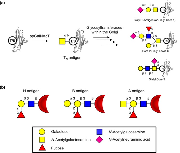
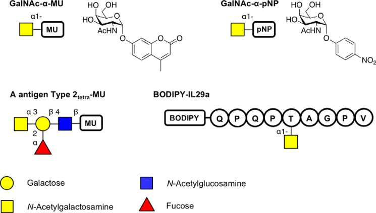
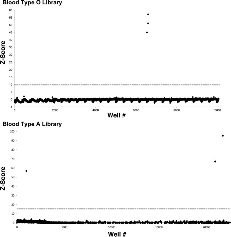
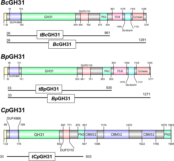
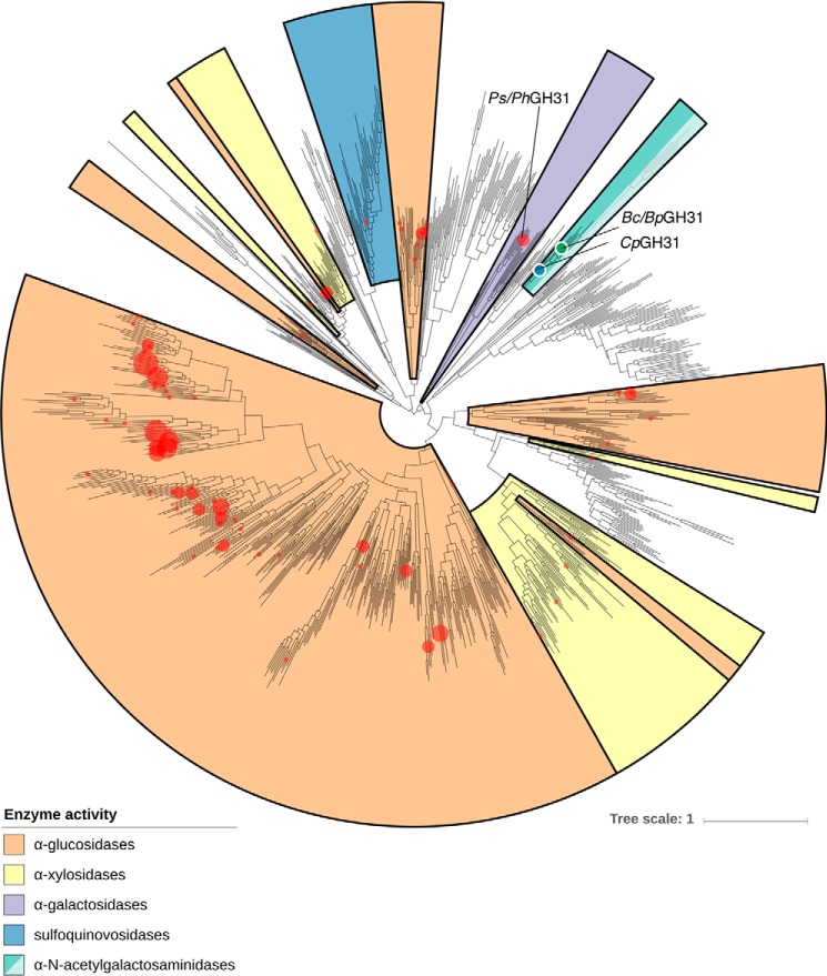
α-Linked GalNAc (α-GalNAc) is most notably found at the nonreducing terminus of the blood type-determining A-antigen and as the initial point of attachment to the peptide backbone in mucin-type O-glycans. However, despite their ubiquity in saccharolytic microbe-rich environments such as the human gut, relatively few α-N-acetylgalactosaminidases are known. Here, to discover and characterize novel microbial enzymes that hydrolyze α-GalNAc, we screened small-insert libraries containing metagenomic DNA from the human gut microbiome. Using a simple fluorogenic glycoside substrate, we identified and characterized a glycoside hydrolase 109 (GH109) that is active on blood type A-antigen, along with a new subfamily of glycoside hydrolase 31 (GH31) that specifically cleaves the initial α-GalNAc from mucin-type O-glycans. This represents a new activity in this GH family and a potentially useful new enzyme class for analysis or modification of O-glycans on protein or cell surfaces.

Keywords: N-acetylgalactosamine (alpha-GalNAc); O-glycanase; O-glycoprotein; blood; glycobiology; glycopeptide cleavage; glycoprotein; glycosidase; glycoside hydrolase 109 (GH109); glycoside hydrolase 31 (GH31); human gut microbiome; metagenomic analysis; metagenomics; mucin.


Conflict of interest statement

The authors declare that they have no conflicts of interest with the contents of this article.

Figures

Figure 1.
Figure 1.
O-Linked glycans and blood antigens. a, scheme of mucin-type O-linked glycosylation. O-Linked glycans are shown bound to either serine or threonine on the surface of a protein. Structures shown on the right are a selection of known mucin-type O-glycans. b, structures of ABO blood antigens present on red blood cells. All antigens are shown as bound to the surface of red blood cells though a Type 1 linkage. Sugars are shown using the Consortium for Functional Glycomics notation (76).
Figure 2.
Figure 2.
Overview of the substrates used in this study for screening or for kinetic analysis. The fluorophore used was either MU, pNP, or BODIPY. The sequence of the peptide BODIPY-IL29a is represented in single-letter format. Sugars are shown using the Consortium for Functional Glycomics notation (76).
Figure 3.
Figure 3.
Z-scores of the screens of the Type O and A libraries using 100 μm GalNAc-α-MU substrate. The dotted line shows the chosen cut-off at Z-score > 10. Z-score = (fluorescence − mean value)/S.D.
Figure 4.
Figure 4.
Protein domain structure of GH31 enzymes in this study. The protein domain structures are presented based on the domains identified based on InterPro prediction (77) and the NCBI conserved domain search (78) and are visualized using DOG2.0 (79). Signal peptides are shown in yellow, whereas the identity and length of all other domains are indicated by color and text within the figure. The truncations of the proteins employed in the present study are displayed below each structure. DUF, domain of unknown function; FN3, fibronectin type 3; F5/8, F5/F8 Type C domain.
Figure 5.
Figure 5.
Determination of the stereochemical outcome of enzymatic hydrolysis at the anomeric center by 1H NMR. The hydrolyses of GalNAc-α-DNP (5 mm) by tBpGH31 and tBcGH31 at room temperature were monitored by 1H NMR. A 1H NMR spectrum of the reaction was obtained after 5 min, and the anomeric stereochemistry of the hydrolysis product was determined. Stereochemistry was determined by observation of the product anomeric hydrogen peak appearing at either 5.2 ppm (α-GalNAc) or 4.6 ppm (β-GalNAc). Only α-GalNAc product was observed following rapid hydrolysis of the starting material, indicating net retention of stereochemistry.
Figure 6.
Figure 6.
Alignment of the amino acid sequences around the catalytic nucleophile and general acid/base. GH31s with α-GalNAcase activity examined in this work were aligned against the well-characterized GH31 α-xylosidase YicI from E. coli (GenBankTM accession no. AAC76680.1) (37), as well as a representative GH31 α-galactosidase PhGal31A from P. heparinus (ACU04898.1) (45) using Clustal Omega (80).
Figure 7.
Figure 7.
α-GalNAcase GH31s show exo-cleavage of O-glycans from fetuin. Native fetuin was subjected to overnight enzymatic cleavage of glycans by sequential activity of a neuraminidase (NedA), β-1,3-galactosidase (BgaC), and α-GalNAcases (BcGH31 or BpGH31). Cleavage products were then separated and analyzed via HPAEC-PAD. All GH31 enzymes tested were able to cleave GalNAc from the fetuin. Sugars were identified based on buffer (20 mm HEPES, pH 7.0, 150 mm NaCl) spiked with free Neu5Ac, Gal, and GalNAc. Some variations in the elution time accrued during the HPAEC-PAD runs, so the released sugars have been assigned with allowance for these variations.
Figure 8.
Figure 8.
α-GalNAcase GH31s cleave α-GalNAc from the peptide backbone. a, a peptide containing a GalNAc-α-threonine residue (BODIPY-IL29) was subjected to overnight enzymatic cleavage by the GH31 exo-α-GalNAcases from B. caccae and B. plebeius identified in this study. Cleavage products were then separated and analyzed via HPAEC-PAD. b, the cleavage of GalNAcylated BODIPY-IL29 was additionally followed via TLC at 5 and 45 min.
Figure 9.
Figure 9.
Phylogeny of GH31 family. All available GH31 members from the CAZy database are presented in a phylogeny. Red dots, locations of sequences of characterized GH31 enzymes. Black dot, location of the BcGH31, BpGH31, and CpGH31 within the phylogeny. Colored areas, major enzymatic activities within the different clades of the GH31 family. Lighter-colored turquoise, putative α-GalNAcases that lack the consensus motif present in the GH31s characterized in this study.
Figure 10.
Figure 10.
BcGH31 PUL. Overview of the genes located within the putative PUL containing BcGH31. Genes are annotated based upon domains that could potentially be involved in polysaccharide utilization. Gene loci are noted for the beginning and end of the putative PUL, BcGH31 in B. caccae ATCC 43185, and for the SusC/D pair that is central to PUL organization.

References

    1. Ten Hagen K. G., Fritz T. A., and Tabak L. A. (2003) All in the family: the UDP-GalNAc:polypeptide N-acetylgalactosaminyltransferases. Glycobiology 13, 1R–16R 10.1093/glycob/cwg007 - DOI - PubMed
    1. Tailford L. E., Crost E. H., Kavanaugh D., and Juge N. (2015) Mucin glycan foraging in the human gut microbiome. Front. Genet. 6, 81 10.3389/fgene.2015.00081 - DOI - PMC - PubMed
    1. Clausen H., and Hakomori S. (1989) ABH and related histo-blood group antigens; immunochemical differences in carrier isotypes and their distribution. Vox Sang. 56, 1–20 10.1111/j.1423-0410.1989.tb03040.x - DOI - PubMed
    1. Skovierová H, Larrouy-Maumus G., Pham H., Belanová M., Barilone N., Dasgupta A., Mikusová K., Gicquel B., Gilleron M., Brennan P. J., Puzo G., Nigou J., and Jackson M. (2010) Biosynthetic origin of the galactosamine substituent of arabinogalactan in Mycobacterium tuberculosis. J. Biol. Chem. 285, 41348–41355 10.1074/jbc.M110.188110 - DOI - PMC - PubMed
    1. Lombard V., Golaconda Ramulu H., Drula E., Coutinho P. M., and Henrissat B. (2014) The carbohydrate-active enzymes database (CAZy) in 2013. Nucleic Acids Res. 42, D490–D495 10.1093/nar/gkt1178 - DOI - PMC - PubMed

Publication types

MeSH terms

LinkOut - more resources