. 2019 Aug 18:2019:6146942.
doi: 10.1155/2019/6146942. eCollection 2019.

Protective Effect of Fat Extract on UVB-Induced Photoaging In Vitro and In Vivo

Affiliations

Protective Effect of Fat Extract on UVB-Induced Photoaging In Vitro and In Vivo

Mingwu Deng et al. Oxid Med Cell Longev. .

Abstract

Background: Nanofat can protect against ultraviolet B- (UVB-) induced damage in nude mice. Fat extract (FE) is a cell-free fraction isolated from nanofat that is enriched with a variety of growth factors.

Objective: To determine whether FE can protect against UVB-induced photoaging in cultured dermal fibroblasts and in nude mice.

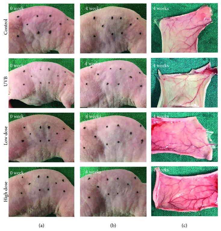
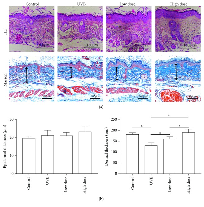
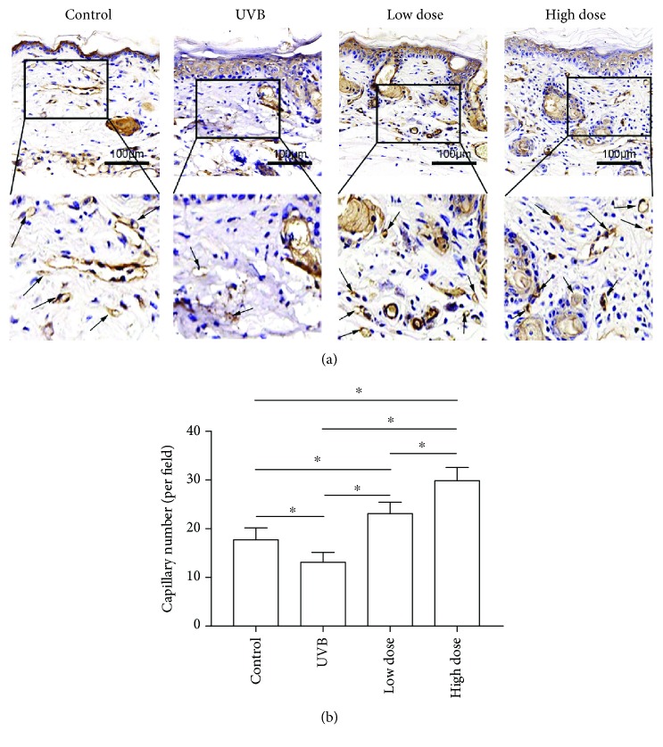
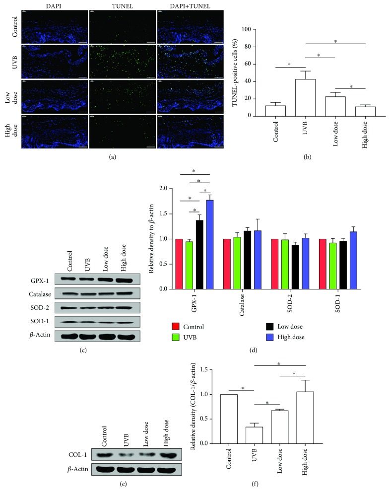
Method: For the in vitro study, human dermal skin fibroblasts were pretreated with FE 24 h prior to UVB irradiation. Generation of reactive oxygen species (ROS) was analyzed immediately following irradiation, while cell cycle analysis was performed 24 h after UVB irradiation. Senescence-associated β-galactosidase (SA-β-gal) expression, cell proliferation, and expression of glutathione peroxidase 1 (GPX-1), catalase, superoxide dismutase-1 (SOD-1), SOD-2, and collagen type 1 (COL-1) were investigated 72 h after UVB irradiation. For the in vivo study, the dorsal skin of nude mice was irradiated with UVB and mice were then treated with FE for 8 weeks. The thickness of the dermis, capillary density, and apoptotic cells in skin tissue sections were investigated after treatment. The expression of GPX-1, catalase, SOD-2, SOD-1, and COL-1 in the tissue was also measured.

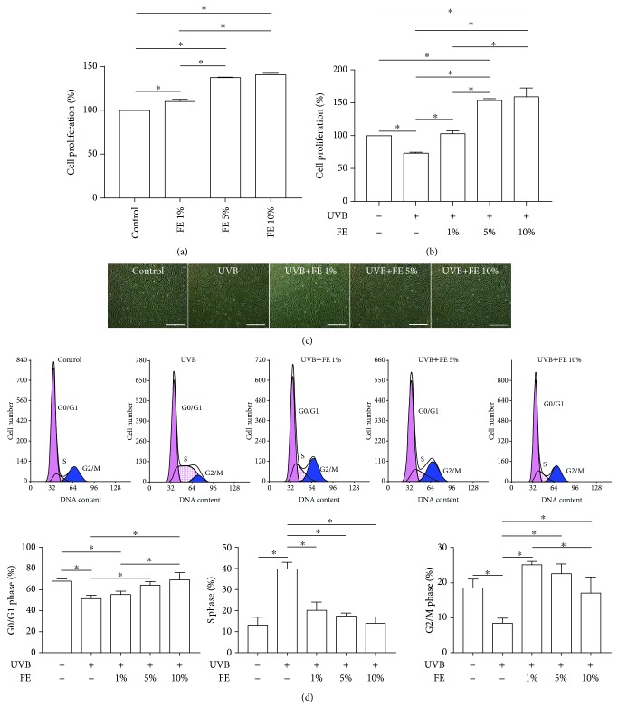
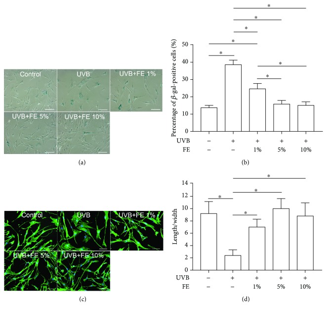
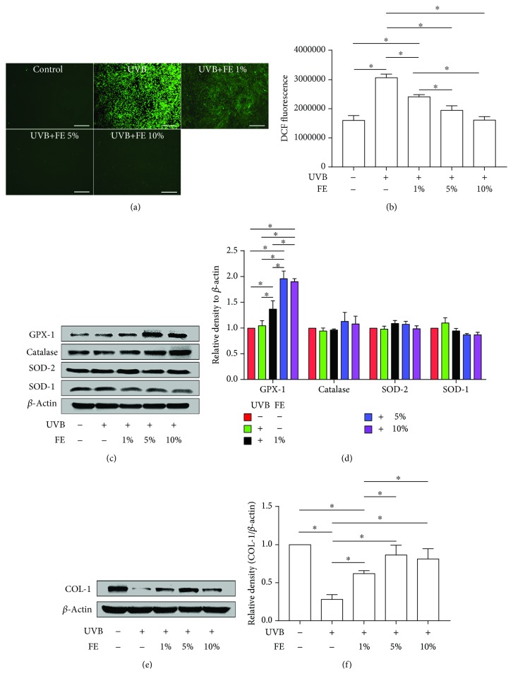
Result: FE significantly increased cell proliferation and protected cells against UVB-induced cell death and cell cycle arrest. FE reduced ROS and the number of aged cells induced by UVB irradiation. FE promoted the expression of COL-1 and GPX-1 in cultured dermal fibroblasts. FE treatment of UVB-irradiated skin increased dermal thickness and capillary density, decreased the number of apoptotic cells, and promoted the expression of COL-1 and GPX-1.

Conclusion: FE protects human dermal fibroblasts and the skin of nude mice from UVB-induced photoaging through its antioxidant, antiapoptotic, and proangiogenic activities.


Conflict of interest statement

The authors have declared that there is no conflict of interest.

Figures

Figure 1
Figure 1
Effect of FE on the proliferation and cell cycle of human skin fibroblasts. (a) Human skin fibroblasts were treated with different concentrations of FE (0%, 1%, 5%, and 10%) for 72 h. Cell proliferation was quantified using the CCK-8 test. (b) The CCK-8 test showed cell proliferation at 72 h after cells were pretreated with or without FE (0%, 1%, 5%, and 10%) for 24 h and then exposed to UVB light. (c) Morphology of human skin fibroblasts at 72 h after UVB irradiation. Scale bar: 150 μm. (d) Cell cycle analysis was carried out 24 h postirradiation. Cell cycle distribution analyzed by flow cytometry. FE treatment significantly increased cell proliferation and abrogated UVB-induced cell cycle arrest (p < 0.05).
Figure 2
Figure 2
Effects of FE on SA-β-gal staining and morphology of human skin fibroblasts. Human skin fibroblasts were pretreated with or without FE (0%, 1%, 5%, and 10%) for 24 h, and the cells were then exposed to UVB light at a total dose of 100 mJ/cm2. (a) SA-β-gal-positive cells were present 72 h after UVB irradiation. Scale bar: 150 μm. (b) The percentage of SA-β-gal-positive aged cells was determined by counting 200 cells per field. (c) Fluorescent labeling of cytoskeletal proteins. Scale bar: 100 μm. (d) The length/width ratio of cells was determined after staining for cytoskeletal proteins. FE treatment significantly reduced the number of SA-β-gal-positive cells induced by UVB irradiation and preserved normal cell morphology (p < 0.05).
Figure 3
Figure 3
FE reduced UVB-induced intracellular ROS and promoted the expression of GPX-1 and COL-1 in human skin fibroblasts. Human skin fibroblasts were pretreated with or without FE (0%, 1%, 5%, and 10%) for 24 h, and the cells were then exposed to UVB light at a total dose of 100 mJ/cm2. (a) Intracellular ROS measured by DCFH2-DA staining 20 min after UVB irradiation. Scale bar: 500 μm. (b) Intracellular ROS detected using DCFH2-DA staining were quantified with flow cytometry. (c, d) Western blotting analysis of GPX-1, catalase, SOD-2, and SOD-1 72 h after UVB irradiation. Representative results from three independent experiments are presented. The density values are expressed in arbitrary units. (e, f) Western blot analysis of COL-1 levels 72 h after UVB irradiation. Representative results from three independent experiments are presented. The density values are expressed in arbitrary units (p < 0.05).
Figure 4
Figure 4
Macroscopic observation after UVB irradiation and FE treatment for 4 weeks. (a, b) There were no differences in the appearance of the dorsal skin between the UVB group and FE treatment groups. (c) No FE residue was observed in the dorsal region where injection was performed, but more blood vessels were observed in the FE treatment group than in the UVB group.
Figure 5
Figure 5
Effect of FE on UVB-induced alterations of the epidermis and dermis in mice. (a) HE and Masson's trichrome staining were performed after FE treatment for 8 weeks. The arrows indicate the dermal portion of the skin. Scale bar: 100 μm. (b) Quantitative analysis of the epidermal and the dermal skin thickness. FE treatment significantly increased the thickness of the dermal portion of the skin (p < 0.05).
Figure 6
Figure 6
Effect of FE on angiogenesis in mice. (a) Anti-CD31 staining was performed after FE treatment for 8 weeks. The arrows indicate blood vessels. Scale bar: 100 μm. (b) FE treatment significantly increased vascular density in the dermis (p < 0.05).
Figure 7
Figure 7
FE reduced ROS-induced cell apoptosis and promoted the expression of GPX-1 and COL-1 in mice. (a) Fluorescence microscopy images and (b) quantification (%) of apoptotic cells following labeling with a TUNEL staining kit. Scale bar: 100 μm. (c, d) Western blotting analysis of GPX-1, catalase, SOD-2, and SOD-1 in mice after FE treatment for 8 weeks. Representative results from three independent experiments are presented. The density values are expressed in arbitrary units. (e, f) Western blotting analysis of COL-1 in mice after FE treatment for 8 weeks. Representative results from three independent experiments are presented. The density values are expressed in arbitrary units (p < 0.05).

References

    1. Gilchrest B. A. Photoaging. The Journal of Investigative Dermatology. 2013;133(Supplement 2):E2–E6. doi: 10.1038/skinbio.2013.176. - DOI - PubMed
    1. Kammeyer A., Luiten R. M. Oxidation events and skin aging. Ageing Research Reviews. 2015;21:16–29. doi: 10.1016/j.arr.2015.01.001. - DOI - PubMed
    1. Lee Y. R., Noh E. M., Jeong E. Y., et al. Cordycepin inhibits UVB-induced matrix metalloproteinase expression by suppressing the NF-κB pathway in human dermal fibroblasts. Experimental & Molecular Medicine. 2009;41(8):548–554. doi: 10.3858/emm.2009.41.8.060. - DOI - PMC - PubMed
    1. Poon F., Kang S., Chien A. L. Mechanisms and treatments of photoaging. Photodermatology, Photoimmunology & Photomedicine. 2015;31(2):65–74. doi: 10.1111/phpp.12145. - DOI - PubMed
    1. Brenneisen P., Sies H., Scharffetter-Kochanek K. Ultraviolet-B irradiation and matrix metalloproteinases: from induction via signaling to initial events. Annals of the New York Academy of Sciences. 2002;973(1):31–43. doi: 10.1111/j.1749-6632.2002.tb04602.x. - DOI - PubMed

MeSH terms