

The preliminary study for three-dimensional alveolar bone morphologic characteristics for alveolar bone restoration
- PMID: 31531306
- PMCID: PMC6726725
- DOI: 10.1186/s40902-019-0216-2
The preliminary study for three-dimensional alveolar bone morphologic characteristics for alveolar bone restoration
Abstract
Background: The concept of the ideal morphology for the alveolar bone form is an important element to reconstruct or restore the in maximizing esthetic profile and functional alveolar bone restoration. The purpose of this preliminary study is to evaluate the normal alveolar bone structure to provide the standard reference and guide template for use in diagnosing for implant placement, determining the correct amount of bone augmentation in actual clinical practice and producing prostheses based on three-dimensional imaging assessment of alveolar bone.
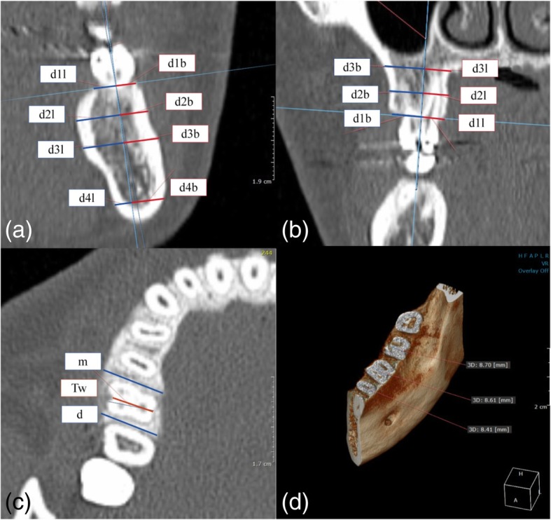
Methods: This study was included 11 men and 11 women (average age, 22.6 and 24.5 years, respectively) selected from among 127 patients. The horizontal widths of alveolar bone of maxilla and mandible were measured at the crestal, mid-root, and root apex level on MDCT (multi-detector computed tomography) images reconstructed by medical imaging software. In addition, tooth dimensions of the central incisors, canines, second premolars, and first molars of maxilla and mandible, including the horizontal width of the interdental alveolar bone crest, were also measured and statistically analyzed.
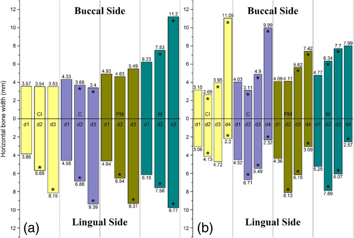
Results: The horizontal alveolar bone width of the palatal side of maxilla showed a distinct increment from the alveolar bone crest to the apical region in both anterior and posterior areas. The average widths of the maxillary alveolar ridge were as follows: central incisor, 7.43 mm; canine, 8.91 mm; second premolar, 9.57 mm; and first molar, 12.38 mm. The average widths of the mandibular alveolar ridge were as follows: central incisor, 6.21 mm; canine, 8.55 mm; second premolar, 8.45 mm; and first molar, 10.02 mm. In the buccal side, the alveolar bone width was not increased from the crest to the apical region. The horizontal alveolar bone width of an apical and mandibular border region was thinner than at the mid-root level.
Conclusions: The results of the preliminary study are useful as a clinical guideline when determining dental implant diameter and position. And also, these measurements can also be useful during the production of prefabricated membranes and customized alveolar bone scaffolds.
Keywords: Alveolar bone morphology; CT; Dental implant; Guided bone regeneration; Width and length of teeth.
Conflict of interest statement
Competing interestsThe authors declare that they have no competing interests.
Figures
Similar articles
-
Anterior maxilla alveolar ridge dimension and morphology measurement by cone beam computerized tomography (CBCT) for immediate implant treatment planning.BMC Oral Health. 2015 Jun 10;15:65. doi: 10.1186/s12903-015-0055-1. BMC Oral Health. 2015. PMID: 26059796 Free PMC article.
-
[Investigation of sagittal root position in relation to the anterior maxillary alveolar bone: a cone-beam CT study in 300 cases with normal occlusion].Zhonghua Kou Qiang Yi Xue Za Zhi. 2017 Oct 9;52(10):631-636. doi: 10.3760/cma.j.issn.1002-0098.2017.10.010. Zhonghua Kou Qiang Yi Xue Za Zhi. 2017. PMID: 29972938 Chinese.
-
Three-dimensional evaluation of interradicular spaces and cortical bone thickness for the placement and initial stability of microimplants in adults.Am J Orthod Dentofacial Orthop. 2009 Sep;136(3):314.e1-12; discussion 314-5. doi: 10.1016/j.ajodo.2009.01.023. Am J Orthod Dentofacial Orthop. 2009. PMID: 19732658
-
Evaluation of cortical bone thickness and root proximity at maxillary interradicular sites for mini-implant placement.Clin Oral Implants Res. 2013 Aug;24 Suppl A100:1-7. doi: 10.1111/j.1600-0501.2011.02354.x. Epub 2011 Oct 20. Clin Oral Implants Res. 2013. PMID: 22092972
-
Buccal Bone Thickness in Anterior and Posterior Teeth-A Systematic Review.Healthcare (Basel). 2021 Nov 30;9(12):1663. doi: 10.3390/healthcare9121663. Healthcare (Basel). 2021. PMID: 34946389 Free PMC article. Review.
Cited by
-
Quantitative assessment of the jawbone quality classification: A meta-analysis study.PLoS One. 2021 Jun 16;16(6):e0253283. doi: 10.1371/journal.pone.0253283. eCollection 2021. PLoS One. 2021. PMID: 34133463 Free PMC article.
-
Residual calcified material volume of β-tricalcium phosphate with platelet-rich fibrin in unilateral alveolar bone graft.Maxillofac Plast Reconstr Surg. 2024 Mar 1;46(1):7. doi: 10.1186/s40902-024-00420-1. Maxillofac Plast Reconstr Surg. 2024. PMID: 38427143 Free PMC article.
-
Assessment of Posterior Maxillary Alveolar Bone for Immediate Implant Placement: A Quantitative and Qualitative Analysis.Eur J Dent. 2024 Jul;18(3):877-882. doi: 10.1055/s-0043-1777049. Epub 2024 Feb 8. Eur J Dent. 2024. PMID: 38331039 Free PMC article.
-
Morphologic analysis of alveolar bone in maxillary and mandibular incisors on sagittal views.Surg Radiol Anat. 2021 Jun;43(6):1009-1018. doi: 10.1007/s00276-020-02640-2. Epub 2021 May 22. Surg Radiol Anat. 2021. PMID: 34023911
-
Towards Polycaprolactone-Based Scaffolds for Alveolar Bone Tissue Engineering: A Biomimetic Approach in a 3D Printing Technique.Int J Mol Sci. 2023 Nov 10;24(22):16180. doi: 10.3390/ijms242216180. Int J Mol Sci. 2023. PMID: 38003368 Free PMC article. Review.
References
-
- Esposito M, Grusovin MG, Felice P, Karatzopoulos G, Worthington HV, Coulthard P. The efficacy of horizontal and vertical bone augmentation procedures for dental implants - a Cochrane systematic review. Eur J Oral Implantol. 2009;2:167–184. - PubMed
-
- Buser D, Martin W, Belser UC. Optimizing esthetics for implant restorations in the anterior maxilla: anatomic and surgical considerations. Int J Oral & Maxillofac Implants. 2004;19 Suppl:43–61. - PubMed
-
- Tardieu PB, Vrielinck L, Escolano E. Computer-assisted implant placement. A case report: treatment of the mandible. Int J Oral Maxillofac Implants. 2003;18:599–604. - PubMed
LinkOut - more resources
Full Text Sources
Miscellaneous