Review
. 2019 Oct 7;11(37):17157-17178.
doi: 10.1039/c9nr05371a. Epub 2019 Sep 18.

The application of nanotechnology in enhancing immunotherapy for cancer treatment: current effects and perspective

Affiliations
Review

The application of nanotechnology in enhancing immunotherapy for cancer treatment: current effects and perspective

Yongjiang Li et al. Nanoscale. .

Abstract

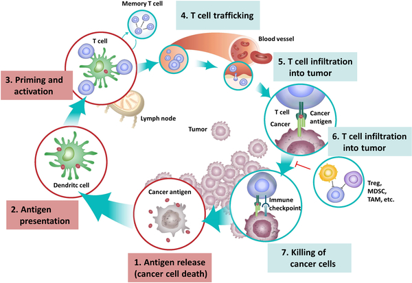
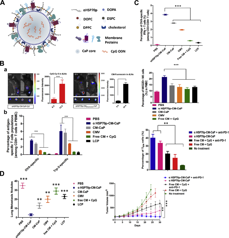
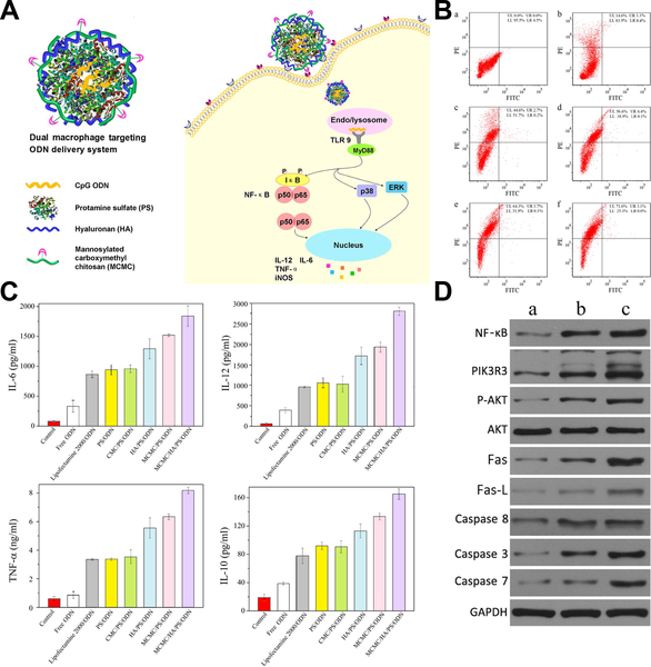
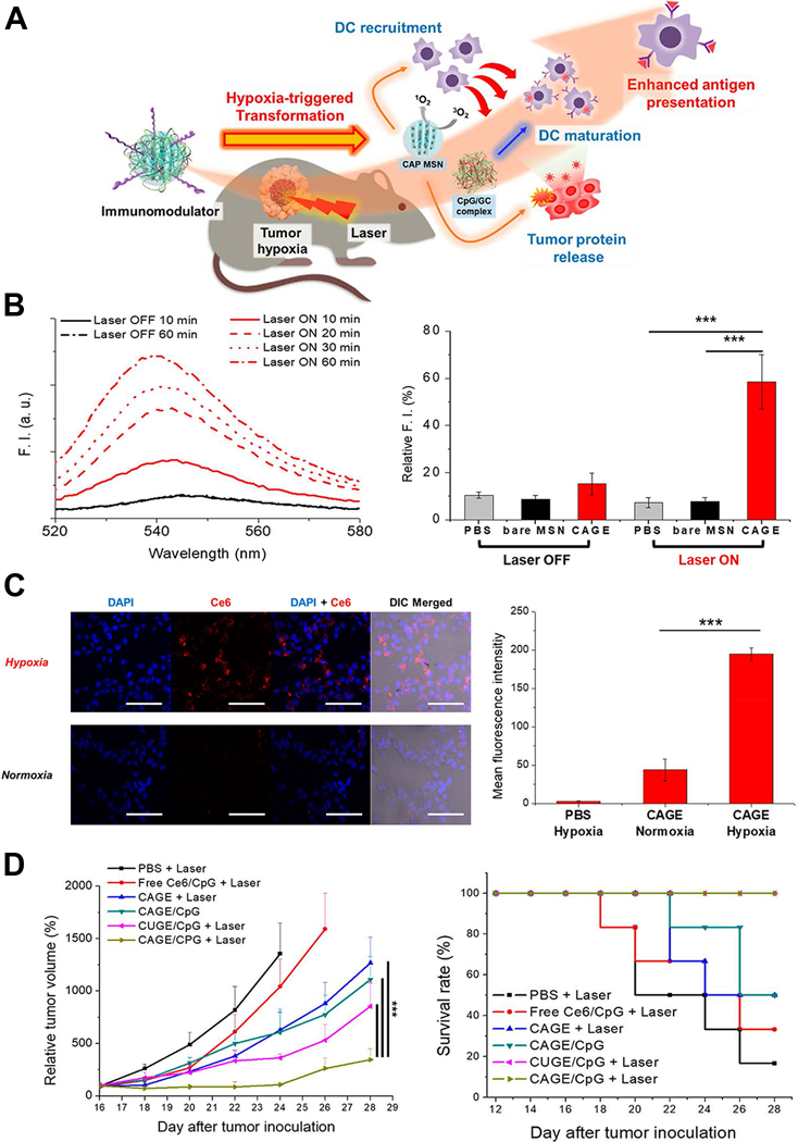
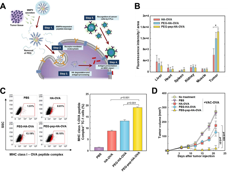
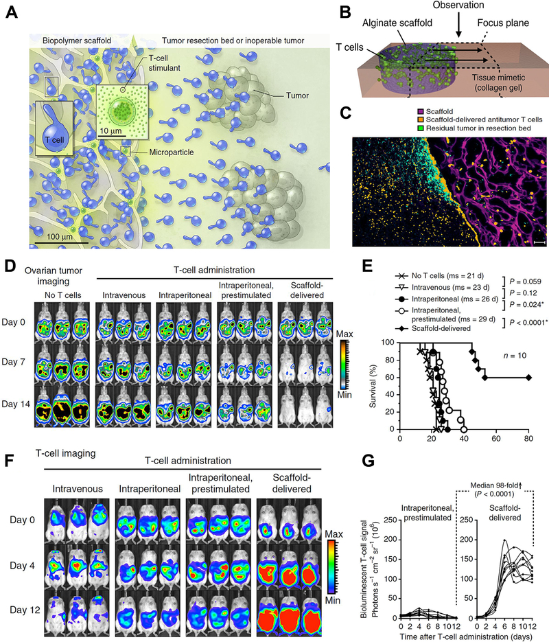
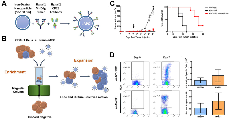
Cancer immunotherapy is emerging as a promising treatment modality that suppresses and eliminates tumors by re-activating and maintaining the tumor-immune cycle, and further enhancing the body's anti-tumor immune response. Despite the impressive therapeutic potential of immunotherapy approaches such as immune checkpoint inhibitors and tumor vaccines in pre-clinical and clinical applications, the effective response is limited by insufficient accumulation in tumor tissues and severe side-effects. Recent years have witnessed the rise of nanotechnology as a solution to improve these technical weaknesses due to its inherent biophysical properties and multifunctional modifying potential. In this review, we summarized and discussed the current status of nanoparticle-enhanced cancer immunotherapy strategies, including intensified delivery of tumor vaccines and immune adjuvants, immune checkpoint inhibitor vehicles, targeting capacity to tumor-draining lymph nodes and immune cells, triggered releasing and regulating specific tumor microenvironments, and adoptive cell therapy enhancement effects.


Conflict of interest statement

Conflicts of Interest: The authors declare no conflicts of interest.

Figures

Figure 1
Figure 1
The cancer-immunity cycle that involves exposure or release of tumor antigens, tumor antigen processing and presentation by APCs, priming and activation of effective immune cells and formation of memory cells, trafficking and infiltration of T cells to tumor tissues, and the recognition and killing of tumor cells. Figure from reference 10 with permission obtained.
Figure 2
Figure 2
Genetic tumor nanovaccine enhanced immunotherapy. (A) Synthesis process and working mechanism of the nanoliposome. (B) Enhanced secretion of TNF-α and IL-12 after treatment. (C) Increased MHC-Ⅰ, MHC-Ⅱ and CD80 expression after treatment. (D) In vivo anti-tumor effects of the genetic liposome nanovaccine. Figures from reference 49 with permission obtained.
Figure 3
Figure 3
Tumor-specific cellular immune response induced by tumor nanovaccine co-delivering bio-adjuvant and immunosuppressive pathway inhibitor. (A) Synthesis and working mechanism of the co-delivery nanovaccine. (B) Enhanced cellular uptake and intracellular localization of siRNA and tumor antigen (OVA) in mouse bone marrow dendritic cells. (C) Increased tumor antigen-specific CD4+ and CD8+ T-cell proliferation, IFN-γ production and CTL response by the nanovaccine. (D) The anti-tumor effects of the co-delivery nanovaccine and survival curves of tumor-bearing mice. Figures from reference 56 with permission obtained.
Figure 4
Figure 4
Elimination of tumor by chemoimmunotherapy with immune checkpoint inhibitor-conjugated nanoparticle. (A) Schematic of immune checkpoint inhibitor-combined nanoparticles for chemo-immunotherapy. (B) Percentage of tumor antigen-specific CD8+ T cells after treatment and the corresponding scatterplots. (C) Whole-animal in vivo imaging of tumor after treatment and quantification of the bioluminescence signal. (D) Tumor growth and survival curves after treatment. Figures from reference 81 with permission obtained.
Figure 5
Figure 5
Tumor draining lymph nodes (TDLNs) targeted nanoparticle boosts tumor-specific T-lymphocyte response, nature killer cells’ immune stimulation and effector memory T cells’ proliferation. (A) Structure of the TDLNs targeted nanoparticle that enables co-delivery of three elements including tumor cell membrane proteins, adjuvant CpG and an a-helix peptide modified HSP70p. (B) Prolonged antigen delivery in TDLNs and multi-epitope T cells priming in vivo: a. Fluorescence showing nanoparticle accumulation in the TDLNs and the corresponding fluorescence intensity; b. Quantitative analysis of the frequency of the nanoparticle positive CD8α+ T cells in the TDLNs after treatment. (C) The nanoparticle activated antigen-specific CD8+ T cells, NKG2D+ NK cells, and induced effector memory T cells proliferation in vivo. (D) Elimination of primary and secondary tumor by the nanoparticle. Figures from reference 94 with permission obtained.
Figure 6
Figure 6
Macrophage dual-targeted nanoparticle for overcoming cancer-associated immunosuppression. (A) Structure of the dual-targeting delivery system inducing immunological stimulation in macrophages. (B) Flow cytometry showing shift of macrophage to activated M1 type that marked by CD80 (a. without treatment, and treated by b. naked nanoparticle, c. nontargeting system, d. mono-targeting system with HA, e. mono-targeting system with MCMC, and f. dual-targeting system with HA and MCMC. (C) Enhanced cytokines secretion of macrophage after being treated. (D) Western blot analysis showing activated NF-κB, PI3K/Akt and Fas/FasL signaling pathways after treatment (a. without treatment, b. treated by naked nanoparticle, and c. treated by dual-targeting system). Figures from reference 105 with permission obtained.
Figure 7
Figure 7
Hypoxia microenvironment-triggered transforming nanoparticles for cancer immunotherapy via photodynamically enhanced antigen presentation. (A) Schematic illustration of working mechanism of the hypoxia-triggered transforming nanoparticle combined with photodynamic therapy. (B) Photoresponsive generation of singlet oxygen and the release profile of tumor proteins. (C) Hypoxia-responsive internalization of the nanoparticles into cells under hypoxic or normoxic conditions and the corresponding fluorescence intensity. (D) In vivo inhibition of tumor growth by the nanosystem and the survival curves. Figures from reference 118 with permission obtained.
Figure 8
Figure 8
MMP9 responsive nanoparticle in enhancing cancer immunotherapy. (A) Schematic illustration depicting working mechanism of the MMP9 responsive polymeric nanoparticle. (B) MMP9 responsive polymeric nanoparticle effectively accumulated in tumor tissue after systemic administration. (C) In vivo OVA antigen presentation in tumor cells after nanoparticle treatment. (D) In vivo therapeutic efficacy of the nanoparticle in tumor bearing mice. Figures from reference 129 with permission obtained.
Figure 9
Figure 9
Nanostructured polyporous scaffold implants enhanced efficacy of adoptive T-cell therapy. (A, B) Schematic diagram of the T cell-loaded scaffold surgically situated at a tumor site. Stimulatory microspheres incorporated into the device triggered cell expansion and promoted their egress into surrounding tissue. (C) Histological analysis of the scaffold and surrounding tissue 3 days after tumor-specific T cells (Orange) were embedded in the scaffold (Purple) and implanted into the tumor resection cavity. (D) Serial in vivo bioluminescence imaging of tumors. (E) Survival curves of tumor bearing mice following T-cell adoptive therapy. (F) In vivo bioluminescent imaging of T cells expressing luciferase. (G) Luciferase signal intensities after T-cell transfer, every line represented one animal and each dot reflected the whole animal photon count. Figures from reference 141 with permission obtained.
Figure 10
Figure 10
Enrichment and expansion with nano-artificial APCs for adoptive cell immunotherapy. (A) Nano-artificial APCs are synthesized by coupling MHC-Ig dimer (Signal 1) and anti-CD28 antibody (Signal 2) to a 50–100 nm iron-dextran nanoparticle. (B) Schematic of magnetic enrichment. Antigen-specific CD8+ T cells (blue) bound to the nano-artificial APC are retained in a magnetic column in the enrichment step, while non-cognate (orange) cells are less likely to bind. Enriched T cells are then activated by the nano-artificial APC and proliferate in the expansion step. (C) Nano-artificial APCs enriched and expanded lymphocytes inhibited melanoma growth after adoptive transfer. (D) Enrichment and expansion with the nano-artificial APC functionalized with human HLA-A2 and induced robust expansion against the tumor antigens. Figures from reference 146 with permission obtained.

References

    1. Mora J, Mertens C, Meier JK, Fuhrmann DC, Brune B and Jung M, Cells, 2019, 8. - PMC - PubMed
    1. Vesely MD, Kershaw MH, Schreiber RD and Smyth MJ, Annu Rev Immunol, 2011, 29, 235–271. - PubMed
    1. Smith HJ, McCaw TR, Londono AI, Katre AA, Meza-Perez S, Yang ES, Forero A, Buchsbaum DJ, Randall TD, Straughn JM Jr., Norian LA and Arend RC, Cancer, 2018, 124, 4657–4666. - PMC - PubMed
    1. Nakasone ES, Hurvitz SA and McCann KE, Drugs Context, 2018, 7, 212520. - PMC - PubMed
    1. Dougan M and Dranoff G, Annu Rev Immunol, 2009, 27, 83–117. - PubMed

MeSH terms