. 2020 Jan;17(1):98-111.
doi: 10.1080/15476286.2019.1669405. Epub 2019 Sep 29.

Targeting of stromal versican by miR-144/199 inhibits multiple myeloma by downregulating FAK/STAT3 signalling

Affiliations

Targeting of stromal versican by miR-144/199 inhibits multiple myeloma by downregulating FAK/STAT3 signalling

Nidhi Gupta et al. RNA Biol. 2020 Jan.

Abstract

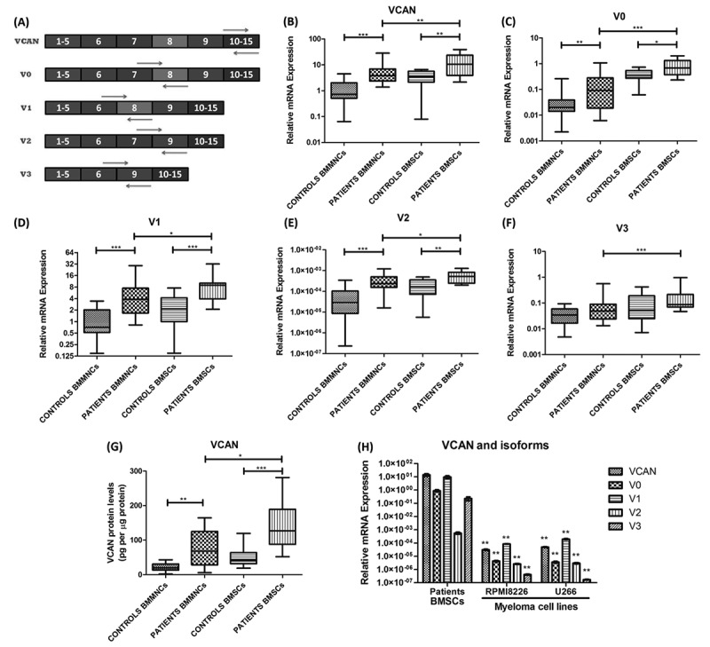
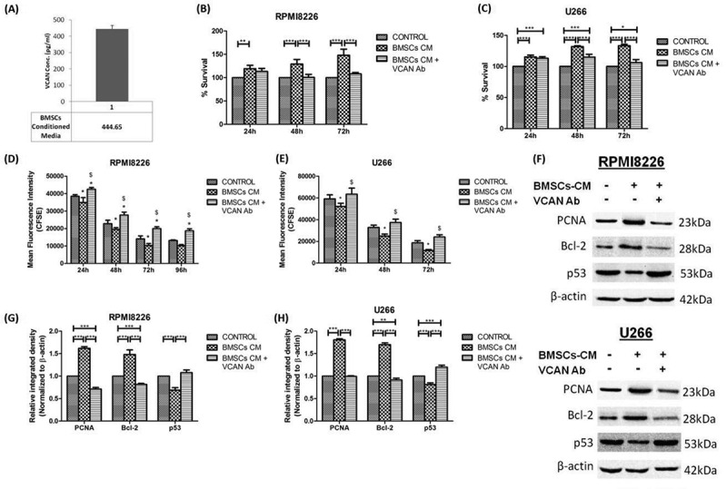
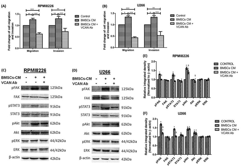
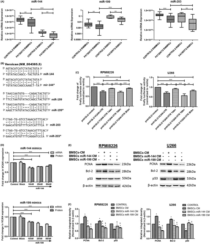
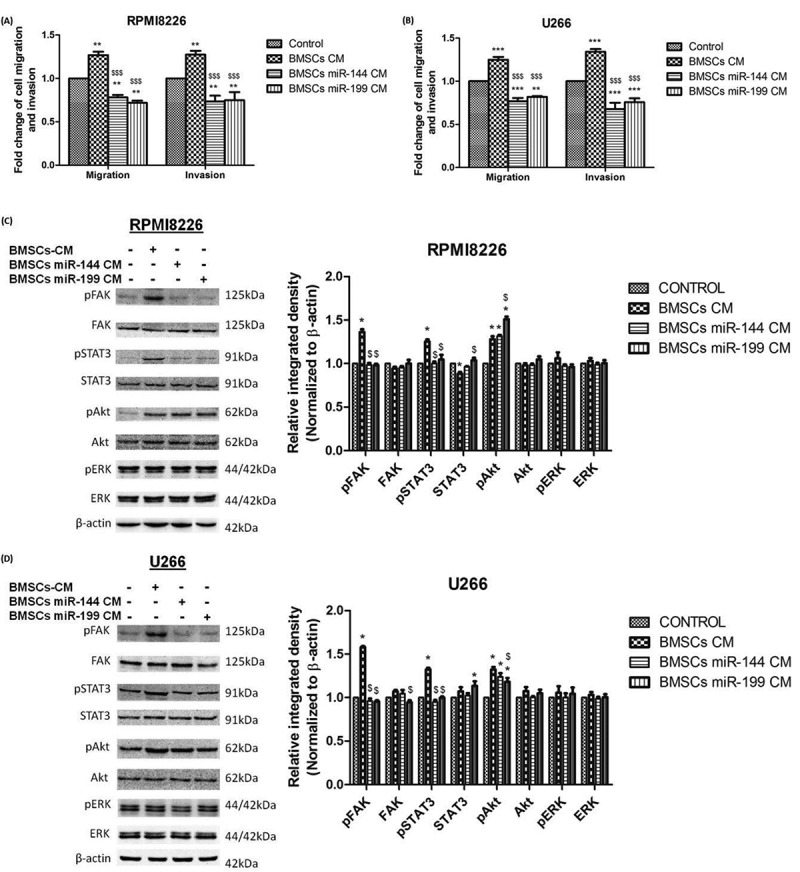
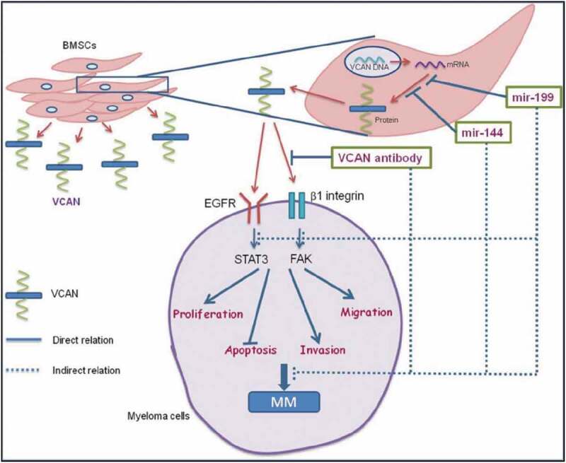
The abnormal growth of malignant plasma cells in Multiple Myeloma (MM) requires bone marrow (BM) niche consisting of proteoglycans, cytokines, etc. Versican (VCAN), a chondroitin sulphate proteoglycan promotes progression in solid tumours but there is dearth of literature in MM. Hence, we studied the involvement of VCAN in MM and its regulation by microRNAs as a therapeutic approach. Thirty MM patients and 20 controls were recruited and BM stromal cells (BMSCs) were isolated by primary culture. Molecular levels of VCAN, miR-144, miR-199 & miR-203 were determined in study subjects and cell lines. The involvement of VCAN in myeloma pathogenesis was studied using BMSCs-conditioned medium (BMSCs-CM) and VCAN-neutralizing antibody or microRNA mimics. Elevated expression of VCAN was observed in patients especially in BM stroma while microRNA expression was significantly lower and showed negative correlation with VCAN. Moreover, BMSCs-CM showed the presence of VCAN which upon supplementing to MM cells alter parameters in favour of myeloma progression, however, this effect was neutralized by VCAN antibody or miR (miR-144 and miR-199) mimics. The downstream signalling of VCAN was found to activate FAK and STAT3 which subsides by using VCAN antibody or miR mimics. The neutralization of oncogenic effect of BMSCs-CM by VCAN blockage affirms its plausible role in progression of MM. VCAN was observed as a paracrine mediator in the cross-talk of BMSCs and myeloma cells in BM microenvironment. Therefore, these findings suggest exploring VCAN as novel therapeutic target and utilization of microRNAs as a therapy to regulate VCAN for better management of MM.

Keywords: Multiple myeloma; bone marrow microenvironment; microRNAs; therapeutics; versican.


Figures

Figure 1.
Figure 1.
Isolation and characterization of primary bone marrow stromal cells (BMSCs). (A) BMSCs were isolated from BMMNCs which were plated in culture flasks. BMSCs have the tendency to adhere, hence, attached to the surface and proliferate with regular change of media. On day 6, some adhered cells were observed which grown further and on day 13–15, these cells attained optimum confluency (70–80%), hence, mentioned as passage 0. Scale bar represents 100 μm; (B) Flow cytometry characterization of BMSCs at passage 2 showing dual positivity for stromal cells marker (CD90 and CD105) while dual negativity for haematopoietic cell marker (CD34 and CD45); (C) percentage purity of BMSCs showing 96%-99% pure culture. [BMSCs: bone marrow stromal cells; BMMNCs: bone marrow mononuclear cells].
Figure 2.
Figure 2.
Molecular expression of VCAN and its isoforms in BMMNCs, BMSCs and cell lines. (A) Primer designing strategy for determining mRNA expression of VCAN and its four different isoforms (V0, V1, V2 and V3). These isoforms resulted by alternative splicing, hence, specific exon-exon junction were used for specific amplification; (B)-(F) box-whisker plot showing relative mRNA expression of VCAN and its four isoforms (V0, V1, V2 & V3) in BMMNCs (n = 30 patients, n = 20 controls) and BMSCs (n = 15 each) of MM patients and controls. GAPDH was used as an endogenous control for normalization; (G) box-whisker plot showing protein expression of VCAN by ELISA in BMMNCs (n = 15 each) and BMSCs (n = 15 each) of MM patients and controls. The levels of VCAN were normalized to the total protein concentration, hence, mentioned as pg per μg of total protein; (H) bar graph showing relative mRNA expression of VCAN and its four isoforms in patients BMSCs and in myeloma cell lines (RPMI8266 and U266). GAPDH was used as an endogenous control for normalization. ** represents significance with respect to patients BMSCs. Data were represented as median (range) for (B)-(G) while mean ± SD for (H). Wilcoxon rank-sum test was applied to determine significance between patients and controls. [BMMNCs: bone marrow mononuclear cells; BMSCs: bone marrow stromal cells; VCAN: versican; *p <0.05; **p <0.01; ***p <0.001].
Figure 3.
Figure 3.
Levels of VCAN in the conditioned medium (CM) of BMSCs and the effect of VCAN on proliferation and apoptosis of myeloma cells in vitro. BMSCs-CM was supplemented in 1:1 ratio in the culture medium of myeloma cells in the presence or absence of VCAN-neutralizing antibody (200 ng/mL) for 48 h. (A) Levels of VCAN in BMSCs-CM as determined by ELISA; (B)-(E) bar graphs showing effect of BMSCs-CM alone or with VCAN-neutralizing antibody on proliferation of myeloma cells (RPMI8226 and U266) as assessed by MTT assay (B) (C) and CFSE assay (D) (E), respectively at different time points. The mean fluorescence intensity of CFSE is inversely proportional to the cell proliferation. In Figure (D) and (E), * represents significance (p <0.05) with respect to control while $ represents significance (p <0.05) with respect to BMSCs-CM; (F) western blot image showing effect of BMSCs-CM on proliferation (PCNA) and apoptosis (Bcl-2 and p53) of RPMI8226 (top) and U266 (bottom) myeloma cells which got reversed by VCAN-neutralizing antibody; (G) (H) Image J densitometry analysis of western blots showing effect of VCAN on proliferation and apoptosis in myeloma cells. β-actin was used for normalization in western blotting. Data were represented as mean ± SD and two-way ANOVA was applied to determine significance between groups. All the experiments were performed in biological triplicates. [BMSCs: bone marrow stromal cells; BMSCs-CM: bone marrow stromal cells-conditioned medium; VCAN: versican; Ab: antibody; PCNA: proliferating cell nuclear antigen; Bcl-2: B-cell lymphoma 2; *p <0.05; **p <0.01; ***p <0.001].
Figure 4.
Figure 4.
Effect of VCAN on the migration and invasion of myeloma cells in vitro along with downstream signalling cascade affected by the action of VCAN. BMSCs-CM was supplemented in 1:1 ratio in the culture medium of myeloma cells with or without VCAN-neutralizing antibody (200 ng/mL) for 48 h. (A)-(B) Bar graphs showing effect of VCAN blockage by neutralizing antibody on cell migration and invasion in RPMI8226 and U266 myeloma cells; (C)-(D) certain signalling cascades involved in myeloma pathogenesis were traced by western blotting and the effect of VCAN on FAK/STAT3 signalling was observed; (E)-(F) Image J densitometry analysis of western blots showing effect of VCAN on downstream signalling cascades in myeloma cells. * represents significance (p <0.05) with respect to control while $ represents significance (p <0.05) with respect to BMSCs-CM. β-actin was used for normalization. Data were represented as mean ± SD and two-way ANOVA was applied to determine significance. All the experiments were performed in biological triplicates. [BMSCs: bone marrow stromal cells; BMSCs-CM: bone marrow stromal cells-conditioned medium; VCAN: versican; Ab: antibody; FAK: focal adhesion kinase; STAT: signal transducer and activator of transcription; *p <0.05; **p <0.01; ***p <0.001].
Figure 5.
Figure 5.
Relative microRNA expression of miR-144, miR-199 and miR-203 in study subjects followed by assessment of VCAN regulation by microRNAs and its effect on myeloma cells. (A) Box-whisker plot showing relative microRNA expression of miR-144, miR-199 and miR-203 in BMMNCs (n = 30 patients, n = 20 controls) and BMSCs (n = 15 each) of MM patients and controls. SNORD48 was used as an endogenous control to normalize microRNA expression; (B) Potential sites in VCAN 3ʹUTR targeted by miR-144, miR-144*, miR-199, miR-199*, miR-203 and miR-203* (* represents site-directed mutagenesis in the binding site of microRNAs); (C) RPMI8226 (left) and U266 (right) were transfected with pmirGLO reporter plasmid containing wild type or mutated VCAN-3′UTR. The firefly luciferase activity was measured normalized to Renilla luciferase. pmirGLO VCAN-UTR 144m/199m/203m represents mutation in particular microRNA binding site. (D) miR-144 (top) and miR-199 (bottom) mimics were transfected in patient-derived BMSCs and alteration in transcript of VCAN (by Q-PCR) along with its secretion in BMSCs-CM (by ELISA) were measured; (E)-(F) The conditioned medium of control BMSCs or microRNA mimics transfected BMSCs were supplemented in 1:1 ratio in the culture medium of myeloma cells and effect was studied after 48 h. Western blot images (E) and Image J densitometry analysis (F) showing the effect of microRNA mimics mediated knockdown of VCAN on proliferation and apoptosis of RPMI8226 (left) and U266 (right) cells. β-actin was used for normalization in western blotting. * represents significance (p <0.05) with respect to control while $ represents significance (p <0.05) with respect to BMSCs-CM. Data were represented as median (range) for (A) and Wilcoxon rank-sum test was applied to determine significance. Data were represented as mean ± SD for (C)-(F) and two-way ANOVA was applied. All the experiments were performed in biological triplicates. [BMMNCs: bone marrow mononuclear cells; BMSCs: bone marrow stromal cells; VCAN: versican; PCNA: proliferating cell nuclear antigen; Bcl-2: B-cell lymphoma 2; *p <0.05; **p <0.01; ***p <0.001].
Figure 6.
Figure 6.
Effect of microRNA mimics mediated knockdown of VCAN on migration and invasion of myeloma cells in addition to the effect on downstream signalling cascade in MM. Control BMSCs-CM or microRNA mimics transfected BMSCs-CM was supplemented in 1:1 ratio in the culture medium of myeloma cells for 48 h. (A)-(B) bar graphs showing the effect of miR-144 and miR-199 mimics mediated knockdown of VCAN on migration and invasion of RPMI8226 and U266 myeloma cells. Data were represented as mean ± SD and two-way ANOVA was applied to determine significance between groups. * (**p <0.01; ***p <0.001) represents significance with respect to control while $ ($$$p <0.001) represents significance with respect to BMSCs-CM; (C)-(D) certain signalling cascades involved in myeloma pathogenesis were traced by western blotting (left) along with their Image J densitometry analysis (right) and the effect of VCAN knockdown on FAK/STAT3 signalling was observed in RPMI8226 (C) and U266 (D). β-actin was used for normalization in western blotting. * represents significance (p <0.05) with respect to control while $ represents significance (p <0.05) with respect to BMSCs-CM. All the experiments were performed in triplicates. [BMSCs: bone marrow stromal cells; BMSCs-CM: bone marrow stromal cells-conditioned medium; FAK: focal adhesion kinase; STAT: signal transducer and activator of transcription; *p <0.05; **p <0.01; ***p <0.001].
Figure 7.
Figure 7.
Graphical abstract showing paracrine regulatory mechanism of VCAN in the cross-talk of BMSCs and myeloma cells. VCAN is secreted by BMSCs in the bone marrow which then interacts with myeloma cells activating FAK & STAT3 signalling cascade favouring myeloma progression. The regulation of VCAN could be achieved at various steps by different sources as shown. microRNAs (miR-144 and miR-199) could inhibit the translation of VCAN protein from mRNA either by transcript degradation or translational repression. Secondly, VCAN-neutralizing antibody could inhibit the interaction of VCAN with the myeloma cells. These inhibitory approaches indirectly impede the growth and proliferation of myeloma cells. Therefore, targeting of VCAN via microRNAs could be employed as a therapy to inhibit myeloma progression. Continuous lines indicate direct relation while dashed lines indicate indirect relation. [MM: multiple myeloma; BMSCs: bone marrow stromal cells; VCAN: versican; FAK: focal adhesion kinase; STAT: signal transducer and activator of transcription; EGFR: Epidermal growth factor receptor].

References

    1. Palumbo A, Anderson K.. Multiple myeloma. N Engl J Med. 2011. March 17;364(11):1046–1060. - PubMed
    1. Bray F, Ferlay J, Soerjomataram I, et al. Global cancer statistics 2018: GLOBOCAN estimates of incidence and mortality worldwide for 36 cancers in 185 countries. CA Cancer J Clin. 2018. November;68(6):394–424. - PubMed
    1. Kyle RA, Rajkumar SV. Multiple myeloma. Blood. 2008. March 15;111(6):2962–2972. - PMC - PubMed
    1. Ghobrial IM. Myeloma as a model for the process of metastasis: implications for therapy. Blood. 2012. July 5;120(1):20–30. - PMC - PubMed
    1. Rahmani M, Wong BW, Ang L, et al. Versican: signaling to transcriptional control pathways. Can J Physiol Pharmacol. 2006. January;84(1):77–92. - PubMed

Publication types