Review
. 2020;47(5):373-386.
doi: 10.1159/000501906. Epub 2019 Sep 18.

The Transitional Heart: From Early Embryonic and Fetal Development to Neonatal Life

Affiliations
Review

The Transitional Heart: From Early Embryonic and Fetal Development to Neonatal Life

Cheryl Mei Jun Tan et al. Fetal Diagn Ther. 2020.

Abstract

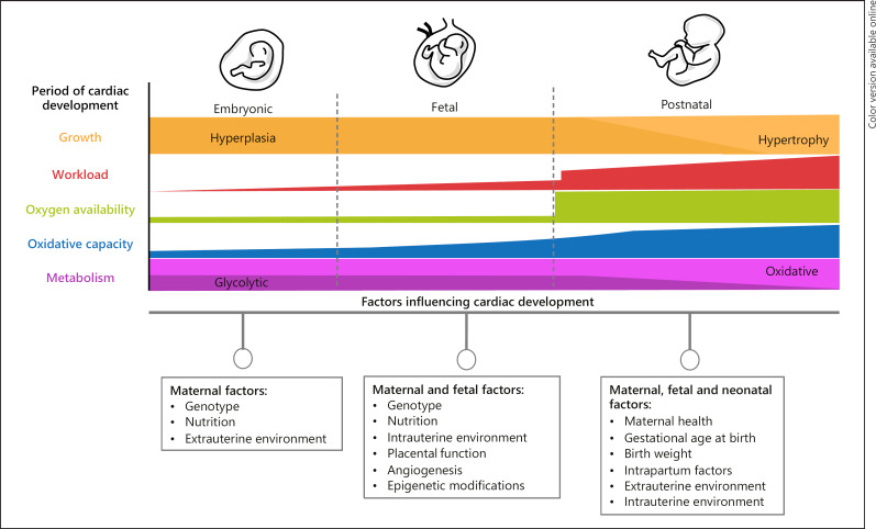
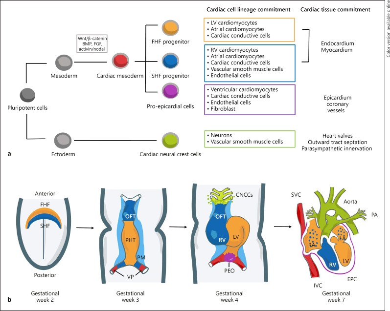
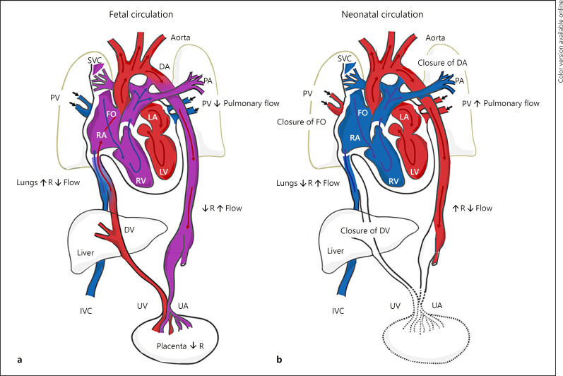
Formation of the human heart involves complex biological signals, interactions, specification of myocardial progenitor cells, and heart tube looping. To facilitate survival in the hypoxemic intrauterine environment, the fetus possesses structural, physiological, and functional cardiovascular adaptations that are fundamentally different from the neonate. At birth, upon separation from the placental circulation, the neonatal cardiovascular system takes over responsibility of vital processes for survival. The transition from the fetal to neonatal circulation is considered to be a period of intricate physiological, anatomical, and biochemical changes in the cardiovascular system. With a successful cardiopulmonary transition to the extrauterine environment, the fetal shunts are functionally modified or eliminated, enabling independent life. Investigations using medical imaging tools such as ultrasound and magnetic resonance imaging have helped to define normal and abnormal patterns of cardiac remodeling both in utero and ex utero. This has not only allowed for a better understanding of how congenital cardiac malformations alter the hemodynamic transition to the extrauterine environment but also how other more common complications during pregnancy including intrauterine growth restriction, preeclampsia, and preterm delivery adversely affect offspring cardiac remodeling during this early transitional period. This review article describes key cardiac progenitors involved in embryonic heart development; the cellular, physiological, and anatomical changes during the transition from fetal to neonatal circulation; as well as the unique impact that different pregnancy complications have on cardiac remodeling.

Keywords: Cardiac development; Developmental biology; Fetal heart; Neonatal heart; Transitional physiology.


Conflict of interest statement

The authors declare no conflicts of interest.

Figures

Fig. 1
Fig. 1
Cardiac development during embryonic, fetal, and neonatal life and the influence of maternal, fetal, and neonatal factors.
Fig. 2
Fig. 2
Early cardiac development. a Cardiac cell lineage and specification during development demonstrating the commitment of pluripotent cells toward mature cardiac cell types within the heart development. b Schematic of cardiac morphogenesis in humans. At the second week of gestation, the cardiogenic mesodermal cells migrate toward the anterior side of the embryo to form the FHF or cardiac crescent and SHF that are specified to form specific segments of the PHT, which is patterned along the anteroposterior axis to form the various regions and chambers of the looped and mature heart during weeks 3 and 4. The FHF gives rise to the beating PHT and will eventually give rise to the LV and parts of the right and left atria (RA and LA, respectively). The SHF, located behind the PHT and within the pharyngeal mesoderm by gestational week 3, will contribute to the formation of the RV, parts of the atria and outflow tract, and later to the base of the aorta and pulmonary artery. At gestational week 3, the cells at the venous pole contribute to the formation of the superior and inferior vena cavas (IVC, respectively). By gestational week 4, the cardiac neural crest cells migrate in from the dorsal neural tube, forming smooth muscle cells within the aortic and pulmonary arteries. In addition, the proepicardial organ formed by the proepicardial progenitor cell clusters later contributes to the formation of the epicardium. The 4 chambers form by the end of week 7. Wnt, Wingles integrated; FGF, fibroblast growth factor; BMP, bone morphogenetic proteins; FHF, first heart field; SHF, second heart field; OFT, outflow tract; PHT, primary heart tube; PM, pharyngeal mesoderm; VP, venous pole; CNCCs, cardiac neural crest cells; LV, left ventricle; RV, right ventricle; PEO, proepicardial organ; SVC, superior vena cavas; IVC, inferior vena cavas; EPC, epicardium; and PA, pulmonary artery.
Fig. 3
Fig. 3
Schematic of normal fetal and neonatal cardiovascular circulation. a During fetal development, oxygenated and nutrient-rich fetal blood from the placenta passes to the fetus via the umbilical vein. Approximately half of this blood bypasses the liver via the DV and enters the IVC. The remainder enters the portal vein to supply the liver with nutrients and oxygen. Blood entering the RA from the IVC bypasses the RV as the lungs are not yet functioning, and then enters the LA via the FO. Blood from the SVC enters the RA, passes to the RV, and moves into the PA trunk. Most of this blood enters the aorta via the DA, a right-to-left shunt. The partially oxygenated blood in the aorta returns to the placenta via the paired UA that arise from the internal iliac arteries. b Under normal physiological conditions, when pulmonary respiration begins at birth, pulmonary blood pressure falls, causing blood from the main PA trunk to enter the left PA and right PA, become oxygenated at the lungs, and then return to the LA via the PV. The FO and DA close, eliminating the fetal right-to-left shunts. The pulmonary and systemic circulations in the heart are now separate. As the infant is separated from the placenta, the UAs occlude (except for the proximal portions), along with the umbilical vein and DV. Blood to be metabolized now passes through the liver. DV, ductus venosus; UV, umbilical vein; IVC, inferior vena cava; RA, right atrium; RV, right ventricle; LA, left atrium; FO, foramen ovale; SVC, superior vena cava; PA, pulmonary artery; DA, ductus arteriosus; UA, umbilical arteries; PV, pulmonary veins.

References

    1. Bulatovic I, Månsson-Broberg A, Sylvén C, Grinnemo KH. Human fetal cardiac progenitors: the role of stem cells and progenitors in the fetal and adult heart. Best Pract Res Clin Obstet Gynaecol. 2016 Feb;31:58–68. - PubMed
    1. Buckingham M, Meilhac S, Zaffran S. Building the mammalian heart from two sources of myocardial cells. Nat Rev Genet. 2005 Nov;6((11)):826–35. - PubMed
    1. Finnemore A, Groves A. Physiology of the fetal and transitional circulation. Semin Fetal Neonatal Med. 2015 Aug;20((4)):210–6. - PubMed
    1. Morton SU, Brodsky D. Fetal Physiology and the Transition to Extrauterine Life. Clin Perinatol. 2016 Sep;43((3)):395–407. - PMC - PubMed
    1. Berhrsin J, Gibson A. Cardiovascular system adaptation at birth. Paediatr Child Health. 2011;21((1)):1–6.