

PGE1-Containing Protocols Generate Mature (Leukemia-Derived) Dendritic Cells Directly from Leukemic Whole Blood
- PMID: 31533251
- PMCID: PMC6769744
- DOI: 10.3390/ijms20184590
PGE1-Containing Protocols Generate Mature (Leukemia-Derived) Dendritic Cells Directly from Leukemic Whole Blood
Abstract
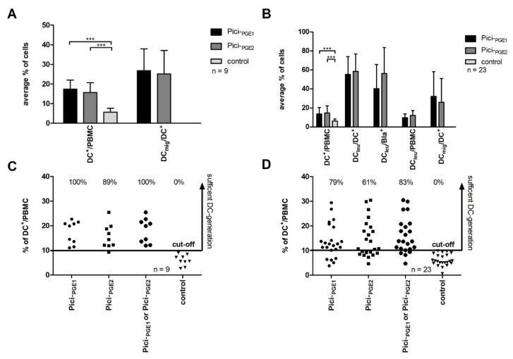
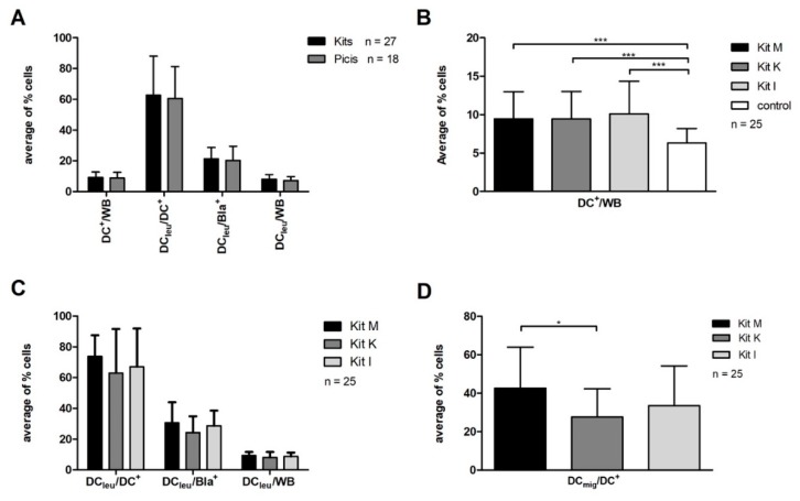
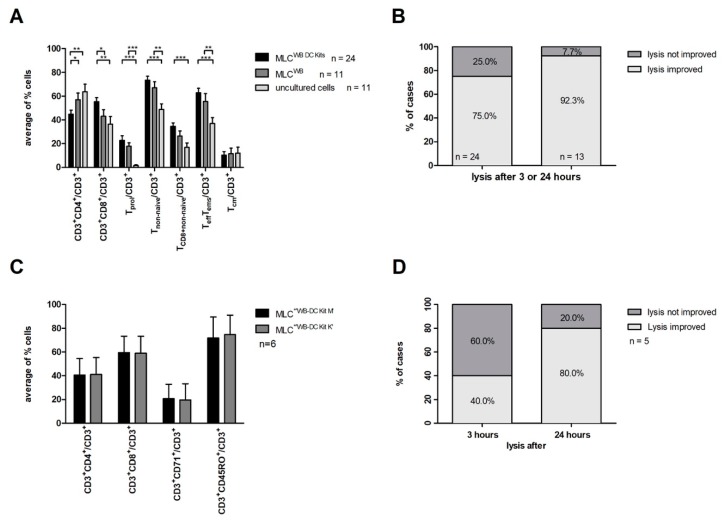
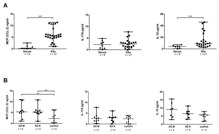
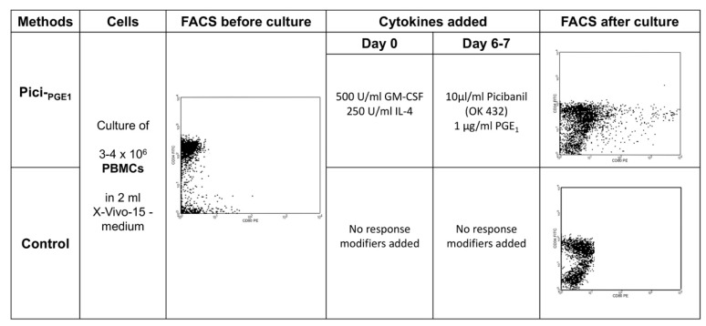
Dendritic cells (DCs) and leukemia-derived DC (DCleu) are potent stimulators of various immunoreactive cells and they play a pivotal role in the (re-) activation of the immune system. As a potential treatment tool for patients with acute myeloid leukemia, we developed and analyzed two new PGE1-containing protocols (Pici-PGE1, Kit M) to generate DC/DCleu ex vivo from leukemic peripheral blood mononuclear cells (PBMCs) or directly from leukemic whole blood (WB) to simulate physiological conditions. Pici-PGE1 generated significantly higher amounts of DCs from leukemic and healthy PBMCs when compared to control and comparable amounts as the already established protocol Pici-PGE2. The proportions of sufficient DC-generation were even higher after DC/DCleu-generation with Pici-PGE1. With Kits, it was possible to generate DCs and DCleu directly from leukemic and healthy WB without induction of blast proliferation. The average amounts of generated DCs and DCleu-subgroups were comparable with all Kits. The PGE1 containing Kit M generated significantly higher amounts of mature DCs when compared to the PGE2-containing Kit K and increased the anti-leukemic-activity. In summary PGE1-containing protocols were suitable for generating DC/DCleu from PBMCs as well as from WB, which reliably (re-) activated immunoreactive cells, improved the overall ex vivo anti-leukemic activity, and influenced cytokine-release-profiles.
Keywords: AML; PGE1; dendritic cells; immunotherapy; leukemia-derived dendritic cells.
Conflict of interest statement
All authors declare, that there are no financial conflicts in regard to this work.
Figures
Similar articles
-
Role of Interferon (IFN)α in "Cocktails" for the Generation of (Leukemia-derived) Dendritic Cells (DCleu) From Blasts in Blood From Patients (pts) With Acute Myeloid Leukemia (AML) and the Induction of Antileukemic Reactions.J Immunother. 2019 Jun;42(5):143-161. doi: 10.1097/CJI.0000000000000266. J Immunother. 2019. PMID: 31090655
-
Concentration-dependent effects of immunomodulatory cocktails on the generation of leukemia-derived dendritic cells, DCleu mediated T-cell activation and on-target/off-tumor toxicity.Front Immunol. 2025 Jan 30;15:1527961. doi: 10.3389/fimmu.2024.1527961. eCollection 2024. Front Immunol. 2025. PMID: 39949718 Free PMC article.
-
Generation of Leukaemia-Derived Dendritic Cells (DCleu) to Improve Anti-Leukaemic Activity in AML: Selection of the Most Efficient Response Modifier Combinations.Int J Mol Sci. 2022 Jul 28;23(15):8333. doi: 10.3390/ijms23158333. Int J Mol Sci. 2022. PMID: 35955486 Free PMC article.
-
Dendritic Cells of Leukemic Origin: Specialized Antigen-Presenting Cells as Potential Treatment Tools for Patients with Myeloid Leukemia.Transfus Med Hemother. 2020 Dec;47(6):432-443. doi: 10.1159/000512452. Epub 2020 Nov 5. Transfus Med Hemother. 2020. PMID: 33442338 Free PMC article. Review.
-
Can leukemia-derived dendritic cells generate antileukemia immunity?Expert Rev Vaccines. 2006 Aug;5(4):467-72. doi: 10.1586/14760584.5.4.467. Expert Rev Vaccines. 2006. PMID: 16989627 Review.
Cited by
-
The potential role of serum extracellular vesicle derived small RNAs in AML research as non-invasive biomarker.Nanoscale Adv. 2023 Feb 20;5(6):1691-1705. doi: 10.1039/d2na00959e. eCollection 2023 Mar 14. Nanoscale Adv. 2023. PMID: 36926576 Free PMC article.
-
In Vivo Induction of Leukemia-Specific Adaptive and Innate Immune Cells by Treatment of AML-Diseased Rats and Therapy-Refractory AML Patients with Blast Modulating Response Modifiers.Int J Mol Sci. 2024 Dec 16;25(24):13469. doi: 10.3390/ijms252413469. Int J Mol Sci. 2024. PMID: 39769232 Free PMC article.
-
Volatile Phases Derived from Serum, DC, or MLC Culture Supernatants to Deduce a VOC-Based Diagnostic Profiling Strategy for Leukemic Diseases.Biomolecules. 2023 Jun 14;13(6):989. doi: 10.3390/biom13060989. Biomolecules. 2023. PMID: 37371569 Free PMC article.
-
Dendritic Cell-Triggered Immune Activation Goes along with Provision of (Leukemia-Specific) Integrin Beta 7-Expressing Immune Cells and Improved Antileukemic Processes.Int J Mol Sci. 2022 Dec 27;24(1):463. doi: 10.3390/ijms24010463. Int J Mol Sci. 2022. PMID: 36613907 Free PMC article.
-
Leukemia-Derived Dendritic Cells Induce Anti-Leukemic Effects Ex Vivo in AML Independently of Patients' Clinical and Biological Features.Int J Mol Sci. 2025 Feb 17;26(4):1700. doi: 10.3390/ijms26041700. Int J Mol Sci. 2025. PMID: 40004163 Free PMC article.
References
-
- National Cancer Institute Cancer Stat Facts. [(accessed on 7 July 2019)]; Available online: https://seer.cancer.gov/statfacts/html/amyl.html.
MeSH terms
Substances
LinkOut - more resources
Full Text Sources
Research Materials
