. 2019 Oct 29;140(18):1491-1505.
doi: 10.1161/CIRCULATIONAHA.119.040676. Epub 2019 Sep 19.

Therapeutic Modulation of the Immune Response in Arrhythmogenic Cardiomyopathy

Affiliations

Therapeutic Modulation of the Immune Response in Arrhythmogenic Cardiomyopathy

Stephen P Chelko et al. Circulation. .

Abstract

Background: Inflammation is a prominent feature of arrhythmogenic cardiomyopathy (ACM), but whether it contributes to the disease phenotype is not known.

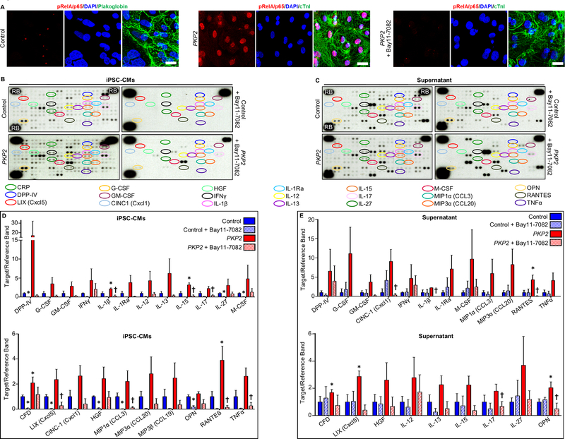
Methods: To define the role of inflammation in the pathogenesis of ACM, we characterized nuclear factor-κB signaling in ACM models in vitro and in vivo and in cardiac myocytes from patient induced pluripotent stem cells.

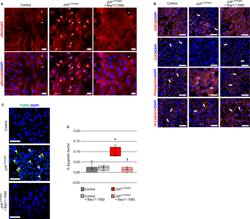
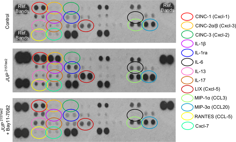
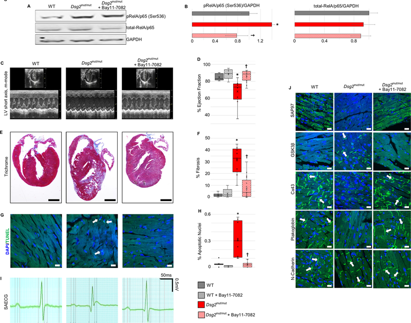
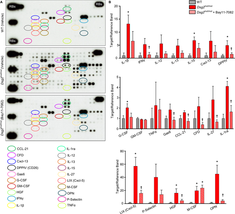
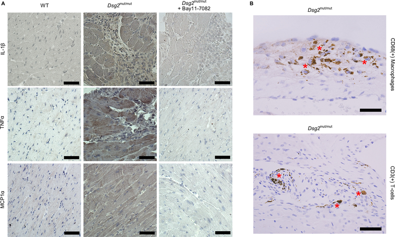
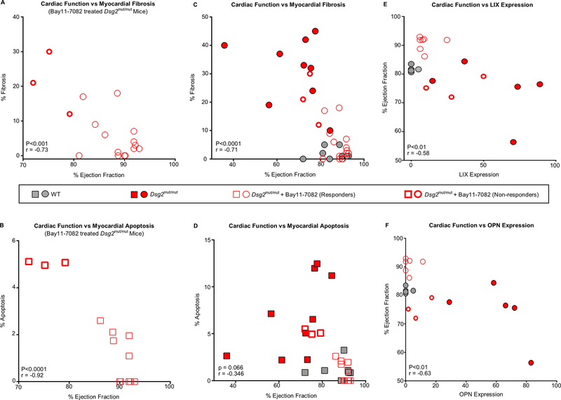
Results: Activation of nuclear factor-κB signaling, indicated by increased expression and nuclear accumulation of phospho-RelA/p65, occurred in both an in vitro model of ACM (expression of JUP2157del2 in neonatal rat ventricular myocytes) and a robust murine model of ACM (homozygous knock-in of mutant desmoglein-2 [Dsg2mut/mut]) that recapitulates the cardiac manifestations seen in patients with ACM. Bay 11-7082, a small-molecule inhibitor of nuclear factor-κB signaling, prevented the development of ACM disease features in vitro (abnormal redistribution of intercalated disk proteins, myocyte apoptosis, release of inflammatory cytokines) and in vivo (myocardial necrosis and fibrosis, left ventricular contractile dysfunction, electrocardiographic abnormalities). Hearts of Dsg2mut/mut mice expressed markedly increased levels of inflammatory cytokines and chemotactic molecules that were attenuated by Bay 11-7082. Salutary effects of Bay 11-7082 correlated with the extent to which production of selected cytokines had been blocked. Nuclear factor-κB signaling was also activated in cardiac myocytes derived from a patient with ACM. These cells produced and secreted abundant inflammatory cytokines under basal conditions, and this was also greatly reduced by Bay 11-7082.

Conclusions: Inflammatory signaling is activated in ACM and drives key features of the disease. Targeting inflammatory pathways may be an effective new mechanism-based therapy for ACM.

Keywords: B; NF-kappa; cardiomyopathies; cytokines; inflammation.


Conflict of interest statement

Conflict of Interest Disclosures

None

Figures

Figure 1.
Figure 1.. Activation of NFκB and reversal of ACM features by Bay 11–7082 in neonatal rat ventricular myocytes (NRVMs) expressing a deletion mutation in the gene encoding plakoglobin (JUP 2157del2).
A. Representative confocal immunofluorescence images from control (non-transfected) NRVMs and NRVMs expressing JUP 2157del2 in the absence or presence of Bay 11–7082 showing the distribution of phospho-RelA/p65 (pRelA/p65). Nuclear accumulation of immunoreactive signal, indicating activation of NFκB, is readily apparent in cells expressing JUP 2157del2 (asterisks) but is absent in such cells after treatment with Bay 11–7082. Scale bar = 50μm. B. Representative confocal immunofluorescence images from control (non-transfected) NRVMs and NRVMs expressing JUP 2157del2 in the absence or presence of Bay 11–7082. Arrows show localization of immunoreactive signal at the cell surface. The normal distribution of N-cadherin in all cells is shown as a positive control. Untreated JUP 2157del2 cells showed abnormal distribution of plakoglobin, Cx43 and GSK3β. The amount of signal for plakoglobin and Cx43 at cell-cell junctions was greatly reduced in these cells whereas signal for GSK3β, which normally resides in the cytoplasm, was seen at the cell surface. Asterisks identify apparent nuclear localization of plakoglobin in JUP 2157del2 cells. The abnormal distribution of plakoglobin, Cx43 and GSK3β was normalized in JUP 2157del2 cells treated with Bay 11–7082. Scale bar = 50μm. C. TUNEL labeling in control NRVMs and NRVMs expressing JUP 2157del2 in the absence or presence of Bay 11–7082. Representative confocal images show increased TUNEL + nuclei (arrow heads) in cultures of cells expressing JUP 2157del2 and normalization after treatment with Bay 11–7082. Scale bar = 50μm. D. Graph showing the percent (%) apoptotic nuclei in 5 microscopic fields from each condition. * P<0.0001 for JUP2157del2 cells vs. control cells; P<0.0001 for Bay 11–7082-treated vs. untreated JUP2157del2 cells determined by one-way ANOVA with Tukey’s multiple comparisons test.
Figure 2.
Figure 2.. Bay 11–7082 reduces cytokines in the culture media in neonatal rat ventricular myocytes (NRVMs) expressing a deletion mutation in the gene for plakoglobin (JUP 2157del2).
Representative cytokine arrays are shown for control (non-transfected) cells and NRVMs expressing JUP 2157del2 in the absence or presence of Bay 11–7082. The spots in the upper right and left and lower left corners are reference markers (Ref Bands) to compare overall exposure levels.
Figure 3.
Figure 3.. Activation of NFκB and reversal of ACM disease features in Dsg2mut/mut mice in vivo by inhibition of NFκB signaling with Bay 11–7082.
A. Western blots of total RelA/p65 and phospho-RelA/p65 in homogenates of hearts from vehicle-treated wildtype (WT) mice, and vehicle- and Bay 11–7082-treated Dsg2mut/mut mice. B. Group data for total-RelA/p65 and pRelA/p65 (Ser536) protein levels in hearts of vehicle-treated WT mice, and vehicle- and Bay 11–7082-treated Dsg2mut/mut mice. C. Representative short-axis m-mode echocardiograms of vehicle-treated wildtype (WT) mice, Dsg2mut/mut mice and Dsg2mut/mut mice treated with Bay 11–7082. D. Group data for % ejection fraction in vehicle- and Bay 11–7082-treated wildtype (WT) mice, and vehicle- and Bay 11–7082-treated Dsg2mut/mut mice. E. Representative long-axis sections of the hearts stained with Masson trichrome from wildtype (WT) mice, Dsg2mut/mut mice and Dsg2mut/mut mice treated with Bay 11–7082. Scale bar = 1mm. F. Group data for % of left ventricular area occupied by fibrosis in Masson trichrome-stained sections of hearts from vehicle- and Bay 11–7082-treated wildtype (WT) mice, and vehicle- and Bay 11–7082-treated Dsg2mut/mut mice. G. Representative images showing TUNEL labeling in sections of hearts from wildtype (WT) mice, Dsg2mut/mut mice and Dsg2mut/mut mice treated with Bay 11–7082. Arrows show TUNEL+ nuclei. Scale bar = 10μm. H. Group data showing % apoptotic nuclei in TUNEL-labeled sections of hearts from vehicle- and Bay 11–7082-treated wildtype (WT) mice, and vehicle- and Bay 11–7082-treated Dsg2mut/mut mice. I. Representative signal-averaged electrocardiograms (SAECGs) from wildtype (WT) mice, Dsg2mut/mut mice and Dsg2mut/mut mice treated with Bay 11–7082. J. Representative confocal images of immunostained hearts from wildtype (WT) mice, Dsg2mut/mut mice and Dsg2mut/mut mice treated with Bay 11–7082. Arrows show localization of immunoreactive signal at the cell surface. The normal distribution of N-cadherin in all cohorts is shown as a positive control. Untreated Dsg2mut/mut mice showed abnormal distribution of plakoglobin, Cx43, GSK3β and SAP97. The amount of signal for plakoglobin, Cx43 and SAP97 at cell-cell junctions was greatly reduced, whereas signal for GSK3β, which normally resides in the cytoplasm, was seen at the cell surface. These abnormal protein distributions were normalized in Dsg2mut/mut mice treated with Bay 11–7082. Scale bar = 10μm. Quantitative data in panels B, D, F and H are shown as mean ± SEM; n=10 for vehicle-treated WT mice; n=5 for Bay 11–7082-treated WT mice; n=9 for vehicle-treated Dsg2mut/mut mice; and n=17 for Bay 11–7082-treated Dsg2mut/mut mice. * P<0.001 for vehicle-treated Dsg2mut/mut mice vs. vehicle-treated WT mice; P<0.001 for Bay 11–7082-treated Dsg2mut/mut mice vs. vehicle-treated Dsg2mut/mut mice as determined by one-way ANOVA with Tukey’s multiple comparisons test.
Figure 4.
Figure 4.. Cytokine expression in the hearts of Dsg2mut/mut mice and its attenuation by Bay 11–7082.
A. Representative cytokine arrays from hearts of vehicle-treated wildtype (WT) mice, Dsg2mut/mut mice and Dsg2mut/mut mice treated with Bay 11–7082. The spots in the upper right and left and lower left corners are reference markers (RBs) to compare overall exposure levels. B. Quantitative data for expression of selected cytokines in hearts of vehicle-treated wildtype (WT) mice, Dsg2mut/mut mice and Dsg2mut/mut mice treated with Bay 11–7082. Data are shown as mean ± SEM; n=5 for each cytokine in vehicle-treated WT and Dsg2mut/mut mice; n=10 for each cytokine in Bay11–7082-treated Dsg2mut/mut mice. * P<0.05 for any cohort vs. vehicle-treated WT mice; P<0.05 for Bay 11–7082-treated Dsg2mut/mut mice vs. vehicle-treated Dsg2mut/mut mice as determined by one-way ANOVA with Tukey’s multiple comparisons test.
Figure 5.
Figure 5.. Cytokine expression in cardiac myocytes and infiltrating inflammatory cells in hearts of Dsg2mut/mut mice.
A. Representative immunoperoxidase stained sections of myocardium from vehicle-treated wildtype (WT) mice, Dsg2mut/mut mice and Dsg2mut/mut mice treated with Bay 11–7082 showing immunoreactive signal distributions for IL-1β, TNFα and MCP1α. Signal intensities for all 3 cytokines were increased in myocardial sections from Dsg2mut/mut mice. Signals for IL-1β and TNFα were seen in both cardiac myocytes and infiltrating inflammatory cells in hearts of Dsg2mut/mut mice. Treatment with Bay 11–7082 reduced signal intensity. B. Immunoperoxidase stained sections of myocardium from Dsg2mut/mut mice showing the presence of both macrophages (CD68 + cells) and T-cells (CD3 + cells) (asterisks). Scale bar = 25 μm.
Figure 6.
Figure 6.. Correlations between cardiac function, myocardial injury and cytokine expression in Dsg2mut/mut mice. A.
Pearson’s correlation between cardiac function (ejection fraction) and myocardial fibrosis in Dsg2mut/mut mice treated with Bay 11–7082 (showing responders and non-responders). B. Pearson’s correlation between cardiac function (ejection fraction) and myocardial apoptosis in Dsg2mut/mut mice treated with Bay 11–7082 (showing responders and non-responders). C. Pearson’s correlation between cardiac function (ejection fraction) and myocardial fibrosis in vehicle-treated WT and Dsg2mut/mut mice, and Dsg2mut/mut mice treated with Bay 11–7082. D. Pearson’s correlation between cardiac function (ejection fraction) and myocardial apoptosis in vehicle-treated WT and Dsg2mut/mut mice, and Dsg2mut/mut mice treated with Bay 11–7082. E, F. Pearson’s correlation between expression levels of LIX (Panel E) and OPN (Panel F) and ejection fraction in each animal in all cohorts (vehicle-treated WT and Dsg2mut/mut mice, and Dsg2mut/mut mice treated with Bay 11–7082). Data in panels A-D are shown as mean ± SEM; n=10 for vehicle-treated WT mice; n=9 for vehicle-treated Dsg2mut/mut mice; and n=17 for Dsg2mut/mut mice treated with Bay 11–7082. Data in panels E, F are shown as mean ± SEM; n=5 for vehicle-treated WT and Dsg2mut/mut mice; n=8 for Dsg2mut/mut mice treated with Bay 11–7082. All correlation coefficients (r) and p-values were calculated using two-tailed Pearson’s correlation analysis.
Figure 7.
Figure 7.. Activation of NFκB and cytokine expression in control and ACM patient derived hiPSC-cardiac myocytes.
A. Representative confocal immunofluorescence images from cultures of cardiac myocytes (CMs) derived from a control hiPSC cell line and a line from an ACM patient with a pathogenic variant in plakophilin-2 (PKP2) grown in the absence or presence of Bay 11–7082. Virtually all PKP2 cells are positive for cardiac troponin-I (cTnI) indicating they are cardiac myocytes. Control hiPSC-CMs showed normal prominent cell surface staining for plakoglobin. Marked nuclear accumulation of immunoreactive signal for phospho-RelA/p65 (pRelA/p65) is seen in PKP2 hiPSC- CMs but not in control cells. Treatment of PKP2 hiPSC-CMs with Bay 11–7082 prevented nuclear accumulation of phospho-RelA/p65 signal. Scale bar = 20μm. B. Representative cytokine arrays prepared from cultures of cardiac myocytes derived from a control hiPSC cell line and a line from a patient with a disease causing variant in plakophillin-2 (PKP2). Arrays are shown for cells grown in the absence (untreated) or presence of Bay 11–7082. The spots in the upper right and left and lower left corners are reference markers (RBs) to compare overall exposure levels. C. Quantitative data (mean ± SEM; n=3 for each cohort and condition) for expression of selected cytokines in control and PKP2 cells with or without Bay 11–7082. * P<0.05 for any cohort vs. control cardiac myocytes; P<0.05 for Bay 11–7082-treated PKP2 cardiac myocytes vs. untreated PKP2 cardiac myocytes. D. Representative cytokine arrays prepared from culture media (supernatant) from cardiac myocytes derived from a control hiPSC cell line and a line from a patient with a disease causing variant in PKP2. Arrays are shown for media isolated from cells grown in the absence or presence of Bay 11–7082. The spots in the upper right and left and lower left corners are reference markers (RBs) to compare overall exposure levels. E. Quantitative data (mean ± SEM; n=3 for each cohort andcondition) for expression of selected cytokines in media from control and PKP2 cells with or without Bay 11–7082. * P<0.05 for any cohort vs. control cardiac myocytes; P<0.05 for Bay 11–7082 treated PKP2 cardiac myocytes vs. untreated PKP2 cardiac myocytes. Statistical analyses in panels C and E were performed using one-way ANOVA with Tukey’s multiple comparisons test.

References

    1. Sen-Chowdhry S, Morgan RD, Chambers JC, McKenna WJ. Arrhythmogenic cardiomyopathy: etiology, diagnosis, and treatment. Annu Rev Med. 2010; 61:233–253. - PubMed
    1. Basso C, Bauce B, Corrado D, Thiene G. Pathophysiology of arrhythmogenic cardiomyopathy. Nat Rev Cardiol. 2012; 9:223–233. - PubMed
    1. Corrado D, Link M, Calkins H. Arrhythmogenic right ventricular cardiomyopathy. New Eng J Med. 2017; 376:61–72. - PubMed
    1. Garcia-Gras E, Lombardi R, Giocondo MR, Willerson JT, Schneider MD, Khoury DS, Marian AJ. Suppression of canonical Wnt/beta-catenin signaling by nuclear plakoglobin recapitulates phenotype of arrhythmogenic right ventricular cardiomyopathy. J Clin Invest. 2006; 116:2012–2021. - PMC - PubMed
    1. Lombardi R, da Graca Cabreira-Hansen M, Bell A, Fromm RR, Willerson JT, Marian AJ. Nuclear plakoglobin is essential for differentiation of cardiac progenitor cells to adipocytes in arrhythmogenic right ventricular cardiomyopathy. Circ Res. 2011;109:1342–1353. - PMC - PubMed

Publication types

MeSH terms