. 2019 Sep 18;7(1):252.
doi: 10.1186/s40425-019-0728-4.

Targeting interferon signaling and CTLA-4 enhance the therapeutic efficacy of anti-PD-1 immunotherapy in preclinical model of HPV+ oral cancer

Affiliations

Targeting interferon signaling and CTLA-4 enhance the therapeutic efficacy of anti-PD-1 immunotherapy in preclinical model of HPV+ oral cancer

Stephanie Dorta-Estremera et al. J Immunother Cancer. .

Abstract

Background: The US is experiencing an epidemic of HPV+ oropharyngeal cancers (OPC), the rates and burden of which now exceed that for cervical cancer. Immunotherapy targeting programmed death 1 (PD-1) on tumor-infiltrating lymphocytes and/or its ligand PD-L1 on tumor cells, which was effective in several cancers has however, showed efficacy in only less than 15% of patients.

Methods: We used a preclinical HPV+ oral tumor model, mEER, consisting of mouse tonsil derived epithelial cells expressing HPV-16 E6 and E7 genes, along with the H-ras oncogene to test strategies for enhancing the efficacy of anti-PD-1 therapy.

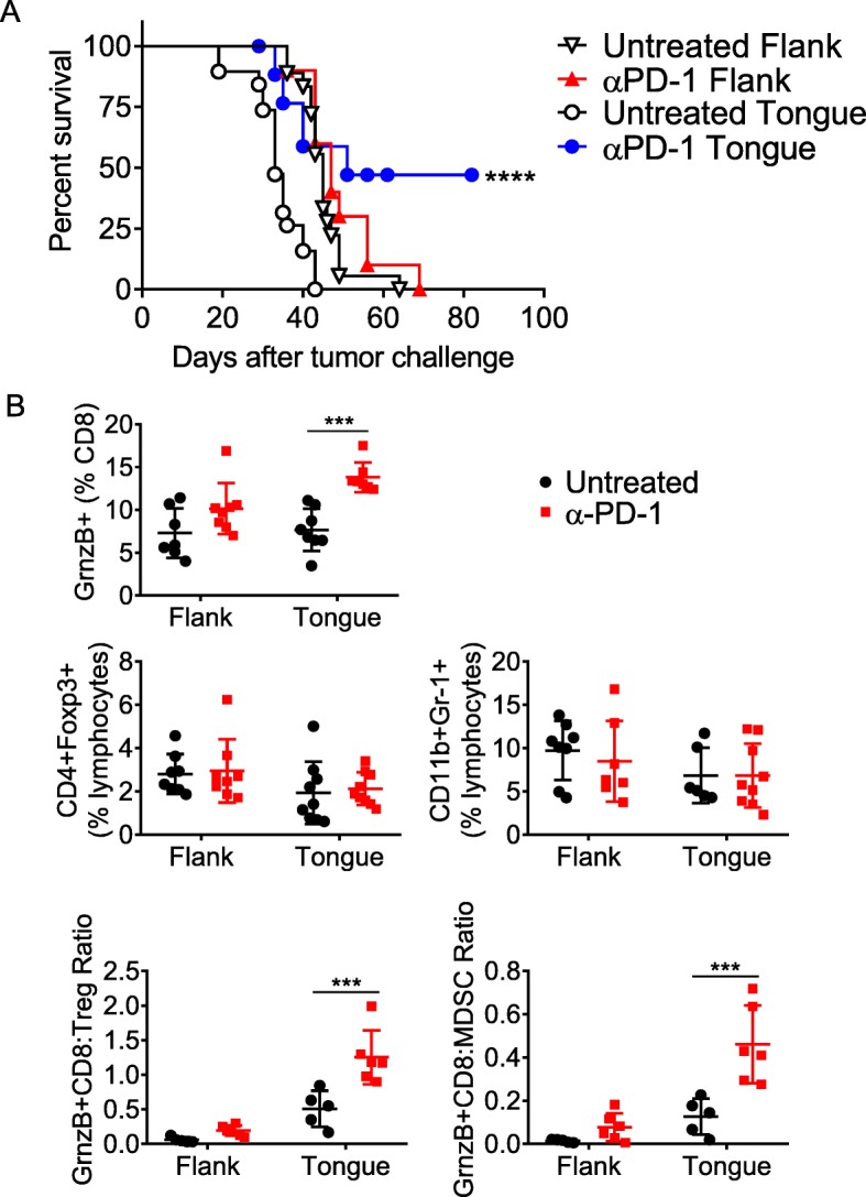
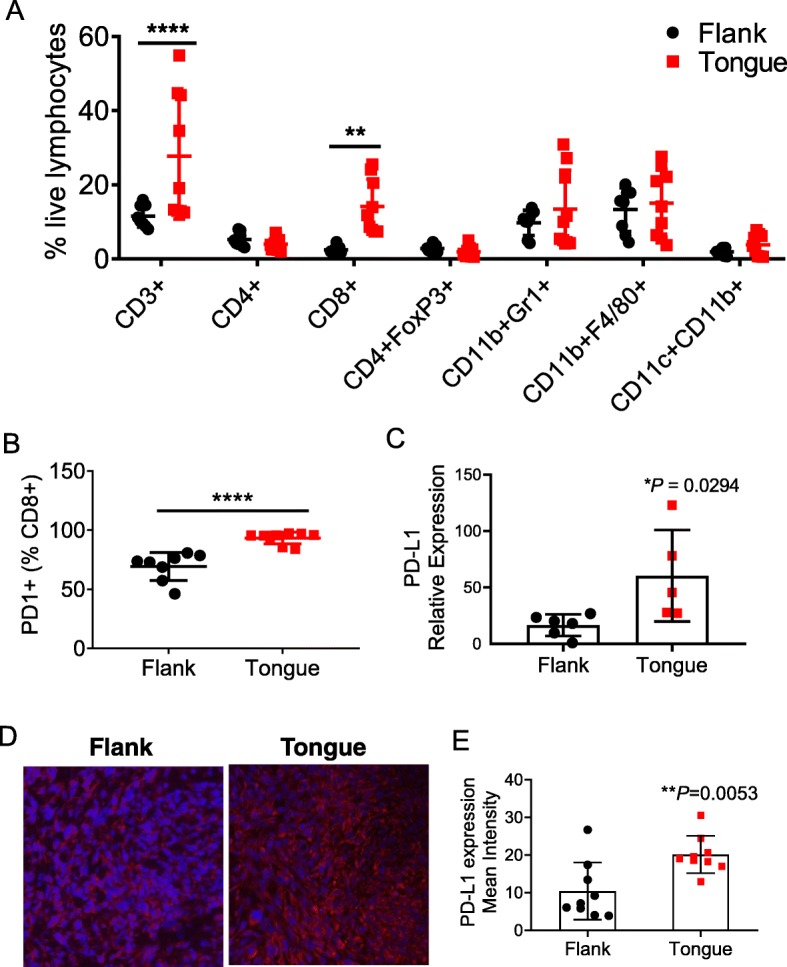
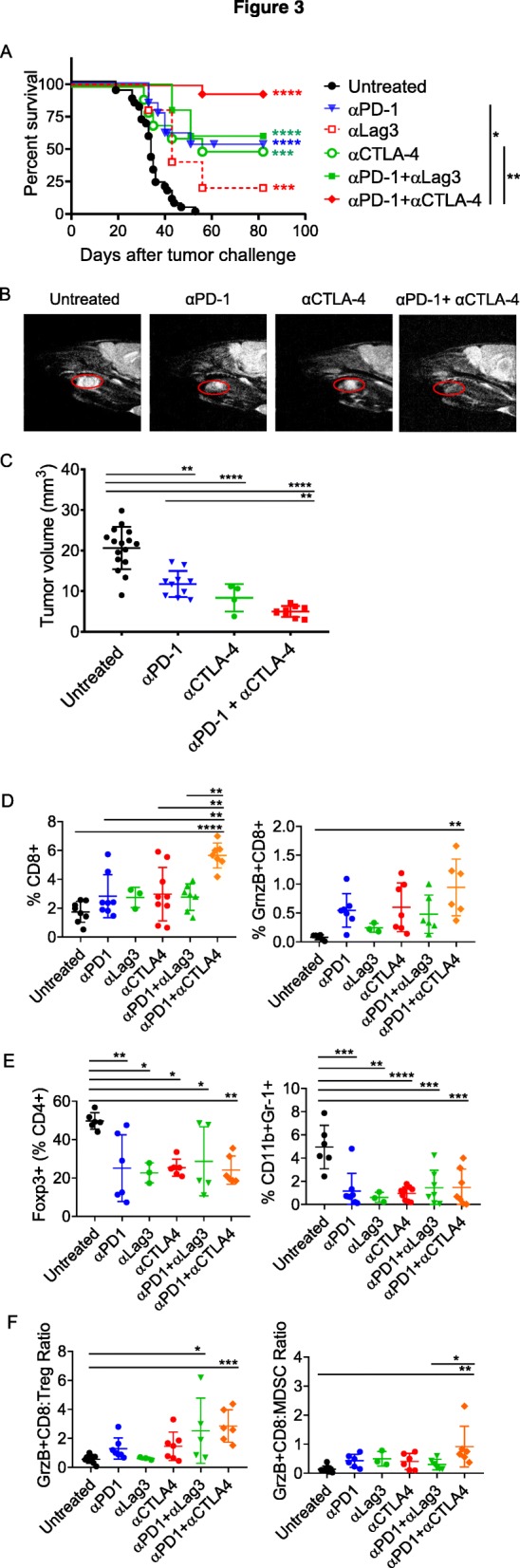
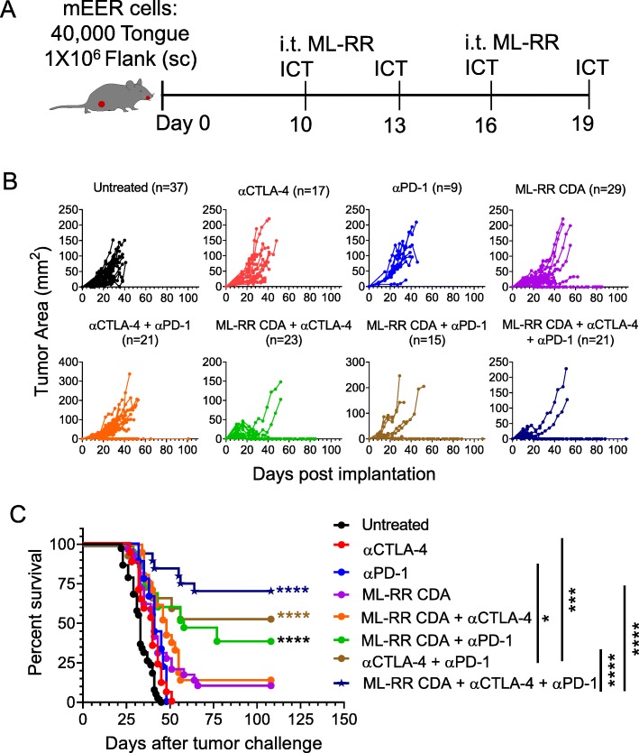
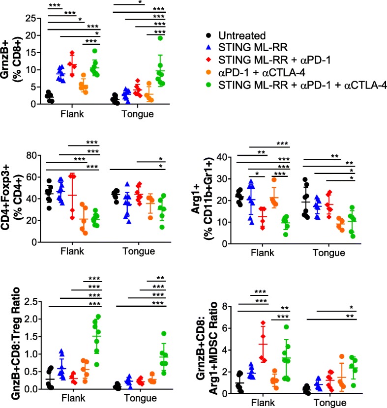
Results: Monotherapy with PD-1 blocking antibody was ineffective against flank-implanted tumors, but induced regression in 54% of mice bearing orthotopic tongue tumors that correlated with higher CD8 T cell responses. Since the CD8+ T cells derived from tongue tumors also showed high levels of the immune checkpoint inhibitory receptor CTLA-4, we tested combination immunotherapy targeting both CTLA-4 and PD-1 together and observed 93.3% survival of mice bearing tumors in the tongue for the duration of our 100-day study. Protective immunity correlated with a significant decrease in immunosuppressive lymphoid and myeloid populations within the tumor microenvironment. Consistent with the reported capacity of interferon-driven PD-L1/PD-1 pathway induction to serve as a biomarker of response to PD-1 blockade, we observed elevated interferon signaling and significantly higher levels of PD-1/PD-L1 in tongue-implanted mEER tumors compared to those growing on the flank correlating with their preferential responsiveness to PD-1 blockade. More importantly, in a pseudometastasic mouse model bearing both flank and tongue tumors to represent metastatic disease, delivery of Stimulator of Interferon Induced Genes (STING) agonist into the flank tumors combined with systemic treatment with α-PD-1 and α-CTLA-4 antibodies resulted in sustained tumor regression in 71% of mice. In this case, productive abscopal anti-tumor immunity was associated with robust increases in the ratios of cytotoxic CD8+ T cells (CTL) versus regulatory T cells (Treg) and versus functional myeloid-derived suppressor cells (MDSC).

Conclusions: These results support combining α-PD-1 therapy with induction of IFN-α/β signaling via provision of STING agonist and/or through CTLA-4 blockade as potential treatment option for HNSCC patients, especially, those not responding to α-PD-1 monotherapy.

Keywords: Abscopal; CD8+ T cells; Checkpoint blockade; HNSCC; HPV; Immunotherapy; MDSC; STING.


Conflict of interest statement

The authors declare that they have no competing interests.

Figures

Fig. 1
Fig. 1
Differential α-PD1 responsiveness of mEER tumors implanted in the flank and tongue. Separate groups of mice were injected with mEER tumor cells in the tongue (4 × 104) or in the flank (1 × 106), and treated with α-PD1 antibodies at days 5, 8 and 11. The percent survival of mice in the different groups is shown (a). Mantel Cox-test was performed to determine the significance of survival for each of the treatment groups relative to respective untreated group ****p < 0.00005. Results represent pooled data from multiple experiments (n = 10–18 mice/group). b At day 15 after tumor implantation mice in the different groups were sacrificed and the TIL were analyzed by flow cytometry to determine the frequencies of Granzyme B expressing functional CD8+ T cell populations, CD4+Foxp3+ Tregs, CD11b+Gr-1+ MDSC as well as ratios of functional Granzyme B expressing CD8+ T cells to Treg and to MDSC
Fig. 2
Fig. 2
Differential infiltration of T cells between oral and subcutaneous mEER tumors. Tumor-infiltrating leukocytes were isolated at day 15 after tumor implantation from mice bearing flank or tongue mEER tumors and analyzed by flow cytometry. Figure shows percentage of different leukocyte subsets among total live lymphocytes (a) and frequencies of PD-1 expressing CD8+ T cells (b). Results represent pooled data from two separate experiments (n = 8–10). Statistical significance was calculated using Two-way ANOVA **p < 0.005, ****p < 0.00005. Flank and Tongue implanted mEER tumors (n = 3–6) were analyzed for PD-L1 expression by real-time qPCR (c) and fluorescence immunohistochemistry (d, e). Representative IHC images (d) and quantification of PD-L1+ cells (e) are shown. **p = 0.0053, Student’s t-test
Fig. 3
Fig. 3
Efficacy of α-PD-1 therapy of tongue-implanted mEER tumors is enhanced by combination treatment with α-CTLA-4 but not α-Lag3. Mice were challenged with mEER tumor cells (4 × 104) in the tongue and treated with antibodies targeting individual checkpoint receptors PD-1, CTLA-4, or Lag3 or using combinations of α-PD-1 and α-CTLA-4 or α-PD-1 and α-Lag-3 antibodies. The percentages of mice surviving in the different groups are shown (a). Statistical significance was calculated using Log-rank (Mantel-Cox) test. The significant difference for each treatment group compared to untreated control group is indicated by colored stars and between groups is shown on the legend; *p < 0.05, **p < 0.01, ***p < 0.001, ****p < 0.0001. Tongue tumor volume was measured by MRI (T2-weighted sagittal image) at day 19 after tumor implantation and representative data is shown for one mouse in each group (b) along with group means ± SD (n = 4–16 mice/group) (c). **p < 0.01, ****p < 0.0001, one-way ANOVA. Flow cytometry analyses of TIL isolated at day 15 from tongue tumor-bearing mice subjected to different treatments showing frequencies of total CD8+ T cells, Granzyme B expressing CD8+ T cells (d), CD4+FoxP3+ Treg, CD11b+Gr-1+ MDSC (e) as well as ratios of GrnzB+CD8+ T cells to Treg and to MDSC (f). Data shown are mean + SD from two experiments (except for anti-Lag3 group) with individual data points representing pooled TILs of 2–3 tumors. Statistical significance was calculated using one-way ANOVA with Turkey post-hoc test; *p < 0.05, **p < 0.01, ***p < 0.001, ****p < 0.0001
Fig. 4
Fig. 4
Abscopal anti-tumor efficacy of intratumoral STING activation in combination with systemic checkpoint antibodies. Mice were inoculated with mEER tumor cells both in the flank (1 × 106) and tongue (4 × 104) and treated with intratumoral (i.t.) administration of STING agonist (ML-RR CDA) on days 10 and 16 along with or without immunotherapy employing individual or combinations of α-PD-1 and α-CTLA-4 antibodies on days 10, 13, 16, and 19 (a). Growth of flank-implanted tumors over time for individual mice in different treatment groups is expressed in terms of tumor area (mm2) in (b). The data is pooled from three separate experiments and the total number of mice in each group is noted. The survival curves for mice in different treatment groups are shown in (c). Statistical significance for differences in survival of mice in different combination treatment groups relative to untreated control group were calculated using Log-rank (Mantel-Cox) test; *p < 0.05, **p < 0.01, ***p < 0.001, ****p < 0.0001
Fig. 5
Fig. 5
Analysis of immune correlates for combination immunotherapy in the mEER flank-tongue pseudometastasic model. Mice were treated as in Fig. 4a except for ICT antibody administrations performed only on days 10, 13 and 16. Leukocytes isolated from both flank and tongue tumors at day 18 after tumor implantation were analyzed by flow cytometry. Frequencies of total CD8+ T cells, Granzyme B+ CD8+ T cells (CTL), Treg, Arg1+ MDSC as well as ratios of CTL to Treg and Arg1+ MDSC are shown. Results represent pooled data from two experiments (n = 8–14). Statistical significance was calculated using two-way ANOVA and post-hoc correction performed with controlling for false discovery rate (FDR); * < 0.05, ** < 0.01, *** < 0.001

References

    1. Gillison ML, et al. Epidemiology of human papillomavirus-positive head and neck squamous cell carcinoma. J Clin Oncol. 2015;33(29):3235–3242. doi: 10.1200/JCO.2015.61.6995. - DOI - PMC - PubMed
    1. Chaturvedi AK, et al. Human papillomavirus and rising oropharyngeal cancer incidence in the United States. J Clin Oncol. 2011;29(32):4294–4301. doi: 10.1200/JCO.2011.36.4596. - DOI - PMC - PubMed
    1. Dogan V, et al. Current studies of immunotherapy in head and neck cancer. Clin Otolaryngol. 2018;43(1):13–21. doi: 10.1111/coa.12895. - DOI - PubMed
    1. Fourcade J, et al. Upregulation of Tim-3 and PD-1 expression is associated with tumor antigen-specific CD8+ T cell dysfunction in melanoma patients. J Exp Med. 2010;207(10):2175–2186. doi: 10.1084/jem.20100637. - DOI - PMC - PubMed
    1. Chauvin JM, et al. TIGIT and PD-1 impair tumor antigen-specific CD8(+) T cells in melanoma patients. J Clin Invest. 2015;125(5):2046–2058. doi: 10.1172/JCI80445. - DOI - PMC - PubMed

Publication types

MeSH terms