. 2019 Sep 18;4(5):e00506-19.
doi: 10.1128/mSphere.00506-19.

Recovirus NS1-2 Has Viroporin Activity That Induces Aberrant Cellular Calcium Signaling To Facilitate Virus Replication

Affiliations

Recovirus NS1-2 Has Viroporin Activity That Induces Aberrant Cellular Calcium Signaling To Facilitate Virus Replication

Alicia C Strtak et al. mSphere. .

Abstract

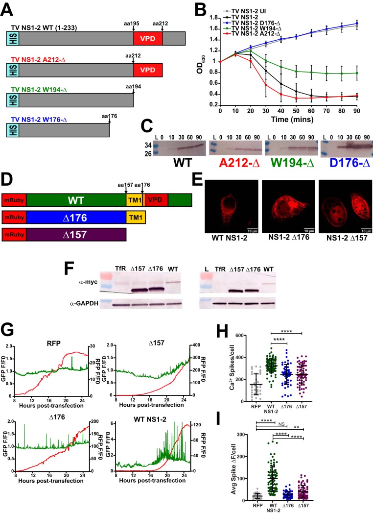
Enteric viruses in the Caliciviridae family cause acute gastroenteritis in humans and animals, but the cellular processes needed for virus replication and disease remain unknown. A common strategy among enteric viruses, including rotaviruses and enteroviruses, is to encode a viral ion channel (i.e., viroporin) that is targeted to the endoplasmic reticulum (ER) and disrupts host calcium (Ca2+) homeostasis. Previous reports have demonstrated genetic and functional similarities between the nonstructural proteins of caliciviruses and enteroviruses, including the calicivirus NS1-2 protein and the 2B viroporin of enteroviruses. However, it is unknown whether caliciviruses alter Ca2+ homeostasis for virus replication or whether the NS1-2 protein has viroporin activity like its enterovirus counterpart. To address these questions, we used Tulane virus (TV), a rhesus enteric calicivirus, to examine Ca2+ signaling during infection and determine whether NS1-2 has viroporin activity that disrupts Ca2+ homeostasis. We found that TV increases Ca2+ signaling during infection and that increased cytoplasmic Ca2+ levels are important for efficient replication. Further, TV NS1-2 localizes to the endoplasmic reticulum, the predominant intracellular Ca2+ store, and the NS2 region has characteristics of a viroporin domain (VPD). NS1-2 had viroporin activity in a classic bacterial functional assay and caused aberrant Ca2+ signaling when expressed in mammalian cells, but truncation of the VPD abrogated these activities. Together, our data provide new mechanistic insights into the function of the NS2 region of NS1-2 and support the premise that enteric viruses, including those within Caliciviridae, exploit host Ca2+ signaling to facilitate their replication.IMPORTANCE Tulane virus is one of many enteric caliciviruses that cause acute gastroenteritis and diarrheal disease. Globally, enteric caliciviruses affect both humans and animals and amass >65 billion dollars per year in treatment and health care-associated costs, thus imposing an enormous economic burden. Recent progress has resulted in several cultivation systems (B cells, enteroids, and zebrafish larvae) to study human noroviruses, but mechanistic insights into the viral factors and host pathways important for enteric calicivirus replication and infection are still largely lacking. Here, we used Tulane virus, a calicivirus that is biologically similar to human noroviruses and can be cultivated by conventional cell culture, to identify and functionally validate NS1-2 as an enteric calicivirus viroporin. Viroporin-mediated calcium signaling may be a broadly utilized pathway for enteric virus replication, and its existence within caliciviruses provides a novel approach to developing antivirals and comprehensive therapeutics for enteric calicivirus diarrheal disease outbreaks.

Keywords: GCaMP; calcium; calicivirus; viroporin.


Figures

FIG 1
FIG 1
TV infection disrupts host calcium signaling kinetics in LLC-MK2 cells. (A) Representative images at early (4 h postinfection [HPI]), onset (8 HPI), and late (12 HPI) stages of mock-infected (top) and TV-infected (bottom) LLC-MK2 GCaMP6s cells. (B) Quantification of GCaMP6s fluorescence from panel A. (C and D) Western blots of TV-infected lysates for nonstructural protein Vpg (C) and structural protein VP1 (D) confirm that aberrant Ca2+ signaling in infected cells coincides with both structural and nonstructural protein synthesis. Mature Vpg in panel C is indicated by a black arrowhead, and the major band (open arrowhead) represents the Vpg-Pro precursor (∼30 kDa). L, lysate; αVpg, anti-Vpg. (E) Western blot of mock lysates for structural protein VP1. (F) One-step growth curve for TV at a low MOI (MOI of 1) shows that virus replication is concomitant with viral protein synthesis (C and D) and with changes in Ca2+ signaling (A). (G) Image from overlay of anti-Vpg staining (red) onto short (10-min) continuous imaging runs of TV-infected cells (MOI of 5) at 12 HPI. Accompanying Ca2+ cell traces (right) show the dynamic increases in cytosolic Ca2+ in infected cells. ROI, region of interest. (H) Compared to mock-infected cells, TV-infected cells have an increased number of Ca2+ spikes per cell that increases in an infectious dose-dependent manner, saturating at an MOI of 5. IRR TV, gamma-irradiated TV. (I) Heatmap data suggest that Ca2+ signaling increases with infectious dose and that a higher MOI disrupts host Ca2+ signaling earlier in infection and sustains this aberrant Ca2+ signaling throughout. Mock-infected and irradiated TV have similar heatmap profiles, suggesting that replication-competent virus is required to drive these changes in Ca2+ signaling. Data are shown as means ± standard deviations (SD) (error bars). Values that are significantly different are indicated by a bar and asterisks as follows: *, P < 0.05; **, P < 0.01; ***, P < 0.001; ****, P < 0.0001. Values that are not significantly different (NS) are also indicated. N ≥ 3 for each experiment, except the one-step growth curve, which was N = 2 with three replicates per experiment.
FIG 2
FIG 2
Intracellular calcium is critical for TV replication. (A) Buffering out extracellular calcium hinders TV replication, significantly reducing the total plaque-forming units (PFU). In contrast, excess extracellular Ca2+ (4 mM Ca2+, right) does not impact replication. (B) Buffering intracellular calcium reduces replication. Depleting ER calcium stores with the SERCA inhibitor thapsigargin (TG), and reducing cytoplasmic Ca2+ with BAPTA-AM significantly reduce TV infectious yield (PFU/ml). (C) Representative images of plaques under normal Ca2+ conditions (2 mM) and reduced Ca2+ (0 mM Ca2+/BAPTA-AM, TG), conditions. The treatment condition is listed above each image, while the dilution each image represents is listed below each image. (D) Diameter of plaques from TV infections cultured in 2 mM Ca2+ (2Ca) or 0 mM Ca2+ (0Ca). (E) Partial one-step growth curve data altering free intracellular (IC) and extracellular (EC) Ca2+. TV replication is stunted in Ca2+-free IC and EC conditions (0 mM Ca2+, 0 mM Ca2+/BAPTA-AM). Inhibiting ER Ca2+ replenishment with thapsigargin (TG) also blunts replication, suggesting that IC Ca2+ stores are critical for TV replication. Data shown are means ± SD. *, P < 0.05; **, P < 0.001; ****, P < 0.0001; NS, not significantly different. N ≥ 3 for each experiment.
FIG 3
FIG 3
TV-induced Ca2+ signaling requires ER Ca2+ stores. (A) Ca2+-free media reduces Ca2+ signaling in TV-infected cells, suggesting that Ca2+ signaling is activated during infection. (B) TV infection in 0 mM Ca2+ phenocopies mock Ca2+ traces in heatmap data, suggesting that extracellular (EC) Ca2+ facilitates TV infection. (C) Intracellular Ca2+ chelator BAPTA-AM abrogated TV-induced Ca2+ signaling. BAPTA-AM-treated TV-infected cells (light green) returns Ca2+ signaling to uninfected levels (gray). (D) Depleting ER Ca2+ with SERCA blocker thapsigargin (TG) significantly reduces TV-induced Ca2+ signaling (pink), suggesting that ER Ca2+ stores are a key source of Ca2+ leveraged during infection. Data shown are means ± SD. ****, P < 0.0001; NS, not significant. N ≥ 3 for each experiment.
FIG 4
FIG 4
Tulane virus NS1-2 is targeted to the ER membrane. (A) Predictive modeling of TV NS1-2 reveals that it has essential features of bona fide viroporins. Kyte-Doolittle hydropathy plots predict an amphipathic moment from amino acids 195 to 212 (aa195-212) (dark green bar), consistent with alpha-helical structure required for channel formation. (B) PSIPred secondary structure algorithms predict that the C terminus of NS1-2 is helical in nature, with the putative viroporin domain (VPD) (dark green bar) contained to helices. (C, top) PSIPred membrane topology predictions suggest that NS1-2 has two transmembrane helices (gray squares). PSIPred algorithms predicting transmembrane helices suggest that NS1-2 transmembrane domains are pore lining (bottom left) and propose a model of membrane insertion and orientation where the putative VPD (aa195-212) comprises the pore-lining helix (bottom right). (D) Helical wheel plot generated from the NS1-2 amphipathic segment (dark green bar) shows clustered basic residues (blue circles) and a hydrophobic moment of 0.522 from aa198-215, coinciding with the putative VPD. (E) Mammalian expressed full-length RFP-NS1-2 and RFP NS1-2 Δ176 are membrane associated, but RFP NS1-2 Δ157 is not. Both the total fraction (T) and membrane pellets (M) extracted with 1% SDS contain RFP-NS1-2 and Δ176, but centrifuged supernatant (S) does not, suggesting that RFP-NS1-2 and Δ176 are membrane-associated proteins. In contrast, the supernatant contains RFP-NS1-2 Δ157. Further, immunoblot assays run under nonreducing conditions show that full-length RFP-NS1-2 and Δ176 oligomerize (black arrowheads). No detection of NS1-2 observed in transfection control lysates. L, lysate; moαmyc, anti-myc monoclonal antibody. (F) Cotransfection experiments using intracellular markers for predominant intracellular Ca2+ stores mitochondria (Mito), Golgi apparatus, and endoplasmic reticulum (ER) to determine whether TV NS1-2 associated with any intracellular organelle(s). Based on deconvolution microscopy data, RFP-NS1-2 localized to the ER (right), but not with the Golgi apparatus (middle). RFP-NS1-2 did not localize to the mitochondria (left) (N ≥ 2). N ≥ 3 for immunoblot experiments.
FIG 5
FIG 5
TV NS1-2 has viroporin activity that disrupts Ca2+ signaling in mammalian cells. (A) Inducing TV NS1-2 in the lysis assay strongly reduces optical density similar to rotavirus NSP4, the positive control for viroporin activity. (B) Western blot data to verify protein expression during the lysis assay for TV NS1-2 (bottom, black arrowhead) and RV NSP4 (top). (C and D) Mammalian recombinant RFP-NS1-2 increases the number (D) and amplitude (C, top row, right) of Ca2+ spikes when transfected into cells similar to RV NSP4 and EV 2B, the viroporin controls for these experiments. Data shown are means ± SD from ≥8 fields of view. *, P < 0.05; ****, P < 0.0001. N ≥ 3 for each experiment.
FIG 6
FIG 6
NS1-2 viroporin mutants do not increase cytoplasmic Ca2+. (A) Schematic of bacterially expressed TV NS1-2 C-terminal truncation mutants to functionally map the viroporin domain. (B) In the lysis assay, truncating the C-terminal domain to amino acid 212 (red) results in wild-type activity (black), but truncating to W194 (green) impairs activity. Truncating to D176 (blue) abrogates viroporin activity, suggesting that the viroporin domain functionally spans from aa177-212. UI, uninduced. (C) Western blots verifying protein expression in the lysis assay. (D) Schematic for the mammalian C-terminal truncation mutant constructs. (E) Immunofluorescence (IF) data for truncation mutants. The Δ157 mutant is cytoplasmic (far right), whereas the Δ176 mutant, which retains one transmembrane segment, is membrane localized (middle). (F) Western blot data confirm the Δ157 and Δ176 mutant constructs. The left blot is run with 20 μl/well to visualize wild-type (WT) NS1-2, whereas the right blot is run with 5 μl/well to resolve the size difference between the Δ157 and Δ176 NS1-2 mutants. TfR, transfection reagent. (G) Representative Ca2+ traces for WT NS1-2 and truncation mutants. (H) Both the Δ157 and Δ176 truncation mutants have significantly fewer Ca2+ spikes/cell compared to wild-type full-length RFP-NS1-2. (I) Compared to full-length RFP-NS1-2, both the Δ157 and Δ176 truncation mutants have significantly reduced Ca2+ spike amplitudes, resulting in a change in cytosolic fluorescence (ΔF) that phenotypically mimics RFP alone. Data shown are means ± SD from ≥8 fields of view. **, P < 0.01; ****, P < 0.0001; NS, not significant. N ≥ 3 for each experiment.
FIG 7
FIG 7
(A) Ca2+ spike analysis for MNV-1 CW-1 infection of GCaMP6s-expressing BV2 cells. Like TV, MNV infection causes aberrant Ca2+ signaling that increases in a dose-dependent manner. (B) Recombinant expression of GII.3 (U201) NS1-2 induces aberrant Ca2+ signaling, similar to TV NS1-2. (C) Representative Ca2+ trace data upon expression of RFP. (D) Representative Ca2+ trace shows that RFP-tagged TV NS1-2 increases the number and amplitude of Ca2+ spikes upon expression. (E) Representative Ca2+ trace for RFP-tagged GII.3 NS1-2 shows that GII.3 NS1-2 also increases the number and amplitude of Ca2+ spikes upon expression. Imaging experiments are quantitated based on ≥30 cells/condition. **, P < 0.01; ****, P < 0.0001. N ≥ 3 for all experiments.

Similar articles

Cited by

References

    1. Wobus CE, Cunha JB, Elftman M, Kolawole AO. 2016. Animal models of norovirus infection, p 397–415. In Svensson L, Desselberger U, Greenberg HB, Estes MK (ed), Viral gastroenteritis: molecular epidemiology and pathogenesis. Academic Press, Cambridge, MA.
    1. Kniel KE. 2014. The makings of a good human norovirus surrogate. Curr Opin Virol 4:85–90. doi:10.1016/j.coviro.2014.01.002. - DOI - PubMed
    1. Desselberger U. 2019. Caliciviridae other than noroviruses. Viruses 11:286. doi:10.3390/v11030286. - DOI - PMC - PubMed
    1. Karst SM, Wobus CE, Goodfellow IG, Green KY, Virgin HW. 2014. Advances in norovirus biology. Cell Host Microbe 15:668–680. doi:10.1016/j.chom.2014.05.015. - DOI - PMC - PubMed
    1. Li J, Predmore A, Divers E, Lou F. 2012. New interventions against human norovirus: progress, opportunities, and challenges. Annu Rev Food Sci Technol 3:331–352. doi:10.1146/annurev-food-022811-101234. - DOI - PubMed

Publication types