. 2019 Nov 26;93(24):e01324-19.
doi: 10.1128/JVI.01324-19. Print 2019 Dec 15.

Tracking the Fate of Endogenous Retrovirus Segregation in Wild and Domestic Cats

Affiliations

Tracking the Fate of Endogenous Retrovirus Segregation in Wild and Domestic Cats

Minh Ha Ngo et al. J Virol. .

Abstract

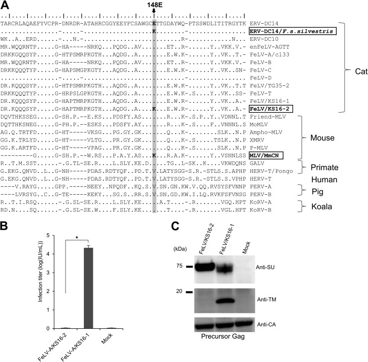
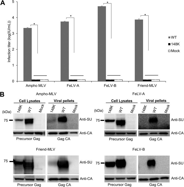
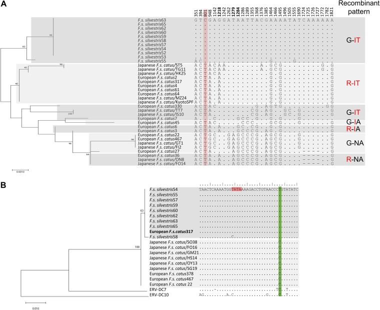
Endogenous retroviruses (ERVs) of domestic cats (ERV-DCs) are one of the youngest feline ERV groups in domestic cats (Felis silvestris catus); some members are replication competent (ERV-DC10, ERV-DC18, and ERV-DC14), produce the antiretroviral soluble factor Refrex-1 (ERV-DC7 and ERV-DC16), or can generate recombinant feline leukemia virus (FeLV). Here, we investigated ERV-DC in European wildcats (Felis silvestris silvestris) and detected four loci: ERV-DC6, ERV-DC7, ERV-DC14, and ERV-DC16. ERV-DC14 was detected at a high frequency in European wildcats; however, it was replication defective due to a single G → A nucleotide substitution, resulting in an E148K substitution in the ERV-DC14 envelope (Env). This mutation results in a cleavage-defective Env that is not incorporated into viral particles. Introduction of the same mutation into feline and murine infectious gammaretroviruses resulted in a similar Env dysfunction. Interestingly, the same mutation was found in an FeLV isolate from naturally occurring thymic lymphoma and a mouse ERV, suggesting a common mechanism of virus inactivation. Refrex-1 was present in European wildcats; however, ERV-DC16, but not ERV-DC7, was unfixed in European wildcats. Thus, Refrex-1 has had an antiviral role throughout the evolution of the genus Felis, predating cat exposure to feline retroviruses. ERV-DC sequence diversity was present across wild and domestic cats but was locus dependent. In conclusion, ERVs have evolved species-specific phenotypes through the interplay between ERVs and their hosts. The mechanism of viral inactivation may be similar irrespective of the evolutionary history of retroviruses. The tracking of ancestral retroviruses can shed light on their roles in pathogenesis and host-virus evolution.IMPORTANCE Domestic cats (Felis silvestris catus) were domesticated from wildcats approximately 9,000 years ago via close interaction between humans and cats. During cat evolution, various exogenous retroviruses infected different cat lineages and generated numerous ERVs in the host genome, some of which remain replication competent. Here, we detected several ERV-DC loci in Felis silvestris silvestris Notably, a species-specific single nucleotide polymorphism in the ERV-DC14 env gene, which results in a replication-defective product, is highly prevalent in European wildcats, unlike the replication-competent ERV-DC14 that is commonly present in domestic cats. The presence of the same lethal mutation in the env genes of both FeLV and murine ERV provides a common mechanism shared by endogenous and exogenous retroviruses by which ERVs can be inactivated after endogenization. The antiviral role of Refrex-1 predates cat exposure to feline retroviruses. The existence of two ERV-DC14 phenotypes provides a unique model for understanding both ERV fate and cat domestication.

Keywords: ERV-DC; FeLV; Felis; Fv-4; MuLV; domestic cat; domestication; endogenous retrovirus; evolution; wildcat.


Figures

FIG 1
FIG 1
Detection of ERV-DC proviruses in domestic cat and wildcat genomes. (A) A phylogenetic tree of the ERV-DC 3′ LTR was constructed using maximum likelihood methods. The percentages at the branch junctions indicate bootstrap values (1,000 replicates). Thirteen ERV-DC loci were classified into three genotypes: genotype I (ERV-DC1, -DC2, -DC3, -DC4, -DC8, -DC14, -DC17, and -DC19), genotype II (ERV-DC7 and -DC16), and genotype III (ERV-DC6, -DC10, and -DC18). (B to D) Insertional polymorphisms of 13 ERV-DCs in Japanese domestic cats (B), European wildcats (C), and European domestic cats (D). Green and +, provirus detected; red and +/−, heterozygous (the copy is present on one of two chromosomes); blue and +/+, homozygous (the copy is present on both chromosomes). (E) Comparison of genotype frequencies for three loci (ERV-DC14, -DC16, and -DC7) among different cat populations. (F) PCR detection of ERV-DC genotypes in European wildcats (F. s. silvestris). Statistical analyses were conducted using Student’s t tests and one-way ANOVAs. *, P < 0.0001.
FIG 2
FIG 2
Characterization and assessment of ERV-DC14/F. s. silvestris. (A) Schematic image of the full-length ERV-DC14/F. s. silvestris provirus. The ERV-DC14 clone SO38 strain was used as the ERV-DC14 reference genome. The gag, pol, and env genes are illustrated together with the 5ʹ and 3ʹ LTRs and the positions of the gag and env translational initiation codons (ATG). Asterisks, stop codons; dark pink box, open reading frame (ORF) of the Gag-Pol polyprotein; light pink box, Env protein; blue and red round circles, single nucleotide polymorphisms (SNPs) between the two proviruses ERV-DC14/SO38 and ERVDC14/F. s. silvestris. Nucleotide substitutions are shown. Flanking 4-bp target duplicate site (TSD) sequences are shown for each provirus. (B) Assessment of the replication-competent activity of ERV-DC14 in European wildcats (F. s. silvestris) and Japanese domestic cats. All tested proviral clones, including ERV-DC14 from different Japanese domestic cats (SO38, GM21, IK19, FO16), ERV-DC14/F. s. silvestris (wildcat 54, and wildcat 63), or the empty vector (mock transfection), were transfected into 293Lac cells, and then their infectivity was tested with fresh HEK293T cells. The viral titers are illustrated as the log number of infectious units (IU) per milliliter with standard deviations. (C) Schematic representation of the chimeric structures of the two proviruses. Chimera1 to Chimera4 were constructed via recombination between the two proviruses using restriction enzyme digestion, and the two mutants were constructed by site-directed mutagenesis. Blue and red round circles indicate single nucleotide polymorphisms (SNPs) between the two proviruses ERV-DC14/SO38 and ERVDC14/F. s. silvestris. (D) Assessment of the replication competence of chimeric ERV-DC14. 293Lac cells were transfected with plasmids containing different chimeric ERV-DC14s or mock transfected (with the empty vector), and the resulting supernatants were collected and used to infect fresh HEK293T cells. The viral titers are illustrated as the log number of infectious units (IU) per milliliter with standard deviations. *, P < 0.0001 (one-way ANOVA).
FIG 3
FIG 3
Determination of the mutation responsible for ERV-DC14/F. s. silvestris Env dysfunction. (A) Amino acid sequence alignment of Env proteins of ERV-DC14 and ERV-DC4/F. s. silvestris. SU, surface subunit; PRR, proline-rich region; TM, transmembrane subunit. RXRR is the cleavage motif. CXXC and CX6CC are sites of covalent interaction. Arrows indicate the positions of amino acids 148 and 273, which differ between these two ERV-DC14 proviruses. (B) Assessment of Env-pseudotyped viruses based on the ERV-DC14/F. s. silvestris wild type (WT), Mutant1 (K148E), and Mutant2 (S273P) or on ERV-DC14. GPLac cells were transfected with the indicated Env expression plasmids. The corresponding Env-pseudotyped viruses were used to infect fresh HEK293T cells. The viral titers are illustrated as the log number of infectious units (IU) per milliliter with standard deviations. *, P < 0.0001 (one-way ANOVA). (C) Western blotting of GPLac cells expressing ERV-DC14/F. s. silvestris Env (K148E, S273P, or WT) or ERV-DC14 Env. The cell lysates and viral pellets from culture supernatants were analyzed. A goat polyclonal anti-FeLV SU (gp70) antibody was used to detect ERV-DC14 SU, and a mouse monoclonal anti-FeLV TM protein (p15E) antibody was used to detect the ERV-DC14 TM protein. The black arrow indicates immature SU; the gray arrow indicates mature SU. Precursor Gag (Pr65) and Gag CA (p30) were both detected with a goat anti-Raucher MLV CA antibody.
FIG 4
FIG 4
Flow cytometry analysis of ERV-DC14/F. s. silvestris cell surface expression. (A and B) Detection of Env on the interior and exterior of cells. HEK293T cells expressing ERV-DC14 Env (A) or ERV-DC14/F. s. silvestris Env (B) were permeabilized with 0.2% Triton X-100 in PBS (right) or were not permeabilized (left), and intracellular (right) and cell surface (left) Env proteins were stained with goat anti-FeLV SU (gp70) and phycoerythrin (PE)-conjugated anti-goat IgG antibody. Fluorescent signals were detected using the FL-2 channel of a flow cytometer. Histograms of Env-expressing cells (red) and mock-transfected cells (black) are overlaid in each graph. The x axis shows the signal intensity in FL-2; the y axis shows the cell counts. (C) Staining of human AKT in permeabilized (right) or nonpermeabilized (left) samples of mock-transfected cells using rabbit anti-human AKT and fluorescein isothiocyanate (FITC)-conjugated goat anti-rabbit IgG antibodies. Histograms of cells treated with anti-human-AKT antibody (red) and not treated with antibody (black) are overlaid in each graph. The x axis shows the signal intensity in FL-1; the y axis shows the cell counts.
FIG 5
FIG 5
Sequence analysis of gammaretrovirus Env proteins and dysfunction of a FeLV variant induced by a single mutation in the SU N-terminal domain. (A) Sequence alignment of Env in gammaretroviruses, constructed by use of the mafft tool (70). The amino acid sequences of the gammaretrovirus SU N-terminal regions are also presented. The critical amino acid position 148E is shaded in light gray. The GenBank accession numbers of the reference sequences are as follows: BBL19108.1 for ERV-DC14/F. s. silvestris, BAM33599.1 for ERV-DC14, BAM33597.1 for ERV-DC10, AY364318.1 for enFeLV-AGTT, BAB63924.2 for FeLV-A clone 33 (cl33), AAA43052.1 for FeLV-B, AAA43049.1 for FeLV-C, BAM33588.1 for FeLV-D, BAU61794.1 for FeLV/TG35-2, AAA43050.1 for FeLV-T, BAK41670.2 for FeLV/KS16-1, BBL19109.1 for FeLV/KS16-2, AAA46480.1 for Friend MLV, NP_057935.1 for MoMLV, AAA46515.1 for Ampho-MLV, ADU55755.1 for XMRV, ARB03464.1 for P-MLV, AMK06448.1 for MLV/MmCN, AAA46811.1 for GALV, CAI15393.1 for HERV-T/Pongo, XP_011526770.1 for HERV-T, AAQ83899.1 for PERV-A, BAM67147.1 for KoRV-A, and AGO86848.1 for KoRV-B. (B) Infection assay using pseudotyped viruses of two FeLV variants (KS16-1 and KS16-2). GPLac cells were transfected with Env expression plasmids for FeLV/KS16-1 and FeLV/KS16-2. The filtered viral supernatants were used to infect fresh HEK293T cells. The viral titers are illustrated as the log number of infectious units (IU) per milliliter with standard deviations. *, P < 0.0001 (one-way ANOVA). (C) Immunoblotting analysis using cell lysates from GPLac cells transfected with the Env expression plasmids which are presented in panel B. Env proteins were detected by use of a mouse anti-FeLV SU (gp70) antibody and an anti-FeLV TM protein (p15E) antibody. Precursor Gag (Pr65) was detected with a goat anti-Raucher MLV CA antibody. The filter exposure time differed for each antibody.
FIG 6
FIG 6
Dysfunction caused by mutations within the SU N-terminal domain in gammaretroviruses. (A) Infection assay using pseudotyped viruses of WT or mutant Env (E148K) of FeLV-A clone 33, FeLV-B GA, Ampho-MLV 4070A, and Friend MLV clone 57. Expression plasmids were constructed for Env mutants and expressed in GPLac cells. After 72 h, the cell lysates and viral pellets were harvested from the culture supernatants. Fresh MDTF cells were inoculated with viral supernatants of Friend MLV, and fresh HEK293T cells were inoculated with viral supernatants of the other pseudotyped viruses. After 48 h, the X-Gal-positive cells were counted, and the viral titers are illustrated as the log number of infectious units (IU) per milliliter with standard deviations; *, P < 0.0001 (one-way ANOVA). (B) Immunoblotting analysis of cell lysates (left) and viral pellets (right) from GPLac cells expressing wild-type (WT) Env or Env mutants. FeLV-A Env and FeLV-B Env were detected with an anti-FeLV SU (gp70) antibody, Ampho-MLV Env was detected with an anti-Ampho-MLV SU (gp70) antibody, and Friend MLV Env was detected with an anti-MLV SU (gp70) antibody. Precursor Gag (Pr65) and Gag CA (p30) were both detected with a goat anti-Raucher MLV CA antibody. The filter exposure times differed between the cell lysates and viral pellets.
FIG 7
FIG 7
Analysis of Refrex-1 in European wildcats. (A) Schematic structure of ERV-DC7 and ERV-DC16 in domestic cat and European wildcats. The gag, deletion of polpol), and env genes are illustrated together with the 5ʹ and 3ʹ LTRs and the positions of the gag and env translational initiation codons (ATG). Asterisks, stop codons; dark pink boxes, predicted Gag; light pink boxes, truncated Env protein; black triangle, insertions; white triangle, deletions. Flanking 4-bp target duplicate site (TSD) sequences are shown for each provirus. (B) Dose-dependent inhibitory effect of Refrex-1 on viral infection. The supernatants of HEK293T cells transfected with each provirus (ERV-DC7, ERV-DC7/F. s. silvestris, ERV-DC16, ERV-DC16/F. s. silvestris, or the empty vector as a control [mock transfection]) were diluted and added to fresh HEK293T cells. After removing the supernatants, those cells were infected with the replication-competent ERV-DC14TA virus. X-Gal-positive cells were counted, and viral titers were calculated as the log number of infectious units (IU) per milliliter with standard deviations. (C) Inhibitory effect on viral infection of the Refrex-1 encoded by truncated env from ERV-DC7 and ERV-DC7/F. s. silvestris. The supernatants of HEK293T cells transfected with the indicated expression vectors encoding ERV-DC7 Env or ERV-DC7/F. s. silvestris Env or with the empty vector (mock transfection) were added to fresh HEK293T cells. After removing the supernatants, the cells were challenged with replication-competent ERV-DC14TA. X-Gal-positive cells were counted, and viral titers were calculated as the log number of infectious units per milliliter with standard deviations. (D and E) Detection of Refrex-1 expression in transfected cells. HEK293T cells were transfected with the provirus of ERV-DC7 or ERV-DC7/F. s. silvestris or with the empty vector (mock transfection). At 72 h posttransfection, the lysates were prepared for Western blotting with a polyclonal goat anti-FeLV SU (gp70) antibody. The Refrex-1 protein was detected as bands of ∼28 kDa for ERV-DC7 and of ∼32 kDa for ERV-DC16. The asterisk indicates the Refrex-1 protein. Human anti-β-actin antibody was used as an internal control.
FIG 8
FIG 8
Sequence diversity of ERV-DC in wildcats and domestic cats. (A) Sequence diversity of ERV-DC7 env in wildcats and domestic cats. The ERV-DC7 env sequences from 11 European wildcats were determined. A phylogenetic tree of the ERV-DC7 env sequences was constructed using maximum likelihood methods (left). The percentages at the branch junctions indicate their bootstrap values (1,000 replicates). Excerpts of polymorphic sites from the ERV-DC7 env sequence are shown in the middle. The numbers indicate nucleotide positions, and bold numbers indicate the nucleotide positions causing amino acid substitutions at positions 407, 427, and 429 (corresponding to R, I, and T, respectively) which cause defective Env cleavage in ERV-DC7fl. The nucleotide change generating a latent stop codon in domestic cats is shaded in pink. On the right, the sequence variations at positions 407 and 427 to 429 are shown. Amino acids suppressing Env cleavage are in red. (B) Phylogenetic and sequence analysis of ERV-DC14 based on the 5ʹ LTR. A phylogenetic tree of the ERV-DC14 5ʹ LTR in three different cat populations was constructed using maximum likelihood methods (left). The percentages at the branch junctions indicate their bootstrap values (1,000 replicates). The nucleotide sequences of the 5ʹ LTR are also presented (right). The position of the TATA box is shaded in pink, and the green shading indicates nucleotide determinants distinguishing wildcats and domestic cats.

Similar articles

Cited by

References

    1. Pontius JU, Mullikin JC, Smith DR, Agencourt Sequencing Team, Lindblad-Toh K, Gnerre S, Clamp M, Chang J, Stephens R, Neelam B, Volfovsky N, Schäffer AA, Agarwala R, Narfström K, Murphy WJ, Giger U, Roca AL, Antunes A, Menotti-Raymond M, Yuhki N, Pecon-Slattery J, Johnson WE, Bourque G, Tesler G, NISC Comparative Sequencing Program, O’Brien SJ. 2007. Initial sequence and comparative analysis of the cat genome. Genome Res 17:1675–1689. doi:10.1101/gr.6380007. - DOI - PMC - PubMed
    1. Waterston RH, Lindblad-Toh K, Birney E, Rogers J, Abril JF, Agarwal P, Agarwala R, Ainscough R, Alexandersson M, An P, Antonarakis SE, Attwood J, Baertsch R, Bailey J, Barlow K, Beck S, Berry E, Birren B, Bloom T, Bork P, Botcherby M, Bray N, Brent MR, Brown DG, Brown SD, Bult C, Burton J, Butler J, Campbell RD, Carninci P, Cawley S, Chiaromonte F, Chinwalla AT, Church DM, Clamp M, Clee C, Collins FS, Cook LL, Copley RR, Coulson A, Couronne O, Cuff J, Curwen V, Cutts T, Daly M, David R, Davies J, Delehaunty KD, Deri J, Dermitzakis ET, et al. . 2002. Initial sequencing and comparative analysis of the mouse genome. Nature 420:520–562. doi:10.1038/nature01262. - DOI - PubMed
    1. Lander ES, Linton LM, Birren B, Nusbaum C, Zody MC, Baldwin J, Devon K, Dewar K, Doyle M, FitzHugh W, Funke R, Gage D, Harris K, Heaford A, Howland J, Kann L, Lehoczky J, LeVine R, McEwan P, McKernan K, Meldrim J, Mesirov JP, Miranda C, Morris W, Naylor J, Raymond C, Rosetti M, Santos R, Sheridan A, Sougnez C, Stange-Thomann Y, Stojanovic N, Subramanian A, Wyman D, Rogers J, Sulston J, Ainscough R, Beck S, Bentley D, Burton J, Clee C, Carter N, Coulson A, Deadman R, Deloukas P, Dunham A, Dunham I, Durbin R, French L, Grafham D, et al. . 2001. Initial sequencing and analysis of the human genome. Nature 409:860–921. doi:10.1038/35057062. - DOI - PubMed
    1. Nakaya Y, Miyazawa T. 2014. Dysfunction of bovine endogenous retrovirus K2 envelope glycoprotein is related to unsuccessful intracellular trafficking. J Virol 88:6896–6905. doi:10.1128/JVI.00288-14. - DOI - PMC - PubMed
    1. Kozak CA. 2014. Origins of the endogenous and infectious laboratory mouse gammaretroviruses. Viruses 7:1–26. doi:10.3390/v7010001. - DOI - PMC - PubMed

Publication types

MeSH terms

Substances

LinkOut - more resources