. 2019 Nov 13;93(23):e00725-19.
doi: 10.1128/JVI.00725-19. Print 2019 Dec 1.

Hepatitis C Virus Infection Is Inhibited by a Noncanonical Antiviral Signaling Pathway Targeted by NS3-NS4A

Affiliations

Hepatitis C Virus Infection Is Inhibited by a Noncanonical Antiviral Signaling Pathway Targeted by NS3-NS4A

Christine Vazquez et al. J Virol. .

Abstract

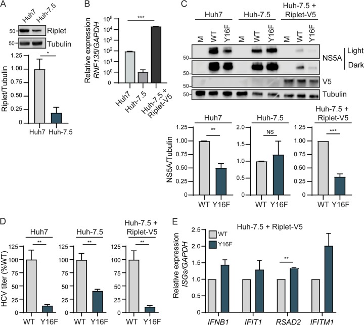
The hepatitis C virus (HCV) NS3-NS4A protease complex is required for viral replication and is the major viral innate immune evasion factor. NS3-NS4A evades antiviral innate immunity by inactivating several proteins, including MAVS, the signaling adaptor for RIG-I and MDA5, and Riplet, an E3 ubiquitin ligase that activates RIG-I. Here, we identified a Tyr-16-Phe (Y16F) change in the NS4A transmembrane domain that prevents NS3-NS4A targeting of Riplet but not MAVS. This Y16F substitution reduces HCV replication in Huh7 cells, but not in Huh-7.5 cells, known to lack RIG-I signaling. Surprisingly, deletion of RIG-I in Huh7 cells did not restore Y16F viral replication. Rather, we found that Huh-7.5 cells lack Riplet expression and that the addition of Riplet to these cells reduced HCV Y16F replication, whereas the addition of Riplet lacking the RING domain restored HCV Y16F replication. In addition, TBK1 inhibition or IRF3 deletion in Huh7 cells was sufficient to restore HCV Y16F replication, and the Y16F protease lacked the ability to prevent IRF3 activation or interferon induction. Taken together, these data reveal that the NS4A Y16 residue regulates a noncanonical Riplet-TBK1-IRF3-dependent, but RIG-I-MAVS-independent, signaling pathway that limits HCV infection.IMPORTANCE The HCV NS3-NS4A protease complex facilitates viral replication by cleaving and inactivating the antiviral innate immune signaling proteins MAVS and Riplet, which are essential for RIG-I activation. NS3-NS4A therefore prevents IRF3 activation and interferon induction during HCV infection. Here, we uncover an amino acid residue within the NS4A transmembrane domain that is essential for inactivation of Riplet but does not affect MAVS cleavage by NS3-NS4A. Our study reveals that Riplet is involved in a RIG-I- and MAVS-independent signaling pathway that activates IRF3 and that this pathway is normally inactivated by NS3-NS4A during HCV infection. Our study selectively uncouples these distinct regulatory mechanisms within NS3-NS4A and defines a new role for Riplet in the antiviral response to HCV. Since Riplet is known to be inhibited by other RNA viruses, such as such influenza A virus, this innate immune signaling pathway may also be important in controlling other RNA virus infections.

Keywords: HCV; MAVS; NS3-NS4A; Riplet; antiviral innate immunity; immune evasion; protease.


Figures

FIG 1
FIG 1
A Y16F substitution in NS4A disrupts replication of an HCV subgenomic replicon in Huh7 cells, but not in Huh-7.5 cells. (A) Amino acid sequence of NS4A, with the Y16 residue starred and indicated in teal. Numbers correspond with the amino acid position within NS4A (amino acids 1 to 54) or the full-length HCV polyprotein (amino acids 1662 to 1715). Strain names are listed as found in the Los Alamos HCV sequence database. Conserved amino acids are indicated with a dot, whereas differences are listed. (B) Representative images of Huh-7.5 or Huh7 cells electroporated with the in vitro-transcribed HCV subgenomic replicon RNA (HP, genotype 1B; WT or Y16F). Cells were plated in serial dilutions (2 × 105, 2 × 104, and 2 × 103) and then stained with crystal violet after 3 weeks of G418 selection. Graphs show the relative transduction efficiency, which denote the percentages of colonies in Y16F-transduced cells relative to the WT. Bars indicate means ± the SEM (n = three to four biological replicates), with data analyzed by Student t test (*, P < 0.05; NS, not significant). (C) Immunoblot analysis of anti-NS4A immunoprecipitated extracts or whole-cell lysate (WCL) from 293T cells transfected with the indicated HCV proteins (genotype 1B) or vector (V). Panels are representative of three independent experiments.
FIG 2
FIG 2
RIG-I deletion in Huh7 cells does not restore HCV NS4A Y16F replication. Huh-7.5 cells (A) or Huh7 cells (B) were infected with HCV, WT, or NS4A Y16F (JFH1, MOI of 0.3). Immunoblot analysis was performed on lysates extracted at the indicated hours postinfection (hpi) or after mock infection (M). Graphs directly to the right of each blot (here and in panel F) show quantification of NS5A protein relative to tubulin at 72 hpi (mean ± the SEM; n = three biological replicates). (C) IFNB1 expression relative to HPRT1 from Huh7 cells infected with HCV, WT, or NS4A Y16F (JFH1, MOI of 1) at 72 hpi as analyzed by RT-qPCR, with data displayed as means ± the SEM (n = three biological replicates). (D) Immunoblot of extracts of Huh7 and Huh7-RIG-I KO cells that were mock or SV infected (20 h). (E) IFN-β promoter reporter luciferase expression of Huh7 and Huh7-RIG-I KO cells expressing either vector or full-length RIG-I that were either mock or SV infected (20 h). Values show means ± the SD (n = three technical replicates) in RLU. (F) Huh7-RIG-I KO cells were infected with HCV, WT, or NS4A Y16F (JFH1, MOI of 0.3). Immunoblot analysis was performed on lysates extracted at the indicated times or mock infected (M). The graph directly to the right of this blot shows the quantification of NS5A protein relative to tubulin at 72 hpi (mean ± the SEM; n = 3 biological replicates). (G to I) Focus-forming assay of supernatants harvested from Huh-7.5 (G), Huh7 (H), and Huh7-RIG-I KO (clones 1 and 2) (I) cells at 72 hpi (MOI of 0.3). The data are presented as the percent HCV titer from the Y16F strain relative to the WT (set at 100%) and show means ± the SEM (n = two or three biological replicates). Data were analyzed by Student t test (*, P < 0.05; **, P < 0.01; ***, P < 0.005; NS, not significant).
FIG 3
FIG 3
HCV NS3-NS4A Y16F retains the ability to cleave MAVS. (A) Immunoblot analysis of lysates harvested from Huh7 cells expressing NS3-NS4A (WT, Y16F, or SA [NS3 active-site mutant S139A]) and vector (V) or Flag-MAVS. Arrows indicate the full-length (FL) and cleaved (C) forms of MAVS. (B) Immunoblot analysis of lysates harvested at 72 hpi from Huh-7.5, Huh7, or Huh7-MAVS KO cells that were either mock infected (M) or infected with HCV, WT, or NS4A Y16F (JFH1, MOI of 0.3). Arrows indicate the full-length (FL) and cleaved (C) forms of MAVS. (C) Intracellular HCV RNA levels in Huh7 cells expressing nontargeting shRNA (shNTV) or MAVS shRNA (shMAVS) infected with HCV, WT, or NS4A Y16F (JFH1, MOI of 0.3) and harvested at 72 hpi, as measured by RT-qPCR. The data are presented as relative HCV copies from Y16F relative to WT (set at 100%) and show the means ± the SEM (n = two biological replicates). Immunoblots to the right of the graph shows MAVS protein expression in the shNTV and shMAVS cell lines. Data were analyzed by Student t test (**, P < 0.01 or as indicated).
FIG 4
FIG 4
IRF3 deletion in Huh7 cells restores HCV Y16F replication to the level of HCV WT. (A) Immunoblot of extracts of Huh7 and Huh7-IRF3 KO cells. (B) IFN-β promoter reporter luciferase expression of Huh7 and Huh7-IRF3 KO cells expressing either vector or IRF3 that were either mock or SV infected (20 h). Values show means ± the SD (n = three technical replicates) in RLU. (C) Immunoblot analysis of lysates harvested at 72 hpi from Huh7 and Huh7-IRF3 KO cells infected with HCV, WT, or NS4A Y16F (JFH1, MOI of 0.3). Graphs below each blot show quantification of NS5A protein relative to GAPDH (means ± the SEM; n = three biological replicates). (D) Focus-forming assay of supernatants harvested at 72 hpi from Huh7 or Huh7-IRF3 KO cells (clones 1 and 2) infected with HCV, WT, or NS4A Y16F (MOI of 0.3). The data are presented as the percent HCV titer from the Y16F strain relative to the WT (set at 100%) and show means ± the SEM (n = two biological replicates). Data were analyzed by Student t test (*, P < 0.05; **, P < 0.01; ***, P < 0.005; NS, not significant).
FIG 5
FIG 5
HCV NS3-NS4A Y16F does not block IRF3 activation. (A) Confocal micrographs of Huh7 cells expressing either NS3-NS4A WT or Y16F (genotype 1B), or vector, that were either mock or SV infected (20 h) and immunostained with anti-IRF3 (green), anti-NS4A (red), or anti-SV (magenta). Nuclei were stained with Hoechst (blue). Scale bar, 10 μm. (B) Quantification of the percent cells positive for SV (and either WT or Y16F NS3-NS4A, as indicated) that had nuclear IRF3. The data are displayed as means ± the SEM (n = two biological replicates of >500 cells counted under each condition and replicate). Data were analyzed by one-way ANOVA (**, P < 0.01; ***, P < 0.005). (C) Immunoblot analysis of lysates from Huh7(–), Huh7-HP WT replicon, or Huh7-HP Y16F replicon cells, and a heat map (below) that shows the mean relative fold induction (SV infected/mock infected, relative to HPRT1) of specific genes, as measured by RT-qPCR analysis of RNA from mock- or SV-infected (20 h) Huh7, Huh7-HP WT replicon, or Huh7-HP Y16F replicon cells from three biological replicates.
FIG 6
FIG 6
TBK1 inhibition restores HCV Y16F viral replication to the level of HCV WT. (A) Immunoblot analysis of lysates harvested from DMSO-treated or BX795 (1 or 10 μM)-treated Huh7 cells that were either mock-infected (M) or infected with SV (20 h). (B) Immunoblot analysis of lysates harvested at 72 hpi from Huh7 cells either pretreated for 1 h with DMSO or with BX795 (10 μM), infected with HCV, WT, or NS4A Y16F (JFH1, MOI of 0.3). Graphs below each blot show quantification of NS5A protein relative to GAPDH (means ± the SEM; n = three biological replicates). Data were analyzed by Student t test (*, P < 0.05; NS, not significant).
FIG 7
FIG 7
Overexpression of Riplet reduces HCV NS4A Y16F replication in Huh-7.5 cells. (A) Immunoblot analysis of lysates harvested from Huh7 and Huh-7.5 cells for endogenous Riplet protein expression. The graph below the blot shows the means ± the SEM (n = three biological replicates) of quantification of Riplet protein expression relative to tubulin. (B) RNF135 (Riplet) expression relative to GAPDH from Huh7, Huh-7.5, and Huh-7.5+Riplet-V5 cells, as analyzed by RT-qPCR, with data displayed as means ± the SD (n = two to three technical replicates). Data were analyzed by one-way ANOVA analysis across the means of the three groups (***, P < 0.005). (C) Immunoblot analysis of lysates harvested from the indicated cell lines infected with HCV, WT, or NS4A Y16F (JFH1, MOI of 0.3) or mock infected (M) at 72 hpi. Two different exposures (light and dark) are shown for NS5A. Graphs below each blot show means ± the SEM (n = three biological replicates) of quantification of NS5A protein relative to tubulin. (D) Focus-forming assay of supernatants harvested at 72 hpi from the indicated cell lines infected with HCV, WT, or NS4A Y16F (MOI of 0.3). The data are presented as the percent HCV titer from the Y16F strain relative to the WT (set at 100%) and show means ± the SEM (n = three biological replicates). (E) RT-qPCR analysis of the indicated IFN-stimulated genes from RNA harvested from Huh-7.5+Riplet-V5 cells infected with HCV, WT, or NS4A Y16F (JFH1, MOI of 1) at 48 hpi. The data are normalized to Huh-7.5+Riplet-V5 mock-infected cells, are presented as the induction of indicated genes (relative to HPRT1 with the WT level set at 1), and display means ± the SEM (n = two biological replicates). Data were analyzed by Student t test (*, P < 0.05; **, P < 0.05; ***, P < 0.005; NS, not significant).
FIG 8
FIG 8
Riplet interaction with HCV NS4A is reduced by the Y16F mutation. (A) Confocal micrographs of Huh7 cells expressing HA-Riplet and either NS3-NS4A WT or Y16F (genotype 1B), or vector, that were immunostained with anti-NS4A (green) and anti-HA (red), with the nuclei stained with Hoechst (blue). The zoom panel is taken from the images in the white boxes. Images are representative of ∼50 cells analyzed. Scale bar, 10 μm. (B) Immunoblot analysis of anti-HA (NS4A) immunoprecipitated extracts or whole-cell lysate (WCL) from Huh7 cells transfected with plasmids expressing Flag-Riplet and NS4A-HA (genotype 1B) WT or Y16F, or vector (–). The graph directly below shows means ± the SEM (n = three biological replicates) of the relative fold change of Flag-Riplet to NS4A-HA in the immunoprecipitated lanes. Data were analyzed by Student t test (*, P < 0.05).
FIG 9
FIG 9
The RING domain of Riplet regulates HCV Y16F virus replication. (A) Immunoblot analysis of Huh-7.5 cells expressing Flag-Riplet full-length (FL) or Flag-Riplet ΔRING. Two different exposures (light and dark) are shown for Flag. (B) Immunoblot analysis of lysates harvested at 72 hpi from Huh-7.5, Huh-7.5+Flag-Riplet FL, and Huh-7.5+Flag-Riplet ΔRING cells infected with HCV, WT, or NS4A Y16F (JFH1, MOI of 0.3). Graphs below the blot show quantification of NS5A protein relative to GAPDH (means ± the SEM; n = three biological replicates).

Similar articles

Cited by

References

    1. World Health Organization. 2017. Global hepatitis report. World Health Organization, Geneva, Switzerland: License: CC BY-NC-SA 3.0 IGO.
    1. Grebely J, Hajarizadeh B, Dore GJ. 2017. Direct-acting antiviral agents for HCV infection affecting people who inject drugs. Nat Rev Gastroenterol Hepatol 14:641–651. doi:10.1038/nrgastro.2017.106. - DOI - PubMed
    1. Bartenschlager R, Baumert TF, Bukh J, Houghton M, Lemon SM, Lindenbach BD, Lohmann V, Moradpour D, Pietschmann T, Rice CM, Thimme R, Wakita T. 2018. Critical challenges and emerging opportunities in hepatitis C virus research in an era of potent antiviral therapy: considerations for scientists and funding agencies. Virus Res 248:53–62. doi:10.1016/j.virusres.2018.02.016. - DOI - PubMed
    1. Horner SM, Gale M Jr. 2013. Regulation of hepatic innate immunity by hepatitis C virus. Nat Med 19:879–888. doi:10.1038/nm.3253. - DOI - PMC - PubMed
    1. Brass V, Berke JM, Montserret R, Blum HE, Penin F, Moradpour D. 2008. Structural determinants for membrane association and dynamic organization of the hepatitis C virus NS3-4A complex. Proc Natl Acad Sci U S A 105:14545–14550. doi:10.1073/pnas.0807298105. - DOI - PMC - PubMed

Publication types

MeSH terms