. 2019 Sep 19;4(18):e128568.
doi: 10.1172/jci.insight.128568.

Inhibition of 4-aminobutyrate aminotransferase protects against injury-induced osteoarthritis in mice

Affiliations

Inhibition of 4-aminobutyrate aminotransferase protects against injury-induced osteoarthritis in mice

Jie Shen et al. JCI Insight. .

Abstract

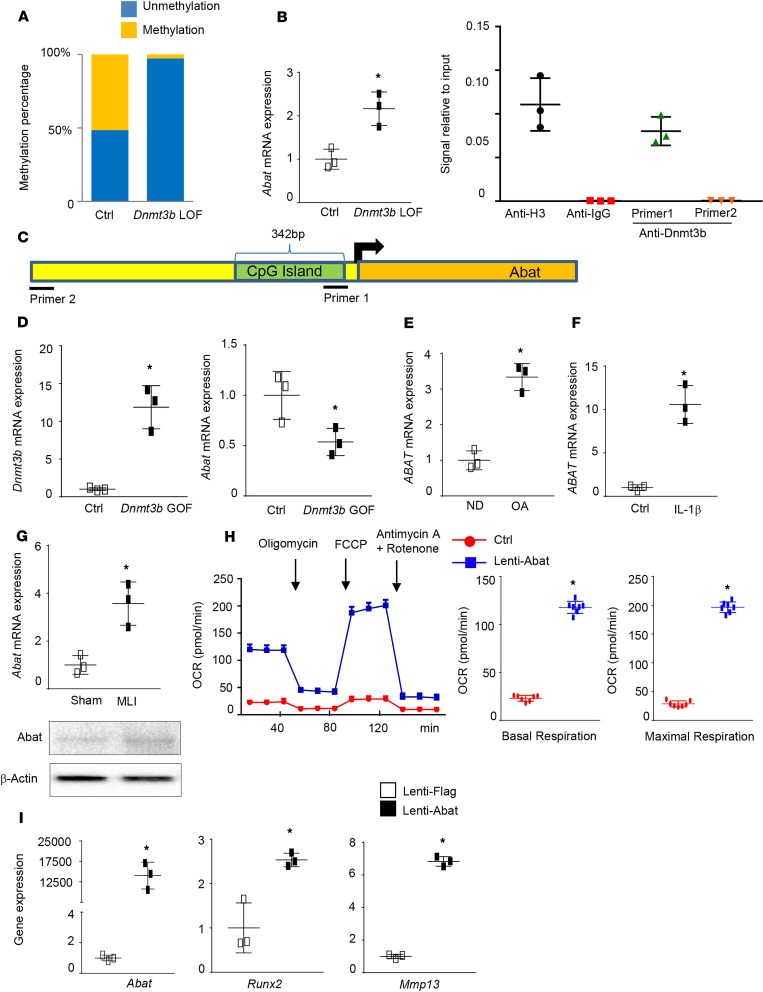
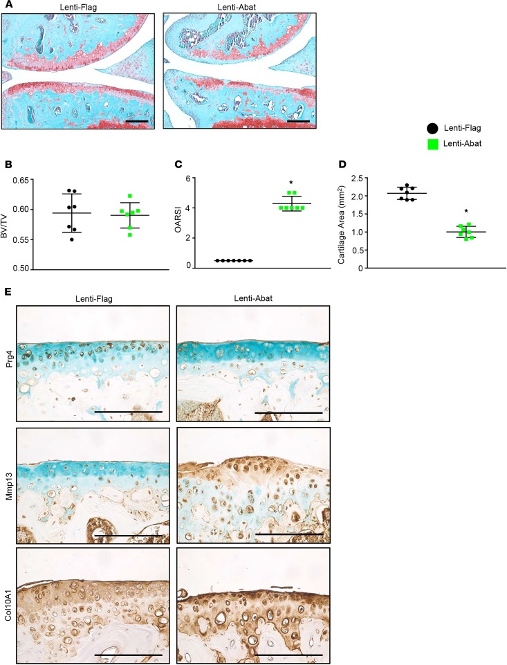
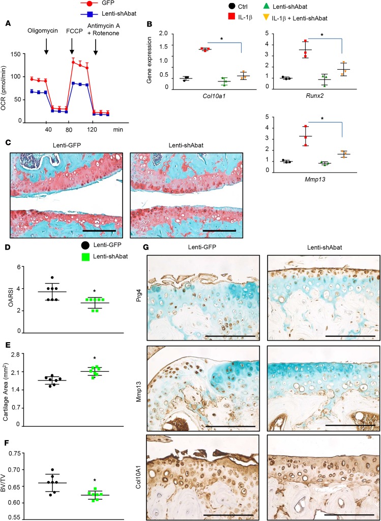
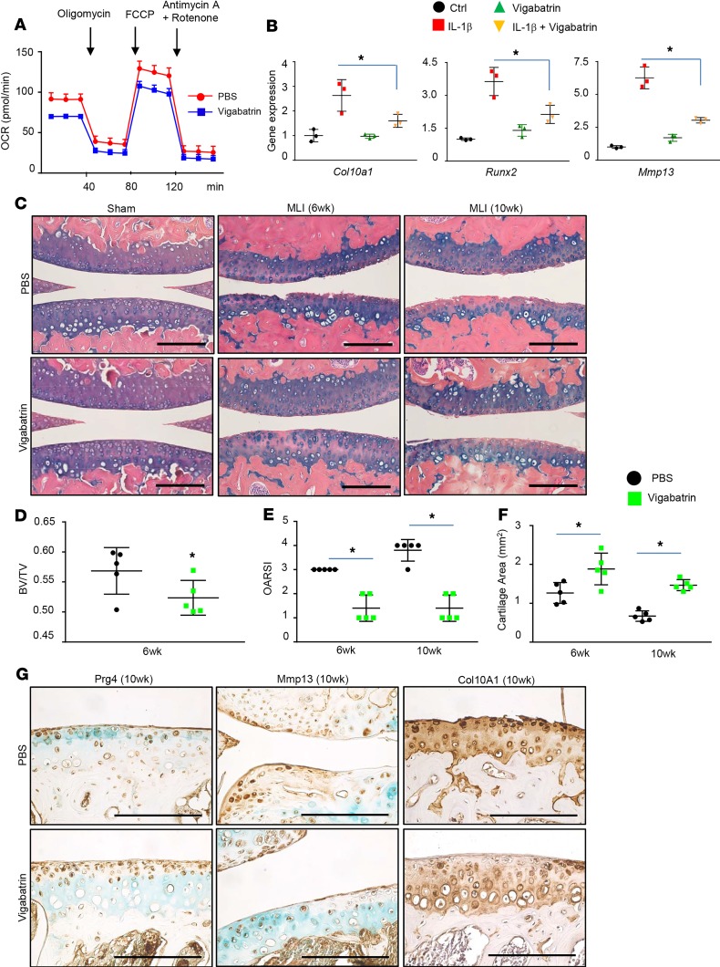
Recently we demonstrated that ablation of the DNA methyltransferase enzyme, Dnmt3b, resulted in catabolism and progression of osteoarthritis (OA) in murine articular cartilage through a mechanism involving increased mitochondrial respiration. In this study, we identify 4-aminobutyrate aminotransferase (Abat) as a downstream target of Dnmt3b. Abat is an enzyme that metabolizes γ-aminobutyric acid to succinate, a key intermediate in the tricarboxylic acid cycle. We show that Dnmt3b binds to the Abat promoter, increases methylation of a conserved CpG sequence just upstream of the transcriptional start site, and inhibits Abat expression. Dnmt3b deletion in articular chondrocytes results in reduced methylation of the CpG sequence in the Abat promoter, which subsequently increases expression of Abat. Increased Abat expression in chondrocytes leads to enhanced mitochondrial respiration and elevated expression of catabolic genes. Overexpression of Abat in murine knee joints via lentiviral injection results in accelerated cartilage degradation following surgical induction of OA. In contrast, lentiviral-based knockdown of Abat attenuates the expression of IL-1β-induced catabolic genes in primary murine articular chondrocytes in vitro and also protects against murine articular cartilage degradation in vivo. Strikingly, treatment with the FDA-approved small-molecule Abat inhibitor, vigabatrin, significantly prevents the development of injury-induced OA in mice. In summary, these studies establish Abat as an important new target for therapies to prevent OA.

Keywords: Arthritis; Bone Biology; Cartilage; Homeostasis.


Conflict of interest statement

Conflict of interest: The authors have declared that no conflict of interest exists.

Figures

Figure 1
Figure 1. Abat is a target of Dnmt3b, and Abat overexpression induces chondrocyte hypertrophy.
Dnmt3b LOF leads to DNA hypomethylation (A) and increased Abat expression (B) in murine chondrocytes (n = 3). (C) Pull-down assay of genomic DNA with a Dnmt3b antibody (ChIP assay) shows interaction of Dnmt3b with its binding site in the Abat promoter region by quantitative PCR (qPCR) using specific primers (n = 3). (D) Real-time qPCR analysis of Dnmt3b and Abat in articular chondrocytes transduced with either lentivirus Dnmt3b (lenti-Dnmt3b) or control virus. (E) Human OA chondrocytes exhibit low ABAT expression compared with nondiseased (ND) chondrocytes (n = 3), and (F) induction of human chondrocytes (n = 3) with IL-1β results in decreased expression of ABAT mRNA. (G) Abat mRNA and protein expression is decreased in MLI cartilage compared with sham surgery cartilage in gene and protein levels (n = 5) 2 weeks after MLI surgery. (H) Mitochondrial respiration was measured by the Seahorse XF Extracellular Flux Analyzer. Basal respiration and maximal respiration, as measured by the oxygen consumption rate (OCR), are shown (n = 8). (I) Real-time qPCR analyses of gene expression from articular chondrocytes transduced with either lenti-Abat or control virus (n = 3). *P < 0.05 by 2-tailed Student’s t test.
Figure 2
Figure 2. Abat overexpression accelerates OA progression in mice.
MLI or sham surgeries were performed on right knee joints followed by intra-articular injection of lenti-Abat (Abat gain of function [GOF]) or lenti-Flag (control). (A) Representative images of histological sections of control and Abat GOF knee joints at 4 weeks following MLI surgery (n = 7). (B) Ratios of bone volume to total volume (BV/TV) of tibial subchondral bone were calculated from the micro-CT images (n = 7). Quantification of histological assessment by Osteoarthritis Research Society International (OARSI) scoring (C) and cartilage area (D) (n = 7). (E) Immunohistochemical analyses for Prg4, Mmp13, and Col10A1 on knee sections of control and Abat GOF cartilage. *P < 0.05 by 2-tailed Student’s t test. Scale bars: 50 μm
Figure 3
Figure 3. Abat inhibition attenuates chondrocyte hypertrophy and protects cartilage degeneration following surgical induction of OA.
(A) Mitochondrial respiration was measured in primary articular chondrocytes treated with lenti-shAbat (Abat LOF) or lenti-GFP (Ctrl) (n = 8). (B) Real-time qPCR analyses of gene expression from articular chondrocytes transduced with either lenti-shAbat (Abat LOF) or control virus following IL-1β treatment (n = 3). (C) Representative images of histological sections from control or Abat LOF knee joints at 10 weeks following MLI surgery (n = 7). Quantification of histological assessment by OARSI scoring (D) and cartilage area (E) (n = 7). (F) BV/TV ratios of tibial subchondral bone were calculated from the micro-CT images (n = 7). (G) Immunohistochemical analyses for Prg4, Mmp13, and Col10A1 on knee sections of control and Abat LOF cartilage. *P < 0.05 by 2-tailed Student’s t test (A and DF) and ANOVA with post hoc test (B). Scale bars: 50 μm.
Figure 4
Figure 4. Vigabatrin attenuates chondrocyte hypertrophy and protects against injury-induced OA progression.
(A) Mitochondrial respiration was measured in primary articular chondrocytes treated with PBS or vigabatrin (n = 8). (B) Real-time qPCR analyses of gene expression from articular chondrocytes pretreated with PBS and vigabatrin (100 μM) following IL-1β treatment (n = 3). (C) Representative images of histological sections of PBS-treated and vigabatrin-treated (200 mg/kg) knee joints at 6 and 10 weeks following MLI surgery (n = 5). (D) BV/TV ratios of tibial subchondral bone were calculated from the micro-CT images (n = 7). Quantification of histological assessment by OARSI scoring (E) and cartilage area (F) (n = 7). (G) Immunohistochemical analyses for Prg4, Mmp13, and Col10A1 on knee sections of PBS- and vigabatrin-treated cartilage 10 weeks after MLI. *P < 0.05 by 2-tailed Student’s t test (A and D) and ANOVA with post hoc test (B, E, and F). Scale bars: 50 μm.

Comment in

  • Taking ABAT to OA.
    Onuora S. Onuora S. Nat Rev Rheumatol. 2019 Dec;15(12):700. doi: 10.1038/s41584-019-0321-8. Nat Rev Rheumatol. 2019. PMID: 31586161 No abstract available.

References

    1. van Baar ME, Dekker J, Lemmens JA, Oostendorp RA, Bijlsma JW. Pain and disability in patients with osteoarthritis of hip or knee: the relationship with articular, kinesiological, and psychological characteristics. J Rheumatol. 1998;25(1):125–133. - PubMed
    1. Woolf AD, Erwin J, March L. The need to address the burden of musculoskeletal conditions. Best Pract Res Clin Rheumatol. 2012;26(2):183–224. doi: 10.1016/j.berh.2012.03.005. - DOI - PubMed
    1. Buckwalter JA, Saltzman C, Brown T. The impact of osteoarthritis: implications for research. Clin Orthop Relat Res. 2004;(suppl 427):S6–15. - PubMed
    1. Cutler DM, Ghosh K. The potential for cost savings through bundled episode payments. N Engl J Med. 2012;366(12):1075–1077. doi: 10.1056/NEJMp1113361. - DOI - PMC - PubMed
    1. Hootman JM, Helmick CG. Projections of US prevalence of arthritis and associated activity limitations. Arthritis Rheum. 2006;54(1):226–229. doi: 10.1002/art.21562. - DOI - PubMed

Publication types

MeSH terms