. 2019 Sep 19;4(18):e129144.
doi: 10.1172/jci.insight.129144.

Lowering circulating apolipoprotein E levels improves aged bone fracture healing

Affiliations

Lowering circulating apolipoprotein E levels improves aged bone fracture healing

Rong Huang et al. JCI Insight. .

Abstract

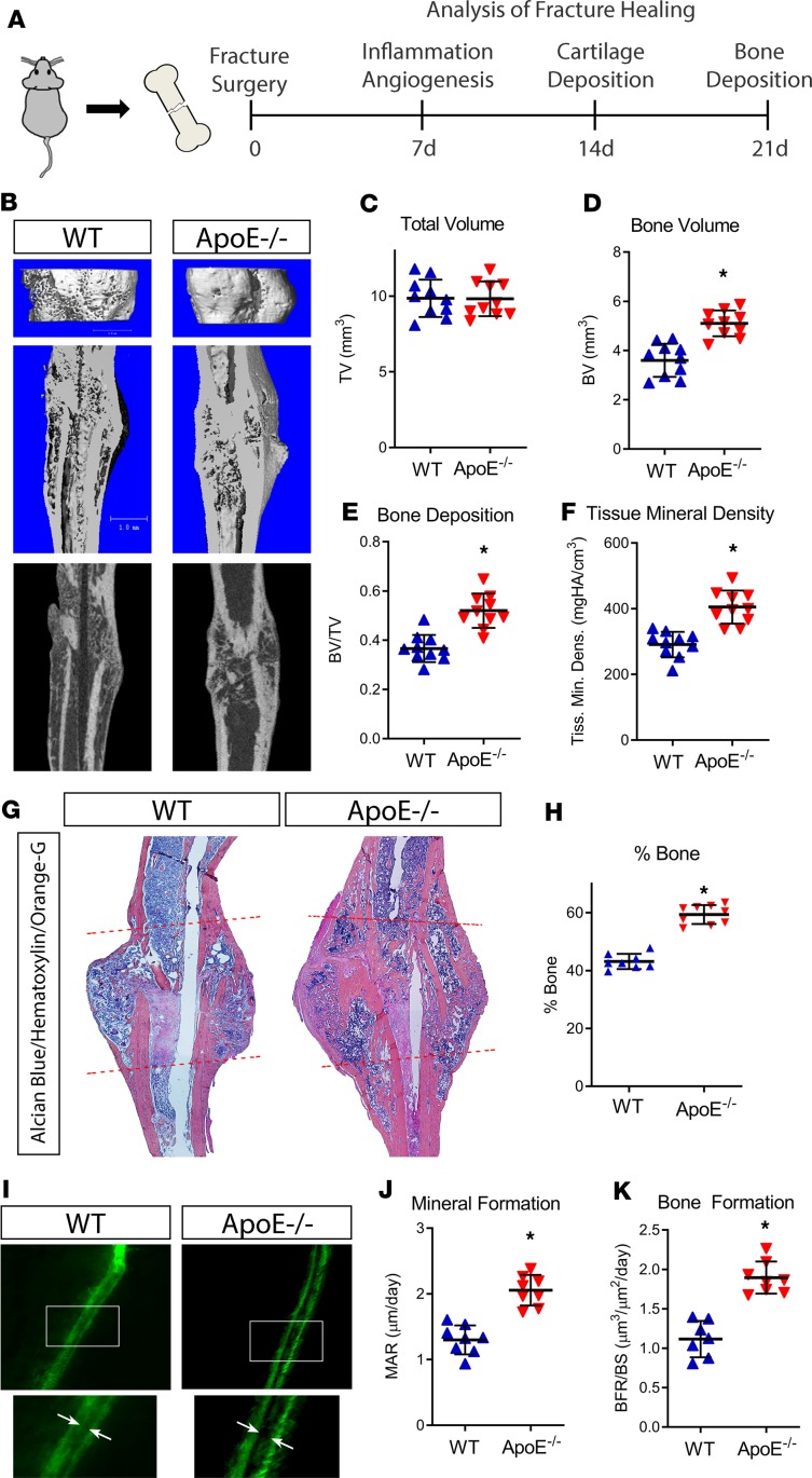
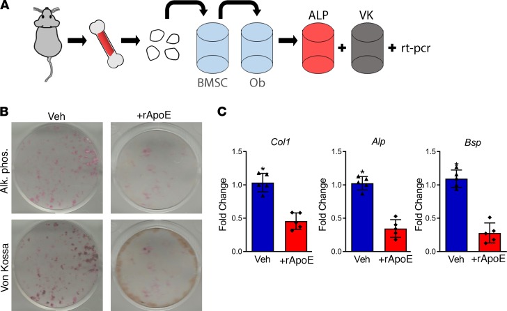
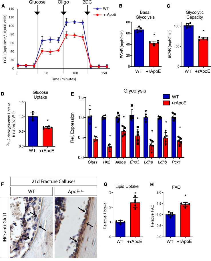
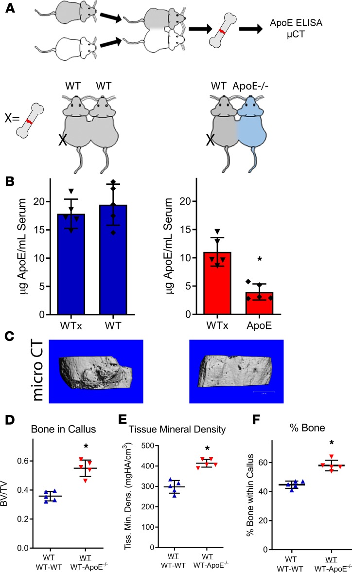
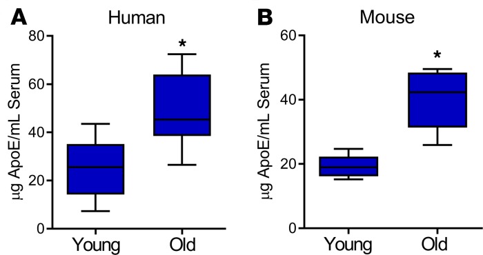
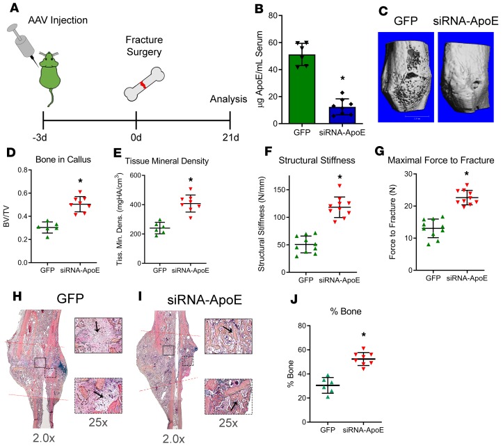
Age is a well-established risk factor for impaired bone fracture healing. Here, we identify a role for apolipoprotein E (ApoE) in age-associated impairment of bone fracture healing and osteoblast differentiation, and we investigate the mechanism by which ApoE alters these processes. We identified that, in both humans and mice, circulating ApoE levels increase with age. We assessed bone healing in WT and ApoE-/- mice after performing tibial fracture surgery: bone deposition was higher within fracture calluses from ApoE-/- mice. In vitro recombinant ApoE (rApoE) treatment of differentiating osteoblasts decreased cellular differentiation and matrix mineralization. Moreover, this rApoE treatment decreased osteoblast glycolytic activity while increasing lipid uptake and fatty acid oxidation. Using parabiosis models, we determined that circulating ApoE plays a strong inhibitory role in bone repair. Using an adeno-associated virus-based siRNA system, we decreased circulating ApoE levels in 24-month-old mice and demonstrated that, as a result, fracture calluses from these aged mice displayed enhanced bone deposition and mechanical strength. Our results demonstrate that circulating ApoE as an aging factor inhibits bone fracture healing by altering osteoblast metabolism, thereby identifying ApoE as a new therapeutic target for improving bone repair in the elderly.

Keywords: Bone Biology; Bone disease; Osteoclast/osteoblast biology.


Conflict of interest statement

Conflict of interest: The authors have declared that no conflict of interest exists.

Figures

Figure 1
Figure 1. Loss of ApoE increases bone deposition during late stages of bone fracture healing.
(A) Schematic diagram of tibial fracture model and subsequent assessment of repair. WT and ApoE–/– mice underwent tibial surgery, and fracture calluses were analyzed 7, 14, and 21 days after injury. (B) Using μCT, 21-day fracture calluses were assessed to determine (C) total callus volume (TV), (D) bone volume (BV), (E) bone content (BV/TV), and (F) tissue mineral density. Scale bar: 1 mm. (G) Histological staining with Alcian blue/hematoxylin/Orange G was used to visualize decalcified tissue (fracture callus region is indicated by dashed lines). (H) Histomorphometric analysis was used to quantify the amount of new bone within the fracture site and related to the total callus size. (I) Calcein double labeling (white arrows) was used to investigate the (J) mineral apposition rate (MAR) and (K) bone formation rate relative to the bone surface (BFR/BS). For μCT, histology, and histomorphometry, WT, n = 10; ApoE–/–, n = 10. For calcein labeling, WT, n = 8; ApoE–/–, n = 8. Data are expressed as mean ± 95% confidence interval. *P < 0.05, 2-tailed t test.
Figure 2
Figure 2. Treatment with rApoE decreases osteoblast differentiation.
(A) Schematic diagram: bone marrow stromal cells (BMSCs) were aspirated from the long bones of WT mice and adhered to tissue culture plastic. After adhesion, cells were passaged to and differentiated to osteoblasts (Ob) by culturing in osteogenic medium for 14 days. ALP, alkaline phosphatase; VK, von Kossa. (B) Fixed, washed wells were assessed for alkaline phosphatase (Alk. Phos.) and for mineralization (Von Kossa). (C) Cell lysates were assessed for osteogenic transcripts (collagen I, Col1; alkaline phosphatase, Alp; and bone sialoprotein, Bsp) using RT-PCR. WT + vehicle, n = 5; WT + rApoE, n = 5. Data are expressed as mean ± 95% confidence interval. *P < 0.05.
Figure 3
Figure 3. rApoE treatment decreases osteoblast glycolytic activity.
WT osteoblasts differentiated in the absence and in the presence of rApoE were assessed for metabolic changes. (A) The Seahorse XF24 extracellular flux metabolic analyzer was used to measure the extracellular acidification rate (ECAR) in response to the addition of glucose, oligomycin (oligo), and 2-deoxyglucose (2DG). These observations were used to determine (B) basal glycolysis and (C) glycolytic capacity. (D) Glucose uptake was measured by pulsing differentiating osteoblasts with 3H-2-deoxyglucose. (E) Transcript levels of key genes involved in glycolytic metabolism were assessed using RT-PCR. (F) Immunohistochemistry was used to investigate the level of Glut1 in healing, 21-day fracture calluses (osteoblasts indicated by arrows). (G) Lipid uptake in cultures was measured in both the absence and presence of rApoE as was (H) fatty acid oxidation (FAO). For metabolic flux experiments, WT + vehicle, n = 5; WT + rApoE, n = 5. For RT-PCR, WT + vehicle, n = 6; WT + rApoE, n = 6. For IHC, WT, n = 4; ApoE–/–, n = 4. For lipid uptake and FAO, WT + vehicle, n = 6, WT + rApoE, n = 6. Data are expressed as mean ± 95% confidence interval. *P < 0.05.
Figure 4
Figure 4. Circulating ApoE inhibits bone fracture healing.
(A) Schematic diagram: WT and ApoE–/– mice were anastomosed together, resulting in a shared blood supply. (B) After 4 weeks of anastomosis, ELISA was used to measure circulating ApoE levels within each mouse. (C) One WT mouse within each pairing underwent tibial fracture surgery, and fracture calluses were assessed 21 days after injury for healing. Scale bar: 1 mm. (D) Bone ratio within the callus and (E) tissue mineral density of the callus were measured using μCT and (F) histomorphometry (decalcified, paraffin embedded, Alcian blue/hematoxylin/Orange G stain) was used to quantify the amount of bone tissue deposited within the fracture callus. For both WT-WT pairs and for WT-ApoE–/– pairs, n = 5. Data are expressed as mean ± 95% confidence interval. *P < 0.05.
Figure 5
Figure 5. Circulating ApoE levels in humans and in mice increase with age.
Serum collected from (A) humans (young, 35–45 years of age; old 75–85 years of age) and from (B) mice (young, 4 months old; old, 24 months old) was assessed for total ApoE protein using ELISA. Young patients, n = 17; old patients, n = 34; young mice, n = 7; old mice, n = 7. Data are expressed as mean ± 95% confidence interval. *P < 0.05.
Figure 6
Figure 6. Decreasing circulating ApoE levels improves bone regeneration in aged mouse models.
(A) Schematic diagram: 24-month-old mice were injected with AAV carrying either GFP or siRNA-ApoE. Mice underwent tibial fracture surgery and were allowed to heal for 21 days. (B) Using ELISA, 24 days after injection, circulating ApoE levels were measured within the serum of mice. (C) Using μCT, 21-day fracture calluses were assessed for (D) bone ratio within the callus and (E) tissue mineral density. Scale bar: 1 mm. Mechanical testing was performed on 28-day fracture calluses to assess (F) structural stiffness and (G) maximal force to fracture. (H and I) Alcian blue/hematoxylin/Orange G staining was performed on decalcified, paraffin-embedded sections. Areas of fibrous tissue (H, GFP, arrows) and areas of bone deposition (I, siRNA-ApoE, arrows) are depicted at indicated original magnifications. (J) Histomorphometry was used to measure the amount of bone tissue deposited within the healing fracture callus. For GFP, n = 6; for siRNA-ApoE, n = 8. Data are expressed as mean ± 95% confidence interval. *P < 0.05.

References

    1. Burge R, Dawson-Hughes B, Solomon DH, Wong JB, King A, Tosteson A. Incidence and economic burden of osteoporosis-related fractures in the United States, 2005–2025. J Bone Miner Res. 2007;22(3):465–475. doi: 10.1359/jbmr.061113. - DOI - PubMed
    1. Baht GS, Vi L, Alman BA. The role of the immune cells in fracture healing. Curr Osteoporos Rep. 2018;16(2):138–145. doi: 10.1007/s11914-018-0423-2. - DOI - PMC - PubMed
    1. Giannoudis PV, Jones E, Einhorn TA. Fracture healing and bone repair. Injury. 2011;42(6):549–550. doi: 10.1016/j.injury.2011.03.037. - DOI - PubMed
    1. Vi L, et al. Macrophage cells secrete factors including LRP1 that orchestrate the rejuvenation of bone repair in mice. Nat Commun. 2018;9(1):5191. doi: 10.1038/s41467-018-07666-0. - DOI - PMC - PubMed
    1. Mahley RW. Apolipoprotein E: cholesterol transport protein with expanding role in cell biology. Science. 1988;240(4852):622–630. doi: 10.1126/science.3283935. - DOI - PubMed

Publication types

MeSH terms