. 2019 Aug 14;9(21):6112-6128.
doi: 10.7150/thno.32982. eCollection 2019.

Noninvasive application of mesenchymal stem cell spheres derived from hESC accelerates wound healing in a CXCL12-CXCR4 axis-dependent manner

Affiliations

Noninvasive application of mesenchymal stem cell spheres derived from hESC accelerates wound healing in a CXCL12-CXCR4 axis-dependent manner

Xiaoyan Wang et al. Theranostics. .

Abstract

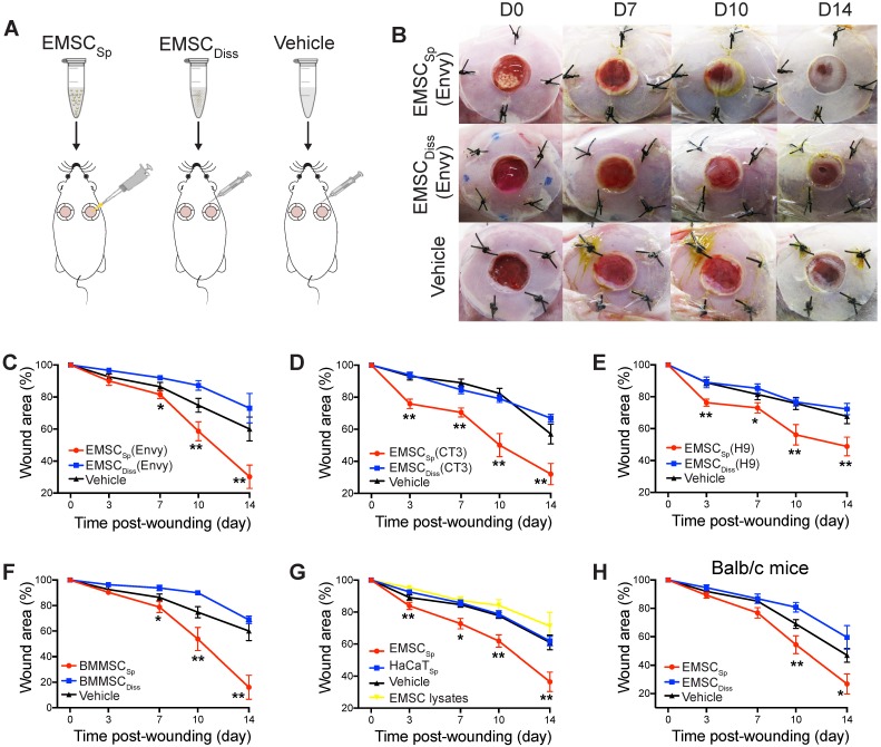
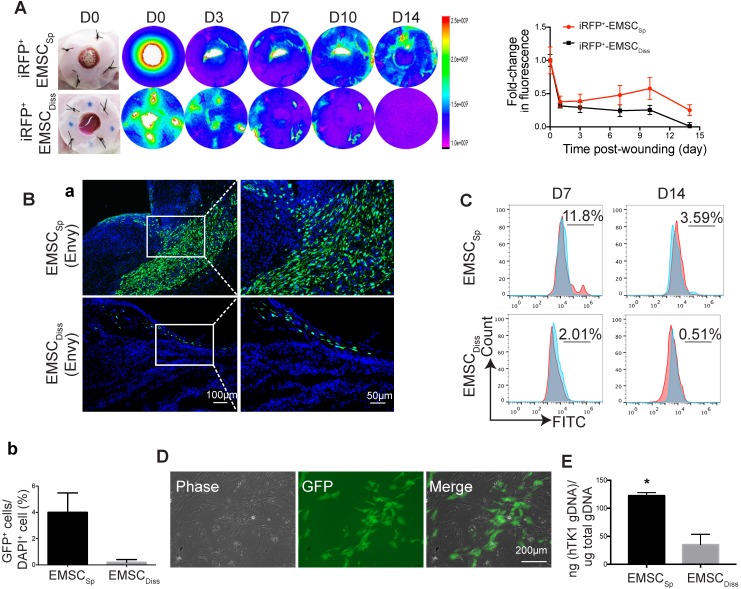
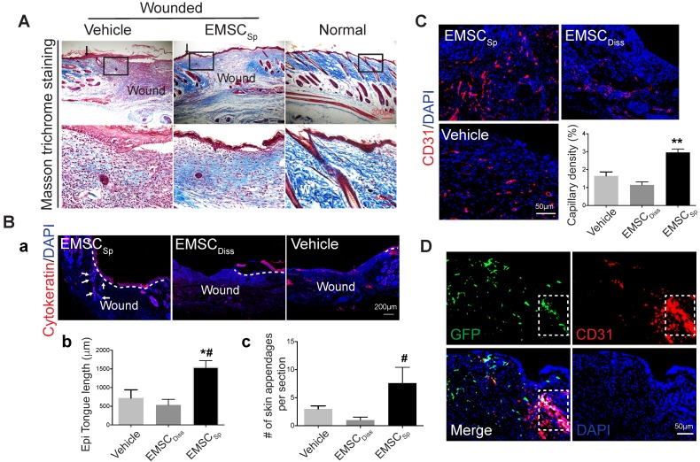
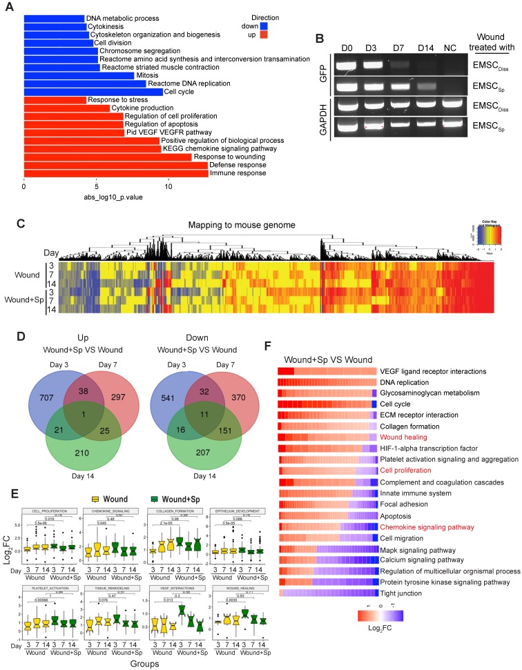
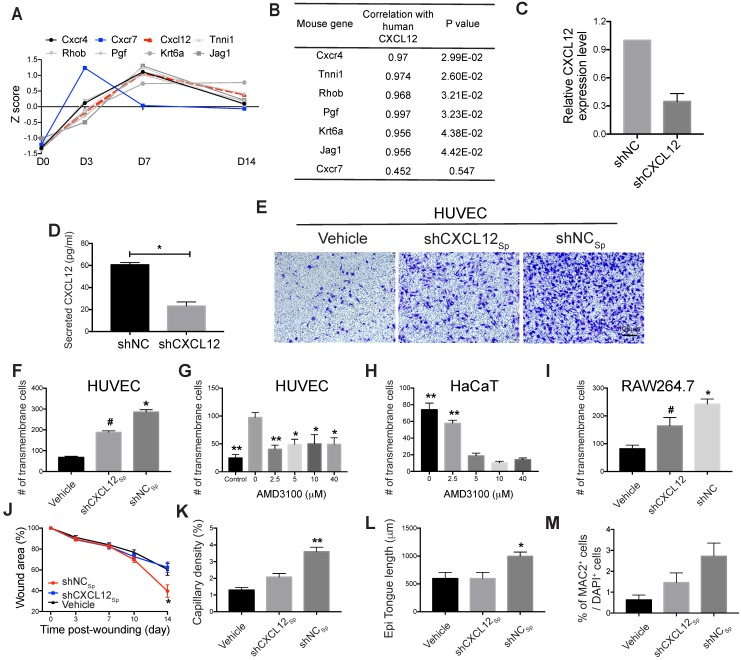
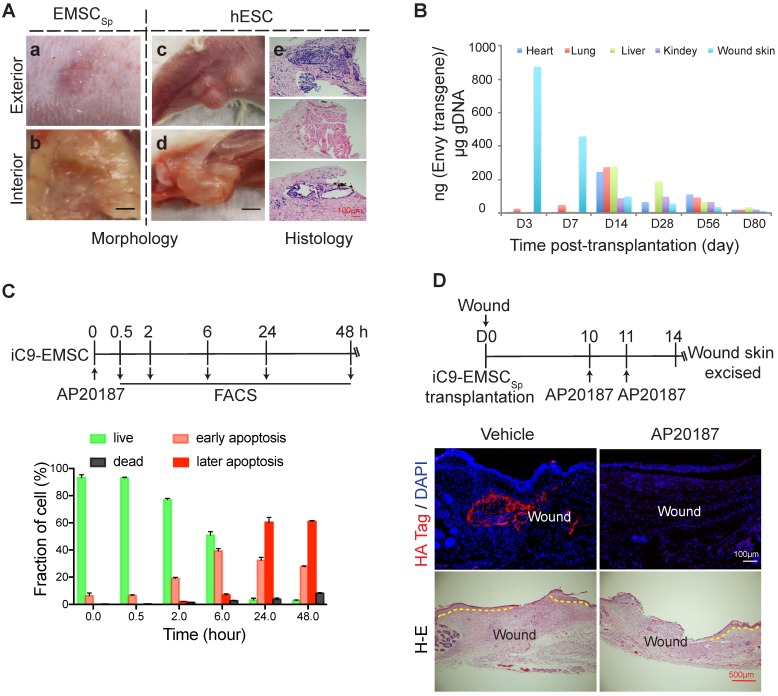
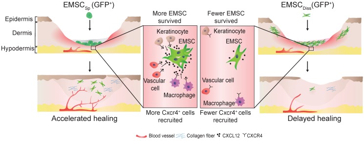
Mesenchymal stem cells (MSC) derived from adult tissues effectively promote wound healing. However, MSC quality varies, and the quantity of MSC is limited, as MSC are acquired through donations. Moreover, the survival and functioning of dissociated MSC delivered to an inflammatory lesion are subject to challenges. Methods: Here, spheres (EMSCSp) generated from human embryonic stem cell-derived MSC (EMSC) were directly dropped onto excised wounds in mice; the effects of EMSCSp were compared to those of dissociated EMSC (EMSCDiss). Following transplantation, we measured the extent of wound closure, dissected the histological features of the wounds, determined transcriptomic changes in cells isolated from the treated and control wounds, and evaluated the molecular mechanism of the effects of EMSC. Results: The application of EMSCSp onto murine dermal wounds substantially increased survival and efficacy of EMSC compared to the topical application of EMSCDiss. RNA sequencing (RNA-Seq) of cells isolated from the wounds highlighted the involvement of CXCL12-CXCR4 signaling in the effects of EMSCSp, which was verified in EMSC via CXCL12 knockdown and in target cells (vascular endothelial cells, epithelial keratinocytes, and macrophages) via CXCR4 inhibition. Finally, we enhanced the biosafety of EMSCSp by engineering cells with an inducible suicide gene. Conclusions: Together, these data suggest the topical application of EMSCSp as an unlimited, quality-assured, safe, and noninvasive therapy for wound healing and the CXCL12-CXCR4 axis as a key player in this treatment.

Keywords: CXCL12/CXCR4; Human embryonic stem cells; mesenchymal stem cells; spheroids; wound healing.


Conflict of interest statement

Competing Interests: R.X. is a founder of ImStem Biotechnology, Inc., a stem cell company. The other authors declare no competing financial interests.

Figures

Figure 1
Figure 1
Effects of EMSC on wound closure. (A) Experimental scheme for the development of an excisional wound splint model in NOD/SCID mice and the transplantation of EMSCSp, EMSCDiss or the vehicle control. (B) Representative images of wounds in mice treated as above at various time points following wound formation. (C-E) The percentage of the wound area in NOD/SCID mice was measured after the transplantation of EMSCs derived from Envy (C), CT3 (D), and H9 (E) hESC lines. n = 5, 8, and 6 biological repeats in C, D, and E, respectively. *P < 0.05 and **P < 0.01 for EMSCSp versus EMSCDiss or the vehicle control per ANOVA followed by Tukey's multiple comparison test. (F) The percentage of the wound area was measured in NOD/SCID mice after transplantation of BM-MSCSp, BM-MSCDiss or the vehicle. n = 5 biological repeats; per ANOVA followed by Tukey's test, *P < 0.05 and **P < 0.01 for BM-MSCSp versus BM-MSCDiss or the vehicle control. (G) The percentage of the wound area was measured in mice after transplantation of EMSCSp, spheres formed by HaCaT cells (HaCaTSp), and EMSC lysates. n = 4 biological repeats, *P < 0.05 and **P < 0.01 for EMSCSp versus HaCaTSp or the EMSC lysates per ANOVA analysis. (H) The percentage of the wound area was measured in Balb/c mice after transplantation of EMSCSp, EMSCDiss or the vehicle. n = 6 biological repeats; per ANOVA followed by Tukey's test, *P < 0.05 and ** P < 0.01 for EMSCSp versus EMSCDiss.
Figure 2
Figure 2
Viability and engraftment of EMSC following their transplantation to wounds. (A) Representative bright-field images of wounds with transplanted iRFP-expressing EMSCSp (upper row) or EMSCDiss (bottom row) were taken on day 0, followed by fluorescent images acquired using the In-Vivo Xtreme System on various days following wound formation. The colored bar indicates the fluorescence intensity. The fold change in the fluorescent intensity is shown in the graph on the right. (B) (a) Immunostaining for GFP+ cells in wounds with transplanted Envy hESC-derived EMSCSp and EMSCDiss 14 days after wound formation. Boxed areas are amplified and shown on the right. (b) The percentage of GFP+ cells (determined via DAPI staining of the nucleus) per wound section. (C) Flow cytometry to detect GFP+ cells in wounds with transplanted Envy hESC-derived EMSCSp and EMSCDiss 7 and 14 days after wound formation. Values represent the percentage of GFP+ cells from 3 biological repeats. (D) Cultured cells were isolated following the digestion of wounds with transplanted EMSCSp (Envy) 7 days after wound formation. Bright-field (left) and fluorescent (middle) images of the cultured cells were taken and merged (right). Scale bar = 200 μm. (E) Detection of human TK1 gDNA in mice 14 days after EMSC transplantation. One x 106 EMSCSp and EMSCDiss cells were transplanted to the skin wound. After 14 days, wounded skin was dissected, and total gDNA was extracted for PCR to detect human TK1. n = 3 biological repeats. * P < 0.05 for EMSCSp versus EMSCDiss per student t-test.
Figure 3
Figure 3
Histological analyses of wounds with transplanted EMSCSp. (A) Masson trichrome staining of normal skin and wounds 14 days after treatment with EMSCSp or vehicle control. Boxed areas are amplified and shown on the bottom row. Arrows in the upper row indicate the junction between the wound and surrounding skin. (B) Re-epithelialization of wounds 14 days after treatment with EMSCSp, EMSCDiss or vehicle. Tissues isolated from the wounds were sectioned and immunostained (a) with anti-pancytokeratin antibody. White dashed lines indicate the detected epidermal keratinocytes, and white arrows indicate skin appendages. The epithelial tongue length (b) and the number of skin appendages per section (c) are shown in the bar charts. n = 5 biological repeats; *P < 0.05 for EMSCSp versus vehicle and #P < 0.05 for EMSCSp versus EMSCDiss per ANOVA followed by Tukey's test. (C) Detection of vascular endothelial cells in wounds 14 days after treatment as described above. Tissues isolated from the wounds were sectioned and immunostained with antibodies against CD31 as a marker for vascular endothelial cells. The results are quantified in a bar graph and displayed as capillary density. n = 5 biological repeats; **P < 0.01 for EMSCSp versus EMSCDiss or vehicle per Kruskal-Wallis test. (D) Differentiation of transplanted EMSCSp into blood vessel endothelial cells. Wounds 14 days after EMSCSp (Envy) transplantation were isolated as described above and immunostained for both GFP (green) and CD31 (red), a marker for blood vessel endothelial cells. The white boxed area indicates double-positive cells.
Figure 4
Figure 4
Transcriptomic analysis of host cells in EMSCSp-treated wounds. (A) RNA sequencing of EMSCSp and EMSCDiss before transplantation. Enrichment analysis of human DEGs in EMSCSp versus EMSCDiss. The bar length represents the -log10 P value. The red and blue bars represent terms enriched for up- and downregulated genes, respectively. (B) Detection of GFP mRNA in the wounds of NOD/SCID mice treated with GFP+ Envy EMSCSp or EMSCDiss. GFP transcript levels were detected via RT-PCR. GFP expression in H9 hESC-derived EMSC was used as a negative control. A GAPDH transcript with a conserved sequence between mice and humans was used as a loading control. (C) Heatmap displaying the transcriptomic changes following the treatment of wounds with EMSCSp (Wound+Sp) and vehicle (Wound) after various time points (3, 7, and 14 days). Expression levels of up- and downregulated genes that differed by more than 1.5-fold are highlighted in red and blue, respectively, as indicated in the color key above. (D) Venn diagram showing the intersections among up- and downregulated genes between the Wound+Sp with Wound samples at the three time points, as described above. (E) Boxplots showing changes in the expression of genes associated with eight wound healing-related pathways in the Wound (yellow) and Wound+Sp (green) samples at the three time points. Each plot stands for a pathway. (F) Heatmaps displaying significantly enriched DEGs categorized based on gene ontology. The expression level of up- and downregulated genes that differed by more than 1.5-fold changes are shown in red and blue, respectively, as indicated in the color key below.
Figure 5
Figure 5
Role of the CXCL12/Cxcr4 axis in EMSCSp-promoted wound healing. (A-B) Pearson's correlation coefficient between the expression of CXCL12 in EMSCSp and the expression of Cxcl12-targeted genes in mouse cells based on RNA-seq data from wounds treated with EMSCSp and vehicle 0, 3, 7, and 14 days after wound formation (A). The X-axis represents the time, and the Y-axis represents the z-score values of the coefficient. The correlations between Cxcl12-targeted genes with CXCL12 expression and their corresponding P values are listed in (B). (C) CXCL12 expression was decreased in EMSC stably transduced with lentivirus expressing shCXCL12 compared to that in EMSC transduced with lentivirus expressing shLacZ. The knockdown efficiency was determined by qPCR with 3 biological repeats. (D) CXCL12 secretion by EMSC transduced with shNC (control) or shCXCL12 was measured via ELISA. *P < 0.05 per the Mann-Whitney U test. (E-F) The migration of HUVECs towards EMSCSp expressing shCXCL12 was reduced compared to HUVEC migration towards EMSCSp expressing shNC in a Transwell assay. HUVECs were seeded on the top of the membrane insert, and no cells (vehicle, left), EMSCSp expressing shNC (shNCSp, right) or shCXCL12 (shCXCL12Sp, middle) were seeded on the bottom of the Transwell insert. HUVECs that migrated across the membrane were stained with crystal violet and photographed under a microscope. The numbers of migrated HUVECs in the three groups are displayed in a bar chart and represent 3 biological repeats. *P < 0.05 for shNCSp versus shCXCL12Sp or vehicle; and #P < 0.05 for shCXCL12Sp versus vehicle per ANOVA followed by Tukey's test. (G) Transwell assay to detect the EMSCSp-oriented migration of HUVECs pretreated with AMD3100 (a CXCR4 antagonist) at various concentrations for 30 min. EMSCSp were seeded on the bottom of the Transwell insert to induce HUVEC migration. After incubation for 24 h, the number of HUVECs that migrated across the membrane was counted. The results were determined in three independent experiments. *P < 0.05 and **P < 0.01 versus 0 μM AMD3100 per ANOVA followed by Tukey's test. (H) Transwell assay to detect the EMSCSp-oriented migration of HaCaT cells pretreated with AMD3100, with the number of cells that crossed the membrane counted as described above. The results were determined in three independent experiments. **P < 0.01 versus 5, 10 or 40 μM AMD3100 per ANOVA followed by Tukey's test. (I) Transwell assay to detect the EMSCSp-oriented migration of RAW264.7 macrophages. The number of cells that crossed the membrane was counted as described above. The results were determined in three independent experiments. *P < 0.05 for shNC versus shCXCL12 or vehicle and #P < 0.05 for shCXCL12 versus vehicle per ANOVA followed by Tukey's test. (J-M) Measurement of the wound area (J), capillary density (number of CD31+ cells among the total cells per wound section) (K), epithelial tongue length (L), and macrophage (MAC2+) percentage (M) in mice after transplantation with shNCSp, shCXCL12Sp or vehicle control. *P < 0.05 and **P < 0.01 for shNCSp versus shCXCL12Sp or vehicle per one-way ANOVA followed by Tukey's or Dunn's posttest.
Figure 6
Figure 6
Evaluation of the biosafety of EMSCSp transplantation and a strategy to establish safety-enhanced EMSC. (A) Teratoma formation assay. Fifty-six days after wound formation, no tumors in the wound area transplanted with EMSCSp were observed based on photographs of the exterior (a) and interior (b) of the healed skin. In contrast, teratomas in the hind leg s.c. injected with hESC were observed in photographs of the exterior (c) and interior (d) of the injection site. Representative tissues from the three germ layers in sections of the teratomas were observed (e). (B) Quantitation of EMSC retained in mouse organs at various times following the transplantation with EMSCSp (Envy) onto wounds. The wounded skin, heart, liver, lung, and kidney were harvested at these time points and processed to isolate gDNA. The amount of GFP (ng) per μg of gDNA was used to determine the number of retained EMSC in each sample. (C) Apoptosis of iC9-expressing EMSC induced by AP20187 in vitro. As shown in the diagram above, after exposure to AP20187, the cells were harvested at the indicated times and processed for flow cytometry via AO/PI staining to determine the ratios of early and late apoptotic cells as well as dead cells. The results of three independent experiments are displayed as the mean ± SEM in a bar chart. (D) AP20187-induced elimination of iC9-EMSC from mice with iC9-EMSCSp transplanted onto the skin wound. As shown in the diagram above, AP20187 was i.p. injected into the mice at 10 and 11 days after wound formation, and the wound skin was dissected at day 14 and subjected to immunostaining for the HA Tag fused with the iC9 gene transduced into EMSC. The samples were counterstained with DAPI to detect cell nuclei. HA+ cells were detected in samples from only mice without AP20187 treatment but not in those treated with the AP20187. The results were determined in four independent experiments. H-E staining in the bottom panel shows the extent of the re-epithelialization of iC9-EMSCSp-transplanted wounds following treatment with the vehicle or CID.
Figure 7
Figure 7
Schematic to summarize the findings of this study.

References

    1. Maxson S, Lopez EA, Yoo D, Danilkovitch-Miagkova A, Leroux MA. Concise review: role of mesenchymal stem cells in wound repair. Stem Cells Transl Med. 2012;1:142–9. - PMC - PubMed
    1. Yan L, Zheng D, Xu RH. Critical role of tumor necrosis factor signaling in mesenchymal stem cell-based therapy for autoimmune and inflammatory diseases. Front Immunol. 2018;9:1658. - PMC - PubMed
    1. Visweswaran M, Pohl S, Arfuso F, Newsholme P, Dilley R, Pervaiz S. et al. Multi-lineage differentiation of mesenchymal stem cells - to Wnt, or not Wnt. Int J Biochem Cell Biol. 2015;68:139–47. - PubMed
    1. Yan L, Jiang B, Niu Y, Wang H, Li E, Yan Y. et al. Intrathecal delivery of human ESC-derived mesenchymal stem cell spheres promotes recovery of a primate multiple sclerosis model. Cell Death Discov. 2018;5:28. - PMC - PubMed
    1. Volarevic V, Gazdic M, Simovic Markovic B, Jovicic N, Djonov V, Arsenijevic N. Mesenchymal stem cell-derived factors: Immuno-modulatory effects and therapeutic potential. Biofactors. 2017;43:633–44. - PubMed

Publication types