. 2019 Aug 26:2019:2432416.
doi: 10.1155/2019/2432416. eCollection 2019.

Sesamin Enhances Nrf2-Mediated Protective Defense against Oxidative Stress and Inflammation in Colitis via AKT and ERK Activation

Affiliations

Sesamin Enhances Nrf2-Mediated Protective Defense against Oxidative Stress and Inflammation in Colitis via AKT and ERK Activation

Xupeng Bai et al. Oxid Med Cell Longev. .

Abstract

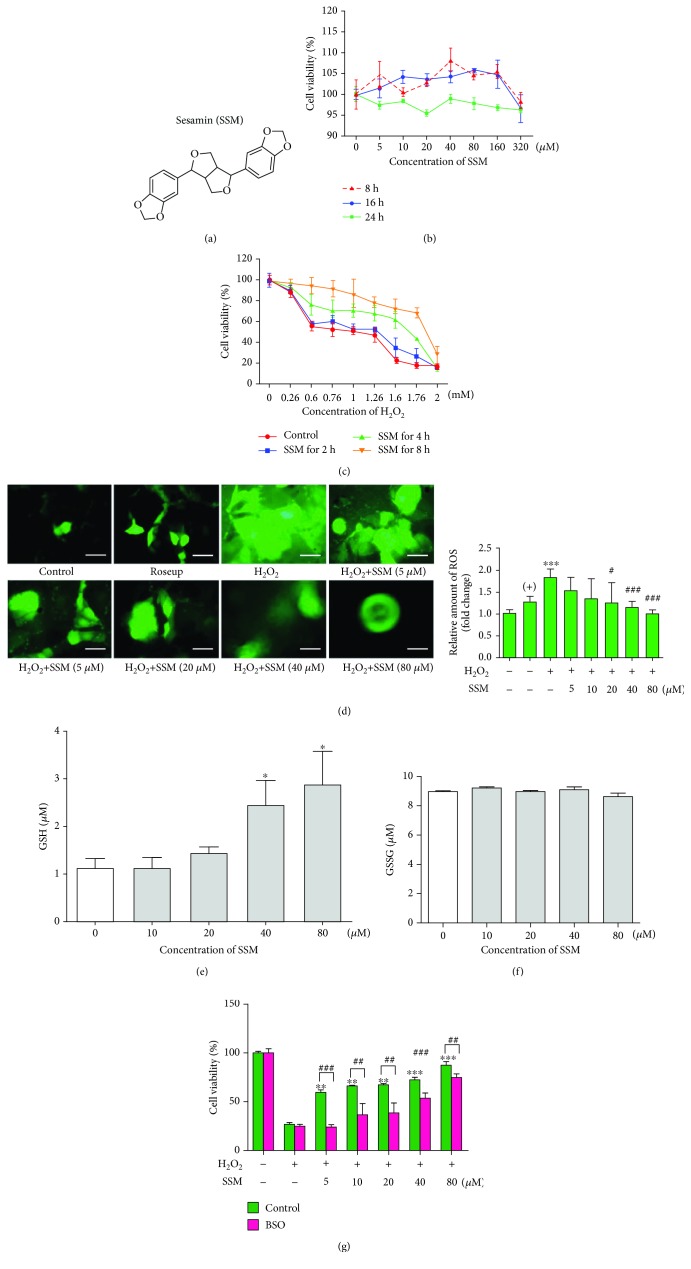
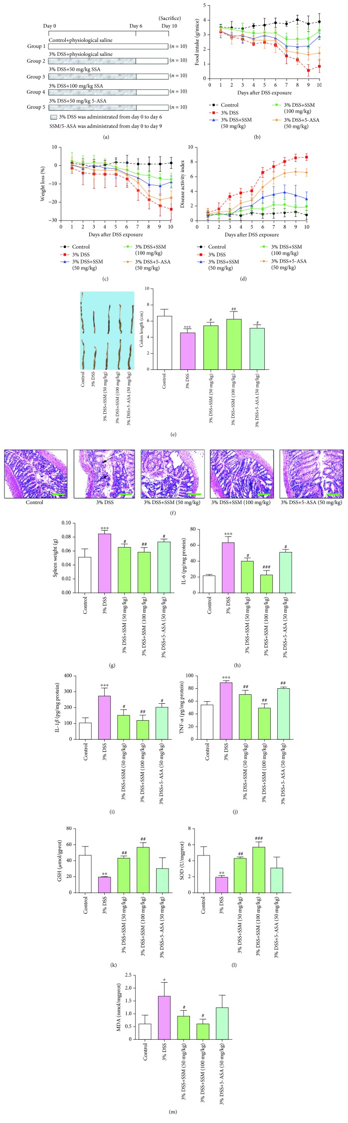
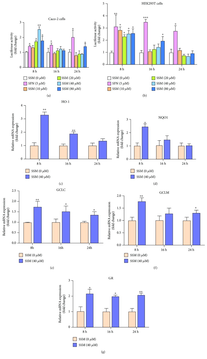
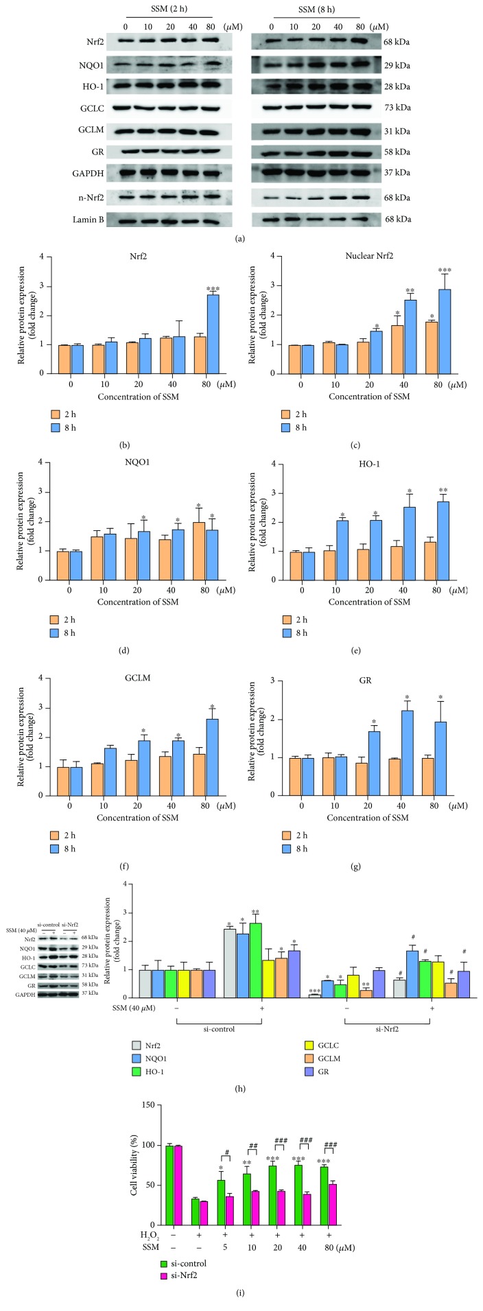
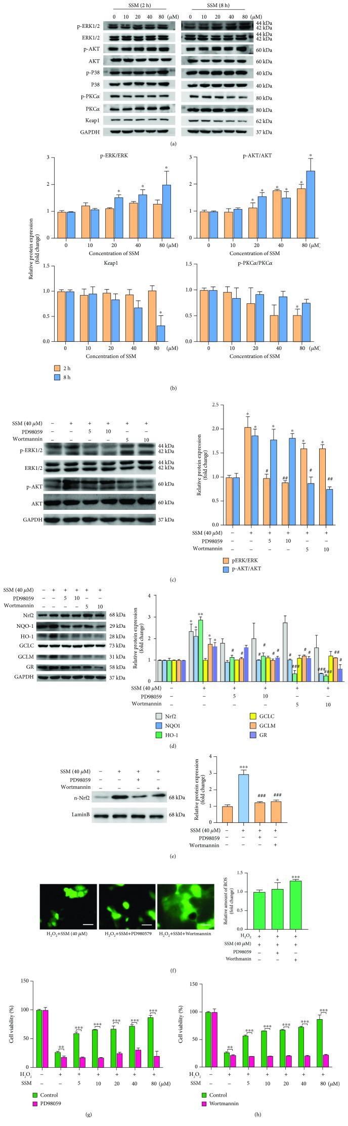
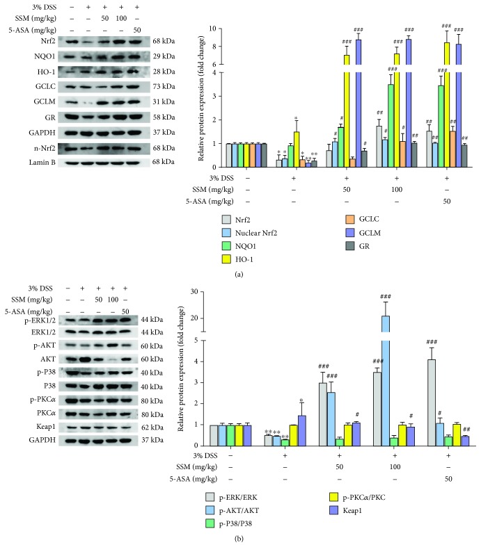
Ulcerative colitis (UC) is a major form of inflammatory bowel disease (IBD) with high incidence and prevalence in many countries. Patients with UC usually suffer from a lifetime of debilitating physical symptoms. Therefore, developing effective therapeutic strategy that can manage this disease better and improve patients' life quality is in urgent need. Sesamin (SSM) is a lignan derived from sesame seeds. In this study, the protective effect of SSM against UC and the underlying mechanism were investigated in vitro and in vivo. Our data showed that SSM protected Caco-2 cells from H2O2-induced oxidative stress injury via GSH-mediated scavenging of reactive oxygen species (ROS). Dual luciferase reporter assay showed that the transcriptional activity of nuclear factor erythroid-related factor 2 (Nrf2) was significantly increased by SSM, and the ability of SSM to activate Nrf2-targeted genes was further confirmed in Caco-2 cells using western blot and quantitative real-time PCR (qRT-PCR). In contrast, Nrf2 knockdown abolished the protective effect of SSM. Additionally, we found that SSM also activated advanced protein kinase B (AKT) and extracellular signal-regulated kinase (ERK) in Caco-2 cells, while either AKT or ERK inhibition can prevent SSM-mediated nuclear translocation of Nrf2. Furthermore, SSM displayed a better protective effect against dextran sulfate sodium- (DSS-) induced UC compared with 5-aminosalicylic acid (5-ASA) in C57BL/6 mice. The enhanced Nrf2 signaling and activated AKT/ERK were also observed in the colon of mice after SSM administration. These results first demonstrate the protective effect of SSM against UC and indicate that the effect is associated with AKT/ERK activation and subsequent Nrf2 signaling enhancement. This study provides a new insight into the medicinal value of SSM and proposes it as a new natural nutrition for better managing the symptoms of UC.


Conflict of interest statement

There are no potential conflicts of interest in this study.

Figures

Figure 1
Figure 1
SSM protected Caco-2 cells from H2O2-induced cytotoxicity via GSH-mediated ROS scavenging. (a) Chemical structure of SSM. (b) Caco-2 cells were treated with 0 μM SSM (0.1% DMSO) and various concentrations of SSM (5, 10, 20, 40, 80, 160, and 320 μM) for 8, 16, and 24 h. Cell viability was measured by MTT assay. (c) Caco-2 cells were pretreated with 0 μM SSM (0.1% DMSO) or 40 μM SSM for 2, 4, and 8 h and then treated with various concentrations of H2O2 (0, 0.26, 0.6, 0.76, 1.0, 1.26, 1.6, 1.76, and 2.0 mM) for 24 h. Cell viability was measured by MTT assay. (d) Caco-2 cells were pretreated with 0 μM SSM (0.1% DMSO) and various concentrations of SSM (5, 10, 20, 40, and 80 μM) for 8 h and then treated with 1.6 mM H2O2 for 24 h. Intracellular ROS were detected using DCFH-DA fluorescence assay. Roseup was used as a positive control (+). Images are displayed at 200x magnification. The quantification of fluorescence was performed with a Flex Station 3 multifunctional microplate reader. Data were expressed as the mean ± SD (n = 5). ∗∗∗p < 0.001 versus the control group. #p < 0.05 and ###p < 0.001 versus the H2O2 treatment group. (e, f) Caco-2 cells were treated with 0 μM SSM (0.1% DMSO) and various concentrations of SSM (10, 20, 40, and 80 μM) for 8 h. Reduced GSH (GSH) and oxidized GSH (GSSG) were detected using commercial kits. Data were expressed as the mean ± SD (n = 5). p < 0.05 versus the DMSO treatment group. (g) Caco-2 cells were pretreated with 0.1% DMSO (control) or 10 μM BSO and 0 μM SSM (0.1% DMSO) or various concentrations of SSM (5, 10, 20, 40, and 80 μM) for 2 and 8 h, respectively, and then treated with 1.6 mM H2O2 for 24 h. Cell viability was measured by MTT assay. Data were expressed as the mean ± SD (n = 5). ∗∗p < 0.01 and ∗∗∗p < 0.001 versus the H2O2 treatment group. ##p < 0.01 and ###p < 0.001 versus the control group.
Figure 2
Figure 2
SSM alleviated DSS-induced acute colitis in mice by reversing proinflammatory cytokine release and antioxidation inhibition. (a) The schematic of the experimental design used in the study. (b) Changes in dietary intake. (c) Changes in body weight. (d) Changes in disease activity index (DAI). (e) Colon length in different treatment groups. (f) The colons collected on day 10 were sectioned and then stained with hematoxylin and eosin (H&E). Arrows indicate the epithelial erosion and crypt distortion and abscess. Images are displayed at 40x magnification using a bright field microscope. (g) Changes in spleen weight. (h–j) The amount of IL-6, IL-1β, and TNF-α in colon tissues of mice was determined using commercial ELISA kits. (k–m) The activity of SOD and the amount of GSH and MDA were determined in colon tissues of mice using commercial ELISA kits. Data were expressed as the mean ± SD (n = 10). p < 0.05, ∗∗p < 0.01, and ∗∗∗p < 0.001 versus the control. #p < 0.05, ##p < 0.01, and ###p < 0.001 versus the 3% DSS treatment group.
Figure 3
Figure 3
SSM promoted the transcriptional activation of Nrf2 in Caco-2 cells. (a, b) Caco-2 and HEK293T cells were transfected with plasmids, including pEF-Nrf2, pGL3-ARE, and pRL-TK, and then treated with 0 μM SSM (0.1% DMSO), 5 μM sulforaphane (SFN) (positive control), and various concentrations of SSM (10, 20, 40, and 80 μM) for 8, 16, and 24 h. The luciferase activities in three cells were detected using a Dual Luciferase Reporter Gene Assay Kit. (c–g) Total RNA was extracted from Caco-2 cells treated with 0 μM SSM (0.1% DMSO) or 40 μM SSM for 8, 16, and 24 h. mRNA expressions of the indicated genes were detected using qPCR and were normalized to GAPDH and β-actin. Data were expressed as the mean ± SD (n = 5). p < 0.05, ∗∗p < 0.01, and ∗∗∗p < 0.001 versus the DMSO treatment group.
Figure 4
Figure 4
SSM increased the nuclear translocation of Nrf2 and induced the expressions of cytoprotective genes in Caco-2 cells. (a) The effect of SSM on the protein expressions of Nrf2 and its target genes was analyzed by western blot in Caco-2 cells treated with 0 μM SSM (0.1% DMSO) and various concentrations of SSM (10, 20, 40, and 80 μM) for 2 and 8 h. (b–g) Quantitative data were obtained from the densitometric quantification of immunoblots using ImageJ software. Data were expressed as the mean ± SD (n = 5). p < 0.05, ∗∗p < 0.01, and ∗∗∗p < 0.001 versus the DMSO treatment group. (h) Caco-2 cells were transfected with si-control or si-Nrf2 for 48 h. Then, the protein expressions of Nrf2 and its target genes were analyzed by western blot in these cells treated with 0.1% DMSO (-) or 40 μM SSM (+) for 8 h. Quantitative data were obtained from the densitometric quantification of immunoblots using ImageJ software. Data were expressed as the mean ± SD (n = 5). p < 0.05, ∗∗p < 0.01, and ∗∗∗p < 0.001 versus the DMSO treatment group. #p < 0.05 versus the SSM treatment group. (i) Caco-2 cells were transfected with si-control or si-Nrf2 for 48 h. Then, the cells were pretreated with 0 μM SSM (0.1% DMSO) or 40 μM SSM for 8 h and 1.6 mM H2O2 for another 24 hours. Cell viability was measured by MTT assay. Data were expressed as the mean ± SD (n = 5). p < 0.05, ∗∗p < 0.01, and ∗∗∗p < 0.001 versus the H2O2 treatment group. #p < 0.05, ##p < 0.01, and ###p < 0.001 versus the si-control group.
Figure 5
Figure 5
SSM activated Nrf2 signaling through promoting ERK and AKT activation in Caco-2 cells. (a) The effect of SSM on ERK, AKT, P38, PKC, and Keap1 was analyzed by western blot in Caco-2 cells treated with 0 μM SSM (0.1% DMSO) and various concentrations of SSM (10, 20, 40, and 80 μM) for 2 and 8 h. (b) Quantitative data were obtained from the densitometric quantification of immunoblots by using ImageJ software. Data were expressed as the mean ± SD (n = 5). p < 0.05 and ∗∗p < 0.01 versus the DMSO treatment group. (c) Caco-2 cells were treated with 0.1% DMSO (control), PD98059 (5 and 10 μM), or wortmannin (5 and 10 μM) for 2 h prior to SSM treatment (40 μM) for 8 h. The protein expressions of AKT and ERK were analyzed by western blot. Quantitative data were obtained from the densitometric quantification of immunoblots using ImageJ software. Data were expressed as the mean ± SD (n = 5). p < 0.05 versus the DMSO treatment group. #p < 0.05 and ###p < 0.001 versus the SSM treatment group. (d) Caco-2 cells were treated with 0.1% DMSO (control), PD98059 (5 and 10 μM), or wortmannin (5 and 10 μM) for 2 h prior to SSM treatment (40 μM) for 8 h. The protein expressions of Nrf2 and its target genes were analyzed by western blot. Quantitative data were obtained from the densitometric quantification of immunoblots using ImageJ software. Data were expressed as the mean ± SD (n = 5). p < 0.05 versus the DMSO treatment group. #p < 0.05, ##p < 0.01, and ###p < 0.001 versus the SSM treatment group. (e) Caco-2 cells were treated with 0.1% DMSO (control), PD98059 (5 and 10 μM), or wortmannin (5 and 10 μM) for 2 h prior to SSM treatment (40 μM) for 8 h. The nuclear protein expression of Nrf2 was analyzed by western blot. Quantitative data were obtained from the densitometric quantification of immunoblots using ImageJ software. Data were expressed as the mean ± SD (n = 5). ∗∗∗p < 0.001 versus the DMSO treatment group. ###p < 0.001 versus the SSM treatment group. (f) Caco-2 cells were treated with 0.1% DMSO (control), PD98059 (10 μM), or wortmannin (10 μM) for 2 h prior to SSM treatment (40 μM) for 8 h and then treated with 1.6 mM H2O2 for another 24 h. Images are displayed at 200x magnification using an OLYMPUS IX73 inverted fluorescence phase-contrast microscope. The quantification of fluorescence was done with a Flex Station 3 multifunctional microplate reader. Data were expressed as the mean ± SD (n = 5). p < 0.05, ∗∗p < 0.01, and ∗∗∗p < 0.001 versus the control group. (g, h) Caco-2 cells were treated with 0.1% DMSO (control), PD98059 (10 μM), or wortmannin (10 μM) for 2 h prior to SSM treatment (40 μM) for 8 h and then treated with 1.6 mM H2O2 for another 24 h. Cell viability was measured by MTT assay. Data were expressed as the mean ± SD (n = 5). p < 0.05, ∗∗p < 0.01, and ∗∗∗p < 0.001 versus the control group.
Figure 6
Figure 6
SSM reactivated Nrf2 signaling via activating ERK and AKT against DSS-induced colitis. (a) The effect of SSM on the ERK, AKT, P38, PKC, and Keap1 in the colon of mice with colitis was detected using western blot. Quantitative data were obtained from the densitometric quantification of immunoblots using ImageJ software. (b) The protein expressions of Nrf2 and its target genes in the colon of mice with colitis were analyzed by western blot. Quantitative data were obtained from the densitometric quantification of immunoblots by using ImageJ software. Data were expressed as the mean ± SD (n = 10). p < 0.05 and ∗∗p < 0.01 versus the control. #p < 0.05, ##p < 0.01, and ###p < 0.001 versus the 3% DSS treatment group.

Similar articles

Cited by

References

    1. Molodecky N. A., Soon I. S., Rabi D. M., et al. Increasing incidence and prevalence of the inflammatory bowel diseases with time, based on systematic review. Gastroenterology. 2012;142(1):46–54.e42. doi: 10.1053/j.gastro.2011.10.001. - DOI - PubMed
    1. Danese S., Fiocchi C. Ulcerative colitis. The New England Journal of Medicine. 2011;365(18):1713–1725. doi: 10.1056/NEJMra1102942. - DOI - PubMed
    1. Moura F. A., de Andrade K. Q., dos Santos J. C. F., Araújo O. R. P., Goulart M. O. F. Antioxidant therapy for treatment of inflammatory bowel disease: does it work? Redox Biology. 2015;6:617–639. doi: 10.1016/j.redox.2015.10.006. - DOI - PMC - PubMed
    1. Kruidenier L., Verspaget H. W. Oxidative stress as a pathogenic factor in inflammatory bowel disease — radicals or ridiculous? Alimentary Pharmacology and Therapeutics. 2002;16(12):1997–2015. doi: 10.1046/j.1365-2036.2002.01378.x. - DOI - PubMed
    1. Wang Z., Li S., Cao Y., et al. Oxidative stress and carbonyl lesions in ulcerative colitis and associated colorectal cancer. Oxidative Medicine and Cellular Longevity. 2016;2016:15. doi: 10.1155/2016/9875298.9875298 - DOI - PMC - PubMed

MeSH terms

LinkOut - more resources