. 2019 Aug 27:2019:5852494.
doi: 10.1155/2019/5852494. eCollection 2019.

Antiobesity, Regulation of Lipid Metabolism, and Attenuation of Liver Oxidative Stress Effects of Hydroxy- α-sanshool Isolated from Zanthoxylum bungeanum on High-Fat Diet-Induced Hyperlipidemic Rats

Affiliations

Antiobesity, Regulation of Lipid Metabolism, and Attenuation of Liver Oxidative Stress Effects of Hydroxy- α-sanshool Isolated from Zanthoxylum bungeanum on High-Fat Diet-Induced Hyperlipidemic Rats

Li Wang et al. Oxid Med Cell Longev. .

Abstract

Zanthoxylum bungeanum is a traditional Chinese medicine (TCM) used to relieve pain, dispel dampness, stop diarrhea, and prevent itching. The aim of this study was to investigate the antiobesity and hypolipidemic effects of hydroxy-α-sanshool (HAS) isolated from Z. bungeanum on hyperlipidemic rats. Wistar rats (n = 48) were randomly divided into six groups: (1) normal diet rats (ND), (2) high-fat diet- (HFD-) treated rats, (3) HFD+fenofibrate-treated rats (HFD+FNB), (4) HFD+low dose of HAS-treated rats (HFD+LD, 9 mg/kg), (5) HFD+middle dose of HAS-treated rats (HFD+MD, 18 mg/kg), and (6) HFD+high dose of HAS-treated rats (HFD+HD, 36 mg/kg). The body weight and food intake of the rats were recorded during the treatment period. After 4 weeks of HAS treatment, abdominal adipose tissues were observed and total cholesterol (T-CHO), triglycerides (TG), high-density lipoprotein (HDL) cholesterol (HDL-C), and low-density lipoprotein (LDL) cholesterol (LDL-C) of serum and liver tissues were determined. Furthermore, histochemical examinations using oil red O and hematoxylin-eosin staining (H&E) were carried out and levels of malondialdehyde (MDA) and glutathione (GSH) and activities of superoxide dismutase (SOD) in the liver were determined. After HFD feeding, the body weight gain and food efficiency ratio of HFD rats were significantly enhanced (p < 0.05vs. ND rats) and HAS treatment (18 and 36 mg/kg) significantly decreased the body weight gain and food efficiency ratio (p < 0.05vs. HFD rats). In addition, HAS treatment could decrease the abdominal adipose tissues and liver adipocytes. Furthermore, HAS treatment significantly decreased the T-CHO, TG, and LDL-C, whereas it increased HDL-C (p < 0.05vs. HFD rats) in serum and the liver. HAS treatment increased the GSH level and SOD activity in the liver (p < 0.05vs. HFD rats), whereas it decreased the levels of MDA (p < 0.05vs. HFD rats). mRNA analyses suggested that HAS treatment increases the expression of Pparg (proliferator-activated receptor γ) and Apoe (peroxisome apolipoprotein E). Immunohistochemistry and Western blotting indicated that HAS stimulation increased the levels of PPARγ and APOE in the liver, as a stress response of the body defense system. These results revealed that HAS exerts antiobesity and hypolipidemic activities in HFD rats by reducing liver oxidative stress and thus could be considered as a potential candidate drug to cure or prevent obesity and hyperlipidemia.


Conflict of interest statement

There are no conflicts of interest associated with this paper.

Figures

Figure 1
Figure 1
Zanthoxylum bungeanum (a) and hydroxy-α-sanshool (b).
Figure 2
Figure 2
High-performance liquid chromatography (HPLC) analysis of the hydroxy-α-sanshool (HAS) extracted from the fruit of Z. bungeanum. (a) HPLC chromatogram of the standard; (b) HPLC chromatogram of the HAS extracted from the fruit of Z. bungeanum.
Figure 3
Figure 3
Total cholesterol (T-CHO) (a), total glycerides (TG) (b), low-density lipoprotein cholesterol (LDL-C) (c), and high-density lipoprotein chol esterol (HDL-C) (d) in rat serum consuming a normal diet (ND) and a high-fat diet (HFD) for two weeks. Values are expressed as the mean ± SD (n = 8), #p < 0.05vs. ND; n.s.: nonsignificant.
Figure 4
Figure 4
Schematic diagram of the experiment.
Figure 5
Figure 5
Effect of hydroxy-α-sanshool (HAS) on body weight (a), body weight gain (b), food intake (c), and food efficiency ratio (d) in rats consuming a high-fat diet. We measured body weight every week for 6 weeks. The food efficiency ratio is the daily weight gain divided by the daily food intake. Values are expressed as the mean ± SD (n = 8); ND: normal diet-treated rats; HFD: high-fat diet-treated rats; HFD+FNB: HFD supplemented with fenofibrate-treated rats; HFD+LD: rats treated with HFD supplemented with a low dose of HAS; HFD+MD: rats treated with HFD supplemented with a middle dose of HAS; HFD+HD: rats treated with HFD supplemented with a high dose of HAS; #p < 0.05vs. ND; p < 0.05vs. HFD; n.s.: nonsignificant.
Figure 6
Figure 6
Effect of HAS on the abdominal adipose tissue in HFD rats. (a–f) represent the amount of abdominal adipose tissues of rats in the ND, HFD, HFD+FNB, HFD+LD, HFD+MD, and HFD+HD groups, respectively. ND: normal diet-treated rats; HFD: high-fat diet-treated rats; HFD+FNB: HFD supplemented with fenofibrate-treated rats; HFD+LD: rats treated with HFD supplemented with a low dose of HAS; HFD+MD: rats treated with HFD supplemented with a middle dose of HAS; HFD+HD: rats treated with HFD supplemented with a high dose of HAS.
Figure 7
Figure 7
Histopathological examinations of liver tissues with oil red O staining. (a–f) represent oil red O-stained liver sections of rats in the ND, HFD, HFD+FNB, HFD+LD, HFD+MD, and HFD+HD groups, respectively. ND: normal diet-treated rats; HFD: high-fat diet-treated rats; HFD+FNB: HFD supplemented with fenofibrate-treated rats; HFD+LD: rats treated with HFD supplemented with a low dose of HAS; HFD+MD: rats treated with HFD supplemented with a middle dose of HAS; HFD+HD: rats treated with HFD supplemented with a high dose of HAS.
Figure 8
Figure 8
Histopathological examinations of liver tissues using H&E staining. (a–f) represent H&E-stained liver sections of rats in the ND, HFD, HFD+FNB, HFD+LD, HFD+MD, and HFD+HD groups, respectively. ND: normal diet-treated rats; HFD: high-fat diet-treated rats; HFD+FNB: HFD supplemented with fenofibrate-treated rats; HFD+LD: rats treated with HFD supplemented with a low dose of HAS; HFD+MD: rats treated with HFD supplemented with a middle dose of HAS; HFD+HD: rats treated with HFD supplemented with a high dose of HAS.
Figure 9
Figure 9
Effect of HAS on the expression of PPARγ and APOE in liver tissues using IHC. ND: normal diet-treated rats; HFD: high-fat diet-treated rats; HFD+FNB: HFD supplemented with fenofibrate-treated rats; HFD+LD: rats treated with HFD supplemented with a low dose of HAS; HFD+MD: rats treated with HFD supplemented with a middle dose of HAS; HFD+HD: rats treated with HFD supplemented with a high dose of HAS; #p < 0.05 vs. ND; p < 0.05 vs. HFD; n.s.: nonsignificant.
Figure 10
Figure 10
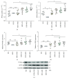
Effect of HAS on the transcription of genes involved in lipid metabolism in liver tissues (a, b); values are expressed as mean ± SD (n = 9). Effect of HAS on the levels of PPARγ and APOE in liver tissue using Western blotting (c–e). Values are expressed as mean ± SD (n = 6). ND: normal diet-treated rats; HFD: high-fat diet-treated rats; HFD+FNB: HFD supplemented with fenofibrate-treated rats; HFD+LD: rats treated with HFD supplemented with a low dose of HAS; HFD+MD: rats treated with HFD supplemented with a middle dose of HAS; HFD+HD: rats treated with HFD supplemented with a high dose of HAS; #p < 0.05vs. ND; p < 0.05vs. HFD; n.s.: nonsignificant.

References

    1. Menees D. S., Bates E. R. Evaluation of patients with suspected coronary artery disease. Coronary Artery Disease. 2010;21(7):386–390. doi: 10.1097/MCA.0b013e32833bfdf7. - DOI - PubMed
    1. Lawton J. S. Sex and gender differences in coronary artery diseases. Seminars in Thoracic and Cardiovascular Surgery. 2011;23(2):126–130. doi: 10.1053/j.semtcvs.2011.07.006. - DOI - PubMed
    1. Durkar A. M., Patil R. R., Naik S. R. Hypolipidemic and antioxidant activity of ethanolic extract of symplocos racemosa roxb. in hyperlipidemic rats: an evidence of participation of oxidative stress in hyperlipidemia. Indian Journal of Experimental Biology. 2014;52(1):36–45. - PubMed
    1. Hu F. B. Globalization of food patterns and cardiovascular disease risk. Circulation. 2008;118(19):1913–1914. doi: 10.1161/CIRCULATIONAHA.108.808493. - DOI - PubMed
    1. Matschke V., Theiss C., Matschke J. Oxidative stress: the lowest common denominator of multiple diseases. Neural Regeneration Research. 2019;14(2):238–241. doi: 10.4103/1673-5374.244780. - DOI - PMC - PubMed