

On the road to structure-based development of anti-virulence therapeutics targeting the type III secretion system injectisome
- PMID: 31534650
- PMCID: PMC6748289
- DOI: 10.1039/c9md00146h
On the road to structure-based development of anti-virulence therapeutics targeting the type III secretion system injectisome
Abstract
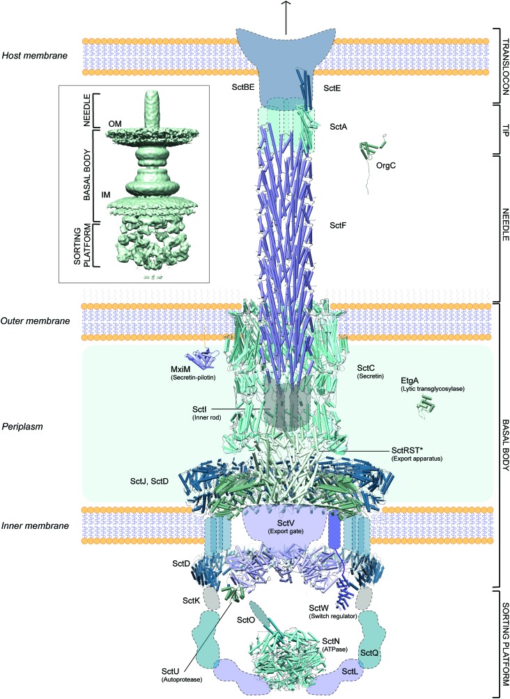
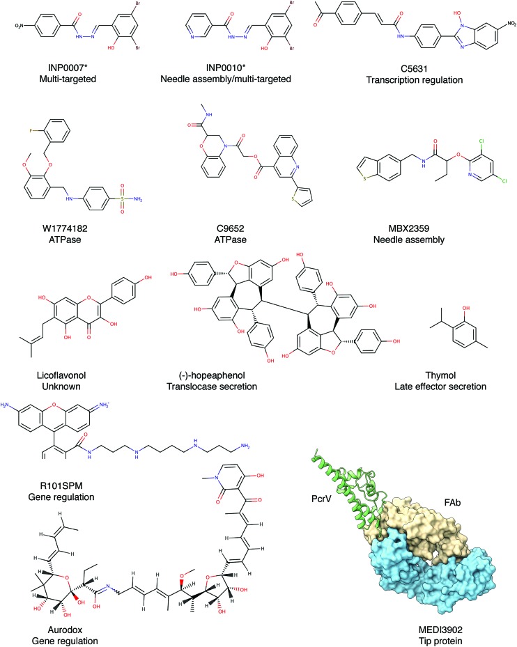
The type III secretion system injectisome is a syringe-like multimembrane spanning nanomachine that is essential to the pathogenicity but not viability of many clinically relevant Gram-negative bacteria, such as enteropathogenic Escherichia coli, Salmonella enterica and Pseudomonas aeruginosa. Due to the rise in antibiotic resistance, new strategies must be developed to treat the growing spectre of drug resistant infections. Targeting the injectisome via an 'anti-virulence strategy' is a promising avenue to pursue as an alternative to the more commonly used bactericidal therapeutics, which have a high propensity for resulting resistance development and often more broad killing profile, including unwanted side effects in eliminating favourable members of the microbiome. Building on more than a decade of crystallographic work of truncated or isolated forms of the more than two dozen components of the secretion apparatus, recent advances in the field of single-particle cryo-electron microscopy have allowed for the elucidation of atomic resolution structures for many of the type III secretion system components in their assembled, oligomerized state including the needle complex, export apparatus and ATPase. Cryo-electron tomography studies have also advanced our understanding of the direct pathogen-host interaction between the type III secretion system translocon and host cell membrane. These new structural works that further our understanding of the myriad of protein-protein interactions that promote injectisome function will be highlighted in this review, with a focus on those that yield promise for future anti-virulence drug discovery and design. Recently developed inhibitors, including both synthetic, natural product and peptide inhibitors, as well as promising new developments of immunotherapeutics will be discussed. As our understanding of this intricate molecular machinery advances, the development of anti-virulence inhibitors can be enhanced through structure-guided drug design.
Figures
Similar articles
-
Near-atomic-resolution cryo-EM analysis of the Salmonella T3S injectisome basal body.Nature. 2016 Dec 22;540(7634):597-601. doi: 10.1038/nature20576. Epub 2016 Dec 14. Nature. 2016. PMID: 27974800
-
Cryo-EM analysis of the T3S injectisome reveals the structure of the needle and open secretin.Nat Commun. 2018 Sep 21;9(1):3840. doi: 10.1038/s41467-018-06298-8. Nat Commun. 2018. PMID: 30242280 Free PMC article.
-
Structural and Functional Analysis of SsaV Cytoplasmic Domain and Variable Linker States in the Context of the InvA-SsaV Chimeric Protein.Microbiol Spectr. 2021 Dec 22;9(3):e0125121. doi: 10.1128/Spectrum.01251-21. Epub 2021 Dec 1. Microbiol Spectr. 2021. PMID: 34851139 Free PMC article.
-
Recent structural advances towards understanding of the bacterial type III secretion injectisome.Trends Biochem Sci. 2022 Sep;47(9):795-809. doi: 10.1016/j.tibs.2022.04.013. Epub 2022 May 30. Trends Biochem Sci. 2022. PMID: 35654690 Review.
-
Structural Insights into Type III Secretion Systems of the Bacterial Flagellum and Injectisome.Annu Rev Microbiol. 2023 Sep 15;77:669-698. doi: 10.1146/annurev-micro-032521-025503. Annu Rev Microbiol. 2023. PMID: 37713458 Review.
Cited by
-
Antibiotic Therapy of Plague: A Review.Biomolecules. 2021 May 12;11(5):724. doi: 10.3390/biom11050724. Biomolecules. 2021. PMID: 34065940 Free PMC article. Review.
-
Pharmacokinetics, tissue distribution, bioavailability and excretion of the anti-virulence drug Fluorothiazinon in rats and rabbits.J Antibiot (Tokyo). 2024 Jun;77(6):382-388. doi: 10.1038/s41429-024-00719-1. Epub 2024 Mar 15. J Antibiot (Tokyo). 2024. PMID: 38491136
-
The Role of Drug Repurposing in the Development of Novel Antimicrobial Drugs: Non-Antibiotic Pharmacological Agents as Quorum Sensing-Inhibitors.Antibiotics (Basel). 2019 Dec 17;8(4):270. doi: 10.3390/antibiotics8040270. Antibiotics (Basel). 2019. PMID: 31861228 Free PMC article.
-
Identification of Small Molecules Blocking the Pseudomonas aeruginosa type III Secretion System Protein PcrV.Biomolecules. 2021 Jan 4;11(1):55. doi: 10.3390/biom11010055. Biomolecules. 2021. PMID: 33406810 Free PMC article.
-
Secretory System Components as Potential Prophylactic Targets for Bacterial Pathogens.Biomolecules. 2021 Jun 15;11(6):892. doi: 10.3390/biom11060892. Biomolecules. 2021. PMID: 34203937 Free PMC article. Review.
References
-
- Global priority list of antibiotic-resistant bacteria to guide research, discovery, and development of new antibiotics, http://www.who.int/medicines/publications/WHO-PPL-Short_Summary_25Feb-ET..., (accessed 20 September 2001).
-
- Roy-Burman A., Savel R. H., Racine S., Swanson B. L., Revadigar N. S., Fujimoto J., Sawa T., Frank D. W., Wiener-Kronish J. P. J. Infect. Dis. 2001;183:1767–1774. - PubMed
-
- Makvana S., Krilov L. R. Pediatr. Rev. 2015;36:167–171. - PubMed
-
- Stefani S., Campana S., Cariani L., Carnovale V., Colombo C., Lleo M. M., Iula V. D., Minicucci L., Morelli P., Pizzamiglio G., Taccetti G. Int. J. Med. Microbiol. 2017;307:353–362. - PubMed
Publication types
LinkOut - more resources
Full Text Sources
Research Materials