

Voxel-based statistical analysis and quantification of amyloid PET in the Japanese Alzheimer's disease neuroimaging initiative (J-ADNI) multi-center study
- PMID: 31535240
- PMCID: PMC6751233
- DOI: 10.1186/s13550-019-0561-2
Voxel-based statistical analysis and quantification of amyloid PET in the Japanese Alzheimer's disease neuroimaging initiative (J-ADNI) multi-center study
Abstract
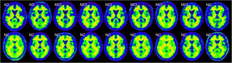
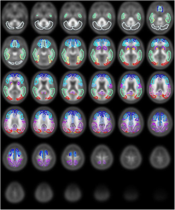
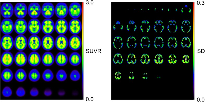
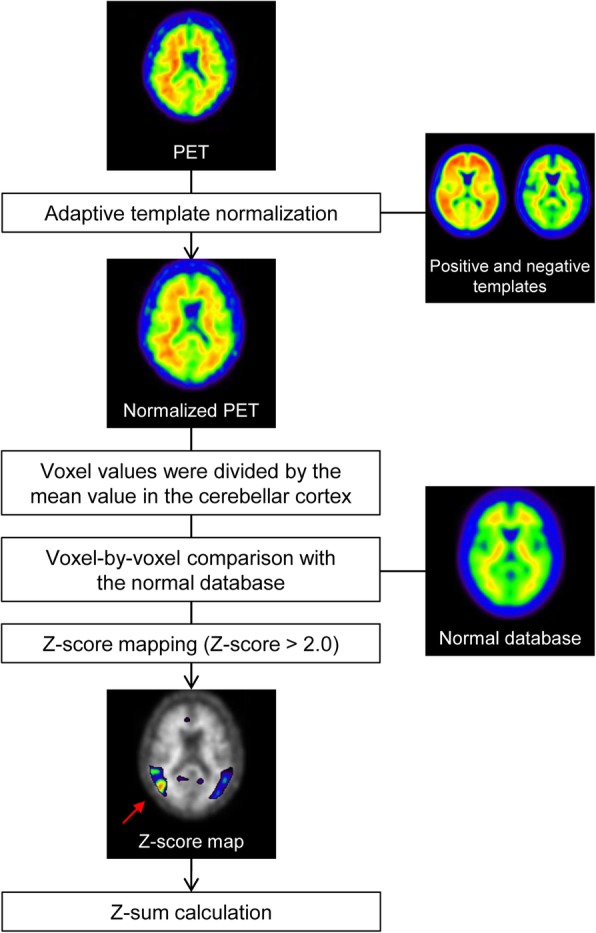
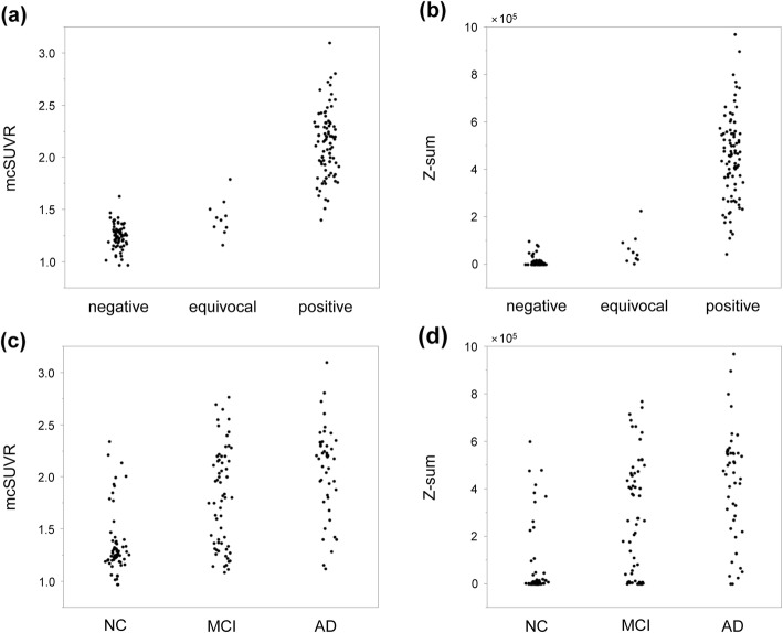
Background: Amyloid PET plays a vital role in detecting the accumulation of in vivo amyloid-β (Aβ). The quantification of Aβ accumulation has been widely performed using the region of interest (ROI)-based mean cortical standardized uptake value ratio (mcSUVR). However, voxel-based statistical analysis has not been well studied. The purpose of this study was to examine the feasibility of analyzing amyloid PET scans by voxel-based statistical analysis. The results were then compared to those with the ROI-based mcSUVR. In total, 166 subjects who underwent 11C-PiB PET in the J-ADNI multi-center study were analyzed. Additionally, 18 Aβ-negative images were collected from other studies to form a normal database. The PET images were spatially normalized to the standard space using an adaptive template method without MRI. The mcSUVR was measured using a pre-defined ROI. Voxel-wise Z-scores within the ROI were calculated using the normal database, after which Z-score maps were generated. A receiver operating characteristic (ROC) analysis was performed to evaluate whether Z-sum (sum of the Z-score) and mcSUVR could be used to classify the scans into positive and negative using the central visual read as the reference standard. PET scans that were equivocal were regarded as positive.
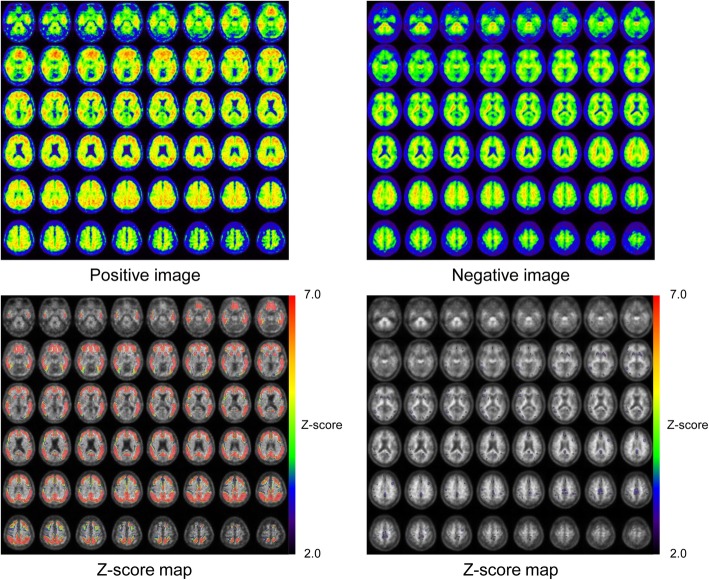
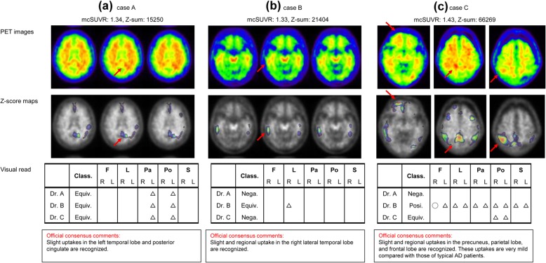
Results: Sensitivity and specificity were respectively 90.8% and 100% by Z-sum and 91.8% and 98.5% by mcSUVR. Most of the equivocal scans were subsequently classified by both Z-sum and mcSUVR as false negatives. Z-score maps correctly delineated abnormal Aβ accumulation over the same regions as the visual read.
Conclusions: We examined the usefulness of voxel-based statistical analysis for amyloid PET. This method provides objective Z-score maps and Z-sum values, which were observed to be helpful as an adjunct to visual interpretation especially for cases with mild or limited Aβ accumulation. This approach could improve the Aβ detection sensitivity, reduce inter-reader variability, and allow for detailed monitoring of Aβ deposition.
Trial registration: The number of the J-ADNI study is UMIN000001374.
Keywords: 11C-PiB; Amyloid; PET; Voxel-based statistical analysis; Z-score.
Conflict of interest statement
Yasuhiko Ikari is an employee of CMIC Co., Ltd., Tokyo, Japan. Hiroyuki Nishida is an employee of Micron, Inc., Tokyo, Japan. All other authors declare that they have no competing interests.
Figures
References
-
- Johnson KA, Minoshima S, Bohnen NI, Donohoe KJ, Foster NL, Herscovitch P, et al. Appropriate use criteria for amyloid PET: a report of the Amyloid Imaging Task Force, the Society of Nuclear Medicine and Molecular Imaging, and the Alzheimer’s Association. J Nucl Med. 2013;54:476–490. doi: 10.2967/jnumed.113.120618. - DOI - PubMed
Grants and funding
LinkOut - more resources
Full Text Sources
Other Literature Sources
