

Efficient quality assurance method with automated data acquisition of a single phantom setup to determine radiation and imaging isocenter congruence
- PMID: 31535781
- PMCID: PMC6806465
- DOI: 10.1002/acm2.12723
Efficient quality assurance method with automated data acquisition of a single phantom setup to determine radiation and imaging isocenter congruence
Abstract
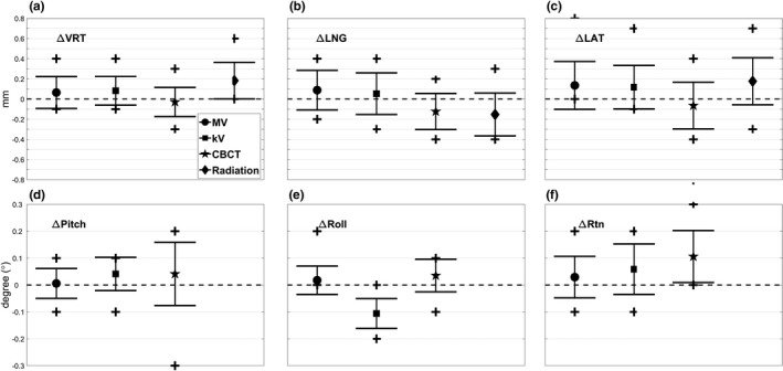
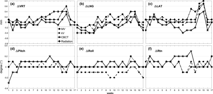
We developed a quality assurance (QA) method to determine the isocenter congruence of Optical Surface Monitoring System (OSMS, Varian, CA, USA), kilovoltage (kV), and megavoltage (MV) imaging, and the radiation isocenter using a single setup of the OSMS phantom for frameless Stereotactic Radiosurgery (SRS) treatment. After aligning the phantom to the OSMS isocenter, a cone-beam computed tomography (CBCT) of the phantom was acquired and registered to a computed tomography (CT) scan of the phantom to determine the CBCT isocenter. Without moving the phantom, MV and kV images were simultaneously acquired at four gantry angles to localize MV and kV isocenters. Then, Winston-Lutz (W-L) test images of the central BB in the phantom were acquired to analyze the radiation isocenter. The gantry and couch were automatically controlled using the TrueBeam Developer Mode during MV, kV, and W-L image acquisition. All the images were acquired weekly for 17 weeks to track the congruence of all the imaging modalities' isocenter in six-dimensional (6D) translations and rotations, and the radiation isocenter in three-dimensional (3D) translations. The shifts of isocenters of all imaging modalities and the radiation isocenter from the OSMS isocenter were within 0.2 mm and 0.2° on average over 17 weeks. The maximum discrepancy between OSMS and other imaging modalities or radiation isocenters was 0.8 mm and 0.3°. However, systematic shifts of radiation isocenter anteriorly and laterally relative to the OSMS isocenter were observed. The measured discrepancies were consistent from week-to-week except for two weeks when the isocenter discrepancies of 0.8 mm were noted due to drifts of the OSMS isocenter. Once recalibration was performed on OSMS, the discrepancy was reduced to 0.3 mm and 0.2°.By performing the proposed QA on a weekly basis, the isocenter congruencies of multiple imaging systems and radiation isocenter were validated for a linear accelerator.
Keywords: automation; imaging quality assurance; optical imaging.
© 2019 The Authors. Journal of Applied Clinical Medical Physics published by Wiley Periodicals, Inc. on behalf of American Association of Physicists in Medicine.
Conflict of interest statement
The authors declare no conflict of interest.
Figures

Similar articles
-
A novel phantom and procedure providing submillimeter accuracy in daily QA tests of accelerators used for stereotactic radiosurgery*.J Appl Clin Med Phys. 2016 Jul 8;17(4):246-253. doi: 10.1120/jacmp.v17i4.6295. J Appl Clin Med Phys. 2016. PMID: 27455506 Free PMC article.
-
Evaluation of the system accuracy of frameless stereotactic radiosurgery using a combination of cone beam CT, six degrees of freedom couch, and surface image-guided systems.J Appl Clin Med Phys. 2025 Jun;26(6):e70082. doi: 10.1002/acm2.70082. Epub 2025 Mar 22. J Appl Clin Med Phys. 2025. PMID: 40119644 Free PMC article.
-
Development of a dedicated phantom for multi-target single-isocentre stereotactic radiosurgery end to end testing.J Appl Clin Med Phys. 2018 Nov;19(6):99-108. doi: 10.1002/acm2.12452. Epub 2018 Sep 16. J Appl Clin Med Phys. 2018. PMID: 30221462 Free PMC article.
-
Modern Radiation Therapy Planning and Delivery.Hematol Oncol Clin North Am. 2019 Dec;33(6):947-962. doi: 10.1016/j.hoc.2019.08.005. Epub 2019 Oct 1. Hematol Oncol Clin North Am. 2019. PMID: 31668213 Review.
-
Imaging for Target Delineation and Treatment Planning in Radiation Oncology: Current and Emerging Techniques.Hematol Oncol Clin North Am. 2019 Dec;33(6):963-975. doi: 10.1016/j.hoc.2019.08.008. Epub 2019 Sep 17. Hematol Oncol Clin North Am. 2019. PMID: 31668214 Free PMC article. Review.
Cited by
-
Efficient quality assurance for isocentric stability in stereotactic body radiation therapy using machine learning.Radiol Phys Technol. 2024 Mar;17(1):219-229. doi: 10.1007/s12194-023-00768-5. Epub 2023 Dec 31. Radiol Phys Technol. 2024. PMID: 38160437
-
Simple quality assurance based on filtered back projection for geometrical/irradiation accuracy in single-isocenter multiple-target stereotactic radiotherapy.Radiol Phys Technol. 2022 Dec;15(4):409-416. doi: 10.1007/s12194-022-00683-1. Epub 2022 Oct 19. Radiol Phys Technol. 2022. PMID: 36261754
-
Development and Longitudinal Analysis of Plan-Based Streamlined Quality Assurance on Multiple Positioning Guidance Systems With Single Phantom Setup.Front Oncol. 2021 Jun 16;11:683733. doi: 10.3389/fonc.2021.683733. eCollection 2021. Front Oncol. 2021. PMID: 34222005 Free PMC article.
-
Analysis of a surface imaging system using a six degree-of-freedom couch.J Appl Clin Med Phys. 2022 Aug;23(8):e13697. doi: 10.1002/acm2.13697. Epub 2022 Jul 12. J Appl Clin Med Phys. 2022. PMID: 35819973 Free PMC article.
References
-
- Benedict SH, Yenice KM, Followill D, et al. Stereotactic body radiation therapy: the report of AAPM Task Group 101: stereotactic body radiation therapy: the report of TG101. Med Phys. 2010;37(8):4078–4101. - PubMed
-
- Klein Eric E, Hanley J, Bayouth J, et al. Task group 142 report: quality assurance of medical accelerators. Med Phys. 2009;36:4197–4212. - PubMed
-
- Schell MC, Bova FJ, Larson DA, et al. “AAPM Reports Task Report 42‐ Stereotactic Radiosurgery”, June 1995, https://www.aapm.org/pubs/reports/detail.asp?docxml:id=53.
-
- Bissonnette J‐P, Balter PA, Dong L, et al. Quality assurance for image‐guided radiation therapy utilizing CT‐based technologies: a report of the AAPM TG‐179. Med Phys. 2012;39:1946–1963. - PubMed
-
- Lutz W, Winston KR, Maleki N. A system for stereotactic radiosurgery with a linear accelerator. Int J Radiat Oncol Biol Phys. 1988;14:373–381. - PubMed
MeSH terms
LinkOut - more resources
Full Text Sources
