. 2019 Nov 1;4(21):e129884.
doi: 10.1172/jci.insight.129884.

Genotype correlates with clinical severity in PIK3CA-associated lymphatic malformations

Affiliations

Genotype correlates with clinical severity in PIK3CA-associated lymphatic malformations

Kaitlyn Zenner et al. JCI Insight. .

Abstract

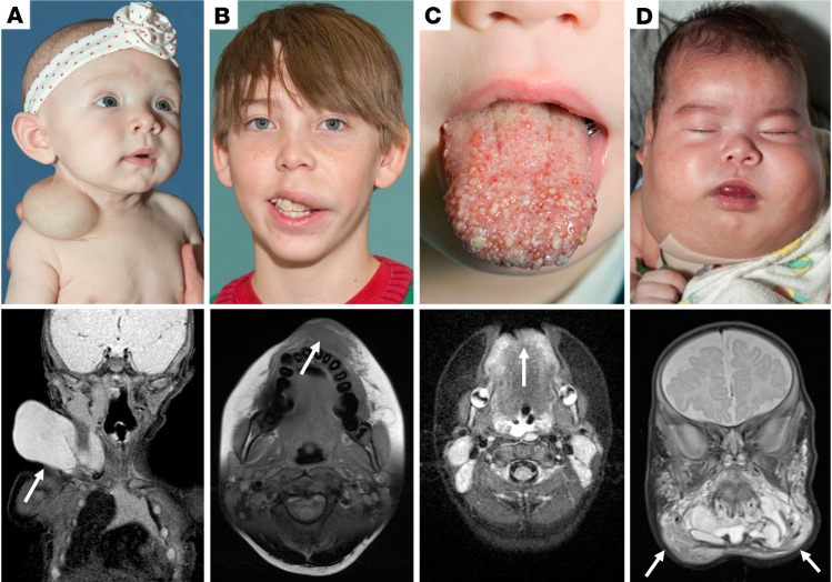
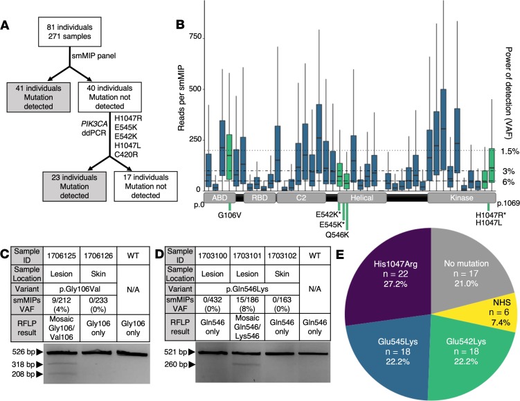
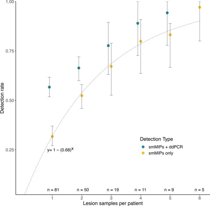
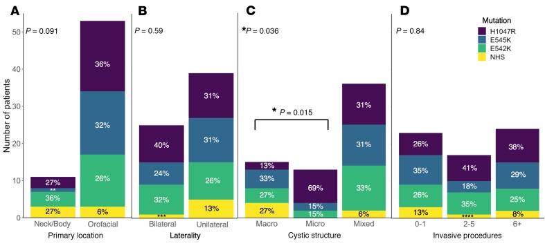
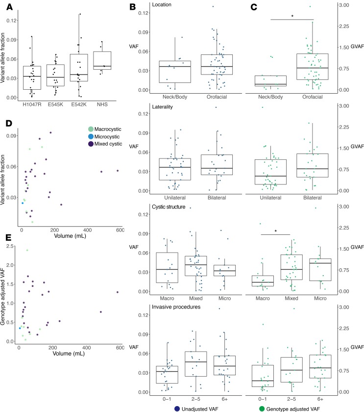
Lymphatic malformations (LMs) are congenital, nonneoplastic vascular malformations associated with postzygotic activating PIK3CA mutations. The mutation spectrum within LMs is narrow, with the majority having 1 of 3 hotspot mutations. Despite this relative genetic homogeneity, clinical presentations differ dramatically. We used molecular inversion probes and droplet digital polymerase chain reaction to perform deep, targeted sequencing of PIK3CA in 271 affected and unaffected tissue samples from 81 individuals with isolated LMs and retrospectively collected clinical data. Pathogenic PIK3CA mutations were identified in affected LM tissue in 64 individuals (79%) with isolated LMs, with variant allele fractions (VAFs) ranging from 0.1% to 13%. Initial analyses revealed no correlation between VAF and phenotype variables. Recognizing that different mutations activate PI3K to varying degrees, we developed a metric, the genotype-adjusted VAF (GVAF), to account for differences in mutation strength, and found significantly higher GVAFs in LMs with more severe clinical characteristics including orofacial location or microcystic structure. In addition to providing insight into LM pathogenesis, we believe GVAF may have broad applicability for genotype-phenotype analyses in mosaic disorders.

Keywords: Genetics; Molecular genetics; Vascular Biology.


Conflict of interest statement

Conflict of interest: RB is cofounder of EigenHealth, Inc., a consultant to SpiWay, LLC, and holds a financial interest of ownership equity with Edus Health, Inc.

Figures

Figure 1
Figure 1. Clinical examples of lymphatic malformations.
Preoperative photos and MRI imaging of individuals with lymphatic malformations of varying severity. Arrows indicate diseased area on MRI. (A) Clinical photo and coronal T2-weighted MRI image of infant with unilateral, macrocystic LM isolated to the right lateral neck. (B) Clinical photo and axial T1-weighted MRI of teenage male with unilateral microcystic LM of the left upper lip. Notably, this lesion is much more challenging to assess on MRI compared with a macrocystic lesion. (C) Clinical photo and axial T2-weighted MRI of bilateral microcystic LM of the anterior tongue. (D) Clinical photo and coronal T2-weighted MRI of an infant with a large, bilateral mixed cystic LM in a “beard distribution.” This severe level of disease burden markedly impacted this patient’s ability to breathe and he required a tracheostomy to maintain a safe airway. LM, lymphatic malformation; MRI, magnetic resonance imaging.
Figure 2
Figure 2. Experimental workflow, smMIP coverage, RFLP results, and mutation results.
(A) Experimental workflow for samples in our cohort. All samples initially went through an smMIP panel, after which 41 individuals had a mutation detected. The remaining 40 individuals underwent screening with 5 ddPCR assays as listed. Following ddPCR screening, 23 more individuals had a mutation detected and 17 remained unsolved. (B) Box plot of reads per smMIP for 271 samples and 42 smMIPs covering the entire coding region of PIK3CA. Boxes are defined by the first quartile inferiorly, median, and third quartile superiorly, with whiskers extending to the farthest non-outlier point (defined as within 1.5 times the interquartile range). Green indicates smMIPs in which a mutation was detected. The right y axis indicates the minimum possible VAF detectable according to read depth. A schematic of PIK3CA cDNA (NM_006218) is along the x axis aligned with the corresponding smMIPs. Mutations detected in our cohort are indicated beneath the cDNA schematic with an asterisk (*) to denote hotspot mutations. (C and D) smMIP and RFLP results for non-hotspot mutations not screened by ddPCR, (C) p.Gly106Val and (D) p.Gln546Lys. Variable mutation detection within an individual is demonstrated in D, as 2 lesion samples were tested but only 1 was positive by both smMIPs and RFLP. (E) Pie chart summarizing mutations detected in the cohort. ddPCR, droplet digital polymerase chain reaction; N/A, not applicable; NHS, non-hotspot mutations; RFLP, restriction fragment length polymorphism; smMIPs, single-molecule molecular inversion probes; VAF, variant allele fraction; WT, wild type.
Figure 3
Figure 3. Number of lesion samples correlates with likelihood of detecting a pathogenic variant.
The results of a bootstrap analysis looking at likelihood of detecting a pathogenic variant given sampling of different number of lesion samples per individual. Mean detection rates for smMIPs only and smMIPs with ddPCR are shown, with error bars indicating the 2.5 and 97.5 percentiles. The number of individuals with adequate sample numbers for each analysis is indicated at the bottom of the chart. For smMIPs alone, the experimental rate of detection corresponded to the cumulative distribution function of the geometric distribution for P = 0.32, y = 1 – (0.68)x, which demonstrates the likelihood of achieving a diagnostic result with a 32% mutation detection rate for 1 lesion sample. ddPCR, droplet digital polymerase chain reaction; smMIPs, single-molecule molecular inversion probes.
Figure 4
Figure 4. Mutation spectrum varies by cystic structure.
Histograms depicting interactions between mutation and phenotype for (A) primary location, (B) laterality, (C) cystic structure, and (D) number of invasive procedures. Percentages are rounded to the nearest whole number. P values in the left upper corner indicate results of Fisher’s exact test when 2 categorical variables were present (location and laterality) or Kruskal-Wallis rank-sum test when 3 categorical variables were present (cystic structure and invasive procedures). *P < 0.05. In C, post hoc Fisher’s exact test showed a significant difference (P = 0.015) in mutations between macro- and microcystic lesions: **9%, ***4%, ****6%. NHS, non-hotspot mutations.
Figure 5
Figure 5. Variant allele fraction (VAF) and genotype-adjusted VAF (GVAF) comparisons by phenotype.
(A) Dot-box plot of VAF by mutation. Wilcoxon’s rank-sum test of hotspots versus NHS showed a trend toward increased VAF with NHS (P = 0.062). (B and C) Dot-box plots with (B) VAF (blue) on the left and (C) GVAF (green) on the right plotted by the 4 phenotype outputs: primary location, laterality, cystic structure, and number of procedures. Boxes are defined by the first quartile inferiorly, median, and third quartile superiorly, with whiskers extending to the farthest non-outlier point (defined as within 1.5 times the interquartile range) (AC). Statistically significant results were seen for GVAF when analyzed by location and macro- versus microcystic structure. *P < 0.05 via Wilcoxon’s rank-sum test. (D and E) Dot plots with dots colored by cystic structure for volume by VAF (D) and GVAF (E) showed no correlation by Pearson’s correlation (n = 31). NHS, non-hotspot mutations.

Similar articles

Cited by

References

    1. Perkins JA. New frontiers in our understanding of lymphatic malformations of the head and neck: natural history and basic research. Otolaryngol Clin North Am. 2018;51(1):147–158. doi: 10.1016/j.otc.2017.09.002. - DOI - PMC - PubMed
    1. Adams MT, Saltzman B, Perkins JA. Head and neck lymphatic malformation treatment: a systematic review. Otolaryngol Head Neck Surg. 2012;147(4):627–639. doi: 10.1177/0194599812453552. - DOI - PubMed
    1. Bhatt N, Perakis H, Watts TL, Borders JC. Traumatic hemorrhage and rapid expansion of a cervical lymphatic malformation. Ear Nose Throat J. 2011;90(1):20–22. doi: 10.1177/014556131109000106. - DOI - PubMed
    1. Perkins JA, Maniglia C, Magit A, Sidhu M, Manning SC, Chen EY. Clinical and radiographic findings in children with spontaneous lymphatic malformation regression. Otolaryngol Head Neck Surg. 2008;138(6):772–777. doi: 10.1016/j.otohns.2008.02.016. - DOI - PubMed
    1. Flint PW, et al. Cummings Otolaryngology, 6th Edition. Amsterdam, The Netherlands: Elsevier; 2015. https://www.elsevier.com/books/cummings-otolaryngology/flint/978-1-4557-....

Publication types

Substances