. 2019 Oct 17;4(20):e93091.
doi: 10.1172/jci.insight.93091.

Exenatide regulates pancreatic islet integrity and insulin sensitivity in the nonhuman primate baboon Papio hamadryas

Affiliations

Exenatide regulates pancreatic islet integrity and insulin sensitivity in the nonhuman primate baboon Papio hamadryas

Teresa Vanessa Fiorentino et al. JCI Insight. .

Abstract

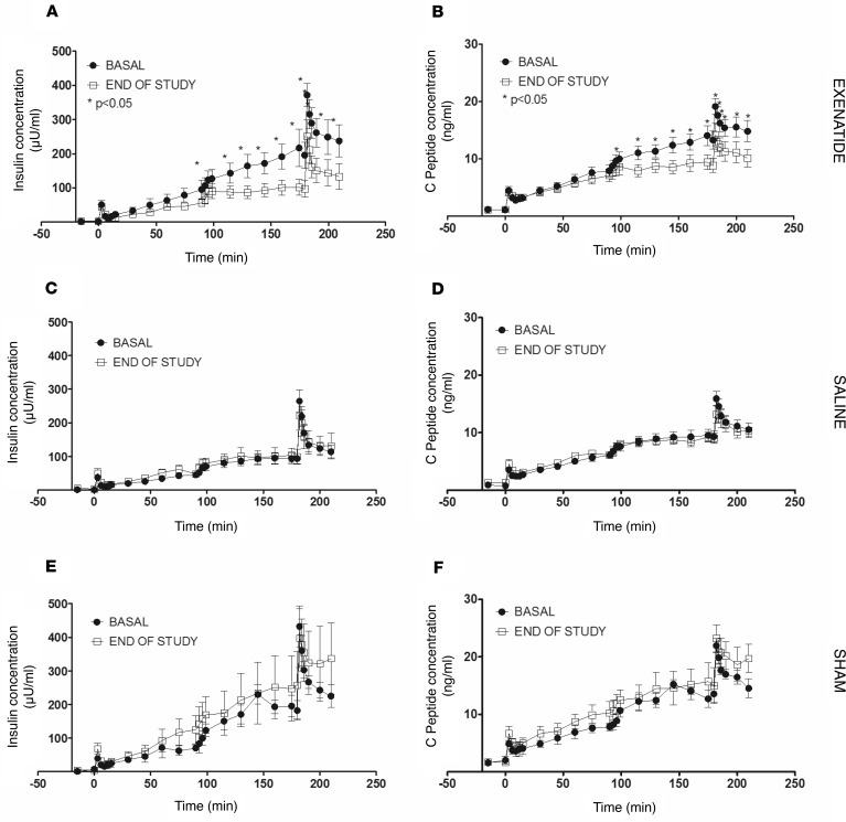
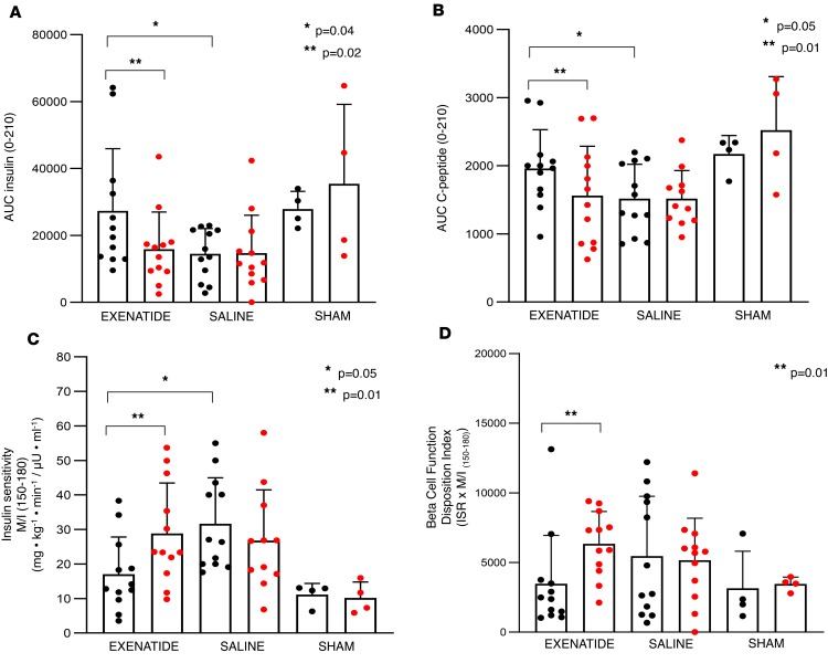
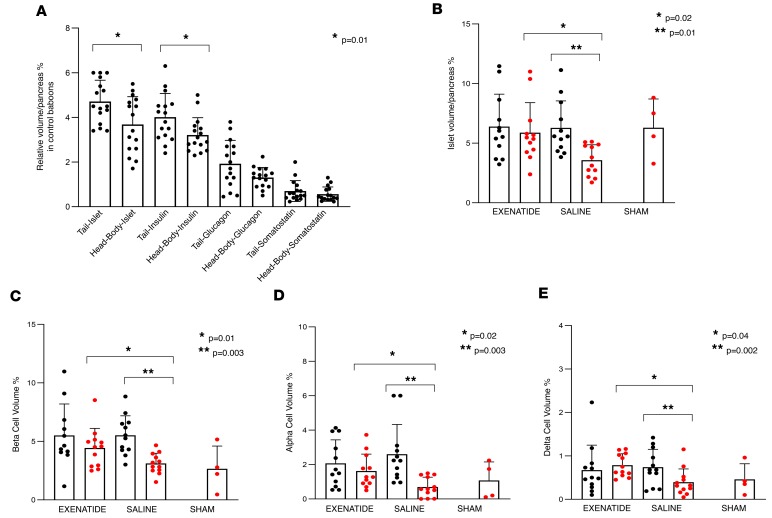
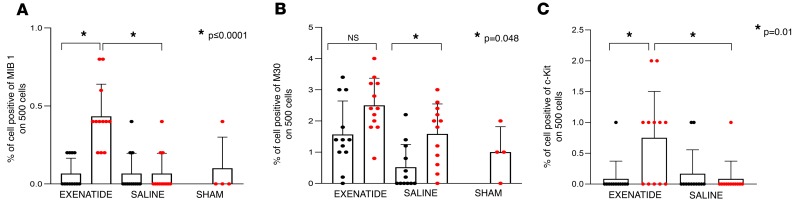
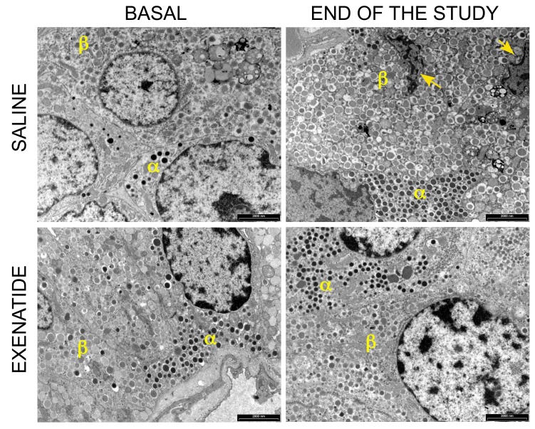
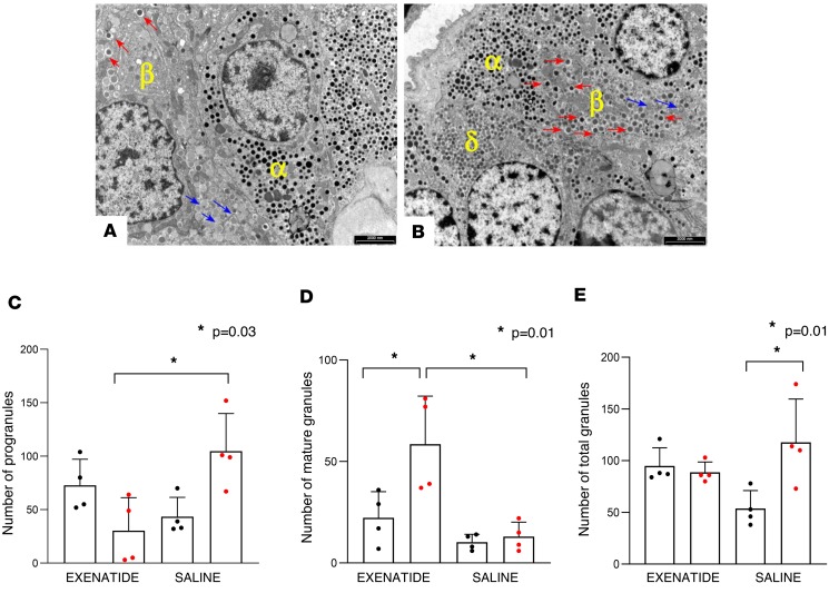
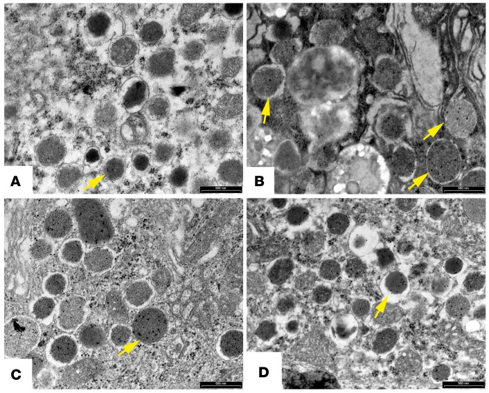
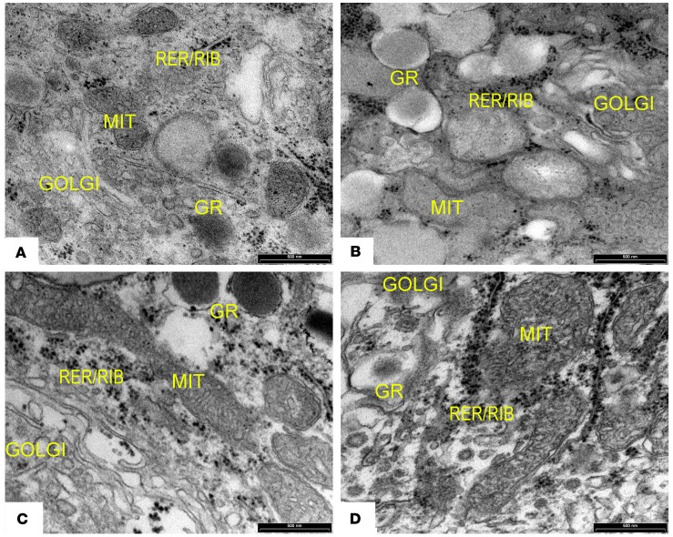
The glucagon-like peptide-1 receptor agonist exenatide improves glycemic control by several and not completely understood mechanisms. Herein, we examined the effects of chronic intravenous exenatide infusion on insulin sensitivity, β cell and α cell function and relative volumes, and islet cell apoptosis and replication in nondiabetic nonhuman primates (baboons). At baseline, baboons received a 2-step hyperglycemic clamp followed by an l-arginine bolus (HC/A). After HC/A, baboons underwent a partial pancreatectomy (tail removal) and received a continuous exenatide (n = 12) or saline (n = 12) infusion for 13 weeks. At the end of treatment, HC/A was repeated, and the remnant pancreas (head-body) was harvested. Insulin sensitivity increased dramatically after exenatide treatment and was accompanied by a decrease in insulin and C-peptide secretion, while the insulin secretion/insulin resistance (disposition) index increased by about 2-fold. β, α, and δ cell relative volumes in exenatide-treated baboons were significantly increased compared with saline-treated controls, primarily as the result of increased islet cell replication. Features of cellular stress and secretory dysfunction were present in islets of saline-treated baboons and absent in islets of exenatide-treated baboons. In conclusion, chronic administration of exenatide exerts proliferative and cytoprotective effects on β, α, and δ cells and produces a robust increase in insulin sensitivity in nonhuman primates.

Keywords: B cells; Endocrinology; Glucose metabolism; Insulin signaling.


Conflict of interest statement

Conflict of interest: The authors have declared that no conflict of interest exists.

Figures

Figure 1
Figure 1. Effect of exenatide on insulin and C-peptide secretion.
Dynamics of insulin (A, C, E) and C-peptide (B, D, F) secretion during the 2-step hyperglycemic clamps performed before (●) and after (□) treatments with exenatide (left), saline (middle), and saline in SHAM-operated (right) baboons. Number of baboons: 12 in exenatide group, 12 in saline group, and 4 in SHAM group. Comparisons between baseline and after-treatment data were performed by Wilcoxon’s test. *P < 0.05.
Figure 2
Figure 2. Effect of exenatide on insulin secretion and sensitivity.
(A) AUC insulin, (B) AUC C-peptide, (C) insulin sensitivity index (M/I), and (D) disposition index during the 2-step hyperglycemic clamp with arginine stimulation performed before (shown in black) and after (shown in red) treatment with exenatide or saline and in SHAM-operated baboons. Number of baboons: 12 in exenatide group, 12 in saline group, and 4 in SHAM group. Comparisons between baseline and after-treatment data were performed by Wilcoxon’s test. Comparison of clinical data between the study groups was performed by using a general linear model for multiple comparisons with adjustment for age and body weight. *P ≤ 0.05; **P ≤ 0.01
Figure 3
Figure 3. Effect of exenatide on pancreatic islet volumes.
(A) The anatomic composition/relative volume of pancreatic islet from different regions of the pancreas obtained in animal/baboon controls. (B) Whole-islet volumes before (black) and after (red) the different treatments are shown. (C–E) The β cell, α cell, and δ cell volumes before (black) and after (red) treatment in the 3 groups. Islet volume and relative islet β, α, and δ cell volume were assessed by using computer assisted stereology toolbox (CAST) and expressed as percentage of total pancreas. Number of sections for each baboon: 10 (5 sections at baseline and 5 sections at the end of the study) evaluated twice. Comparisons between baseline and after-treatment data for each study group were performed by Wilcoxon’s test. A general linear model for multiple comparisons adjusted for age and body weight was used to test differences in relative islet/cell volumes between exenatide- versus saline-treated animals.
Figure 4
Figure 4. Effect of exenatide on islet cell replication and apoptosis.
(A) The percentage of the islet cells positive for the replication marker Ki-67 nuclear protein (antibody MIB-1) before (black) and after the treatments (red). (B) The percentage of the islet cells positive for the apoptosis marker M30 before (black) and after (red) treatment. (C) The percentage of islet cells positive for the hematopoietic stem cell marker c-KIT before (black) and after (red) treatment with exenatide or saline. Number of sections for each baboon: 4 (2 sections at baseline and 2 sections at the end of the study). Comparisons between baseline and after-treatment data for each study group were performed by Wilcoxon’s test. A general linear model for multiple comparisons adjusted for age and body weight was used to test differences between exenatide- and saline-treated animals.
Figure 5
Figure 5. Representative figure of electron microscopy of pancreatic specimens taken before and after treatment with exenatide or saline.
At baseline both β and α cells appeared healthy and well granulated. After exenatide treatment β and α cells continued to appear healthy and well granulated. Conversely, after treatment with saline, both cell types showed degenerative features, including pycnotic nuclei (arrows) and dark cytoplasm, indicative of ongoing apoptosis and poorly granulated β cells. Scale bar: 2000 nm.
Figure 6
Figure 6. Effect of exenatide on the maturation process of insulin granules.
(A) Baboon pancreatic β cell contains classic, electron-dense, mature insulin granules (indicated by red arrows) as well as immature larger and electron-clear (indicated by blue arrows) insulin containing progranules. (B) After PPx and in vivo exenatide treatment, a decrease in immature granules and an increase in the number of mature granules were observed. Mean values from (C) quantitation of insulin progranules and (D) mature insulin granules and (E) total number of granules (progranules + mature granules) before (black) and after (red) exenatide and saline treatment in the pancreas of nonhuman primates. Number of baboons: 4 at baseline and 4 after treatment for both groups. Comparisons between baseline and after-treatment data for each study group were performed by Wilcoxon’s test. A general linear model for multiple comparisons adjusted for age and body weight was used to test differences between exenatide- versus saline-treated animals. *P ≤ 0.01. Scale bar: 2000 nm.
Figure 7
Figure 7. Effect of exenatide on proinsulin and insulin secretory granules.
Electron microscopy of pancreatic specimens before (A and C) and after in vivo treatment with saline (B) or exenatide (D). Images show immunogold labeling for the presence of insulin-containing (12 nm) and proinsulin-containing (18 nm) secretory granules. Proinsulin-labeled granules (arrows) were more numerous in the saline-treated (B) than in the exenatide-treated animals (D).Scale bar: 500 nm.
Figure 8
Figure 8. Exenatide resulted in islet cell ultrastructural changes consistent with protection from cellular stress.
Electron microscopy of pancreatic specimens taken before (A and C) and after treatment with saline (B) or exenatide (D) showing organelle features in islet cells. At the end of the study in the saline-treated group, mitochondrial cristae were disordered, and endoplasmic reticulum and Golgi membranes showed features of degeneration; such changes in mitochondrial, endoplasmic reticulum, and Golgi structure were absent in the exenatide-treated group. MIT: mitochondria; GR: granules; RER/RIB: rough endoplasmic reticulum/ribosomes. Scale bar: 500 nm.

References

    1. Eisenbarth GS. Type I diabetes mellitus. A chronic autoimmune disease. N Engl J Med. 1986;314(21):1360–1368. doi: 10.1056/NEJM198605223142106. - DOI - PubMed
    1. Weir GC, Bonner-Weir S. Five stages of evolving beta-cell dysfunction during progression to diabetes. Diabetes. 2004;53(Suppl 3):S16–S21. - PubMed
    1. Gastaldelli A, Ferrannini E, Miyazaki Y, Matsuda M, DeFronzo RA, San Antonio metabolism study Beta-cell dysfunction and glucose intolerance: results from the San Antonio metabolism (SAM) study. Diabetologia. 2004;47(1):31–39. doi: 10.1007/s00125-003-1263-9. - DOI - PubMed
    1. Butler AE, Janson J, Bonner-Weir S, Ritzel R, Rizza RA, Butler PC. Beta-cell deficit and increased beta-cell apoptosis in humans with type 2 diabetes. Diabetes. 2003;52(1):102–110. doi: 10.2337/diabetes.52.1.102. - DOI - PubMed
    1. Rahier J, Goebbels RM, Henquin JC. Cellular composition of the human diabetic pancreas. Diabetologia. 1983;24(5):366–371. - PubMed

Publication types

MeSH terms