. 2019 Sep 19;14(9):e0222779.
doi: 10.1371/journal.pone.0222779. eCollection 2019.

Proton pump inhibitors attenuate myofibroblast formation associated with thyroid eye disease through the aryl hydrocarbon receptor

Affiliations

Proton pump inhibitors attenuate myofibroblast formation associated with thyroid eye disease through the aryl hydrocarbon receptor

Christine L Hammond et al. PLoS One. .

Abstract

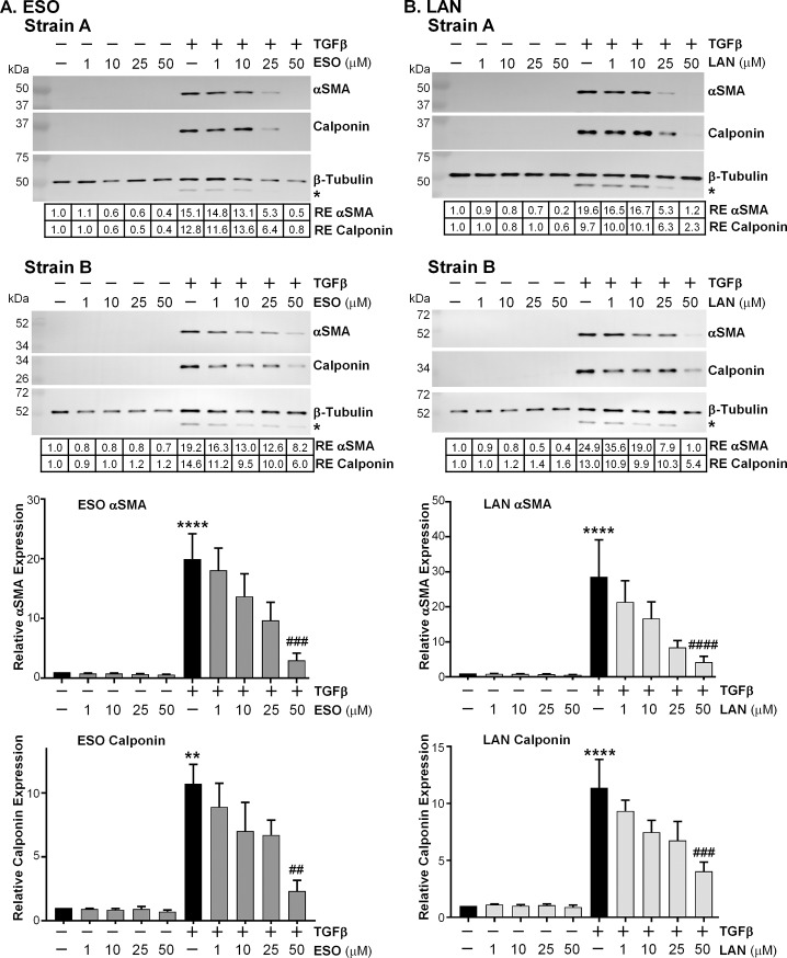
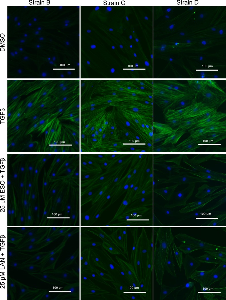
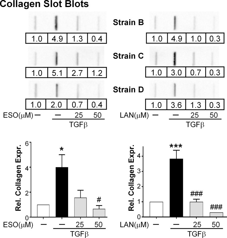
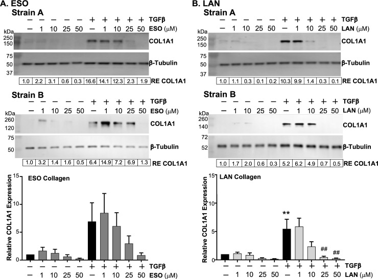
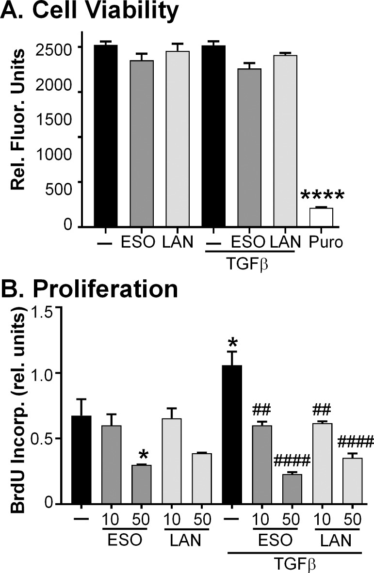
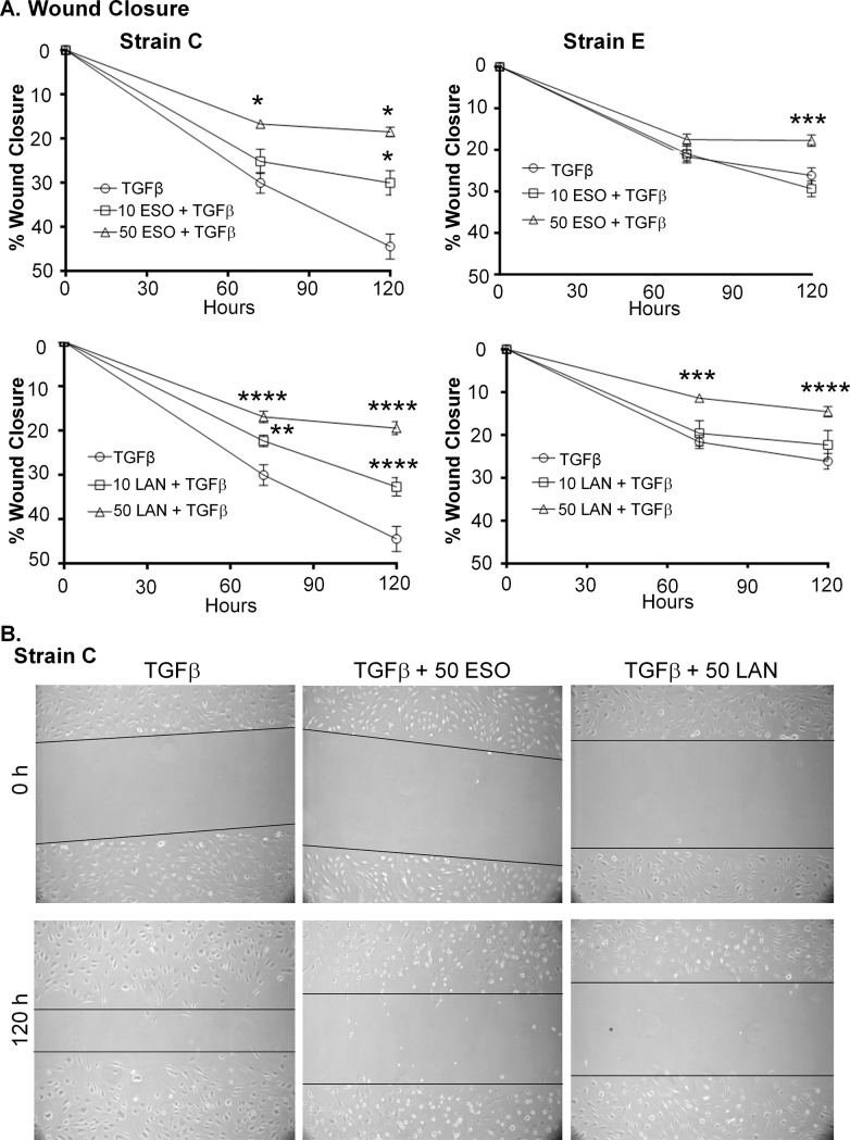
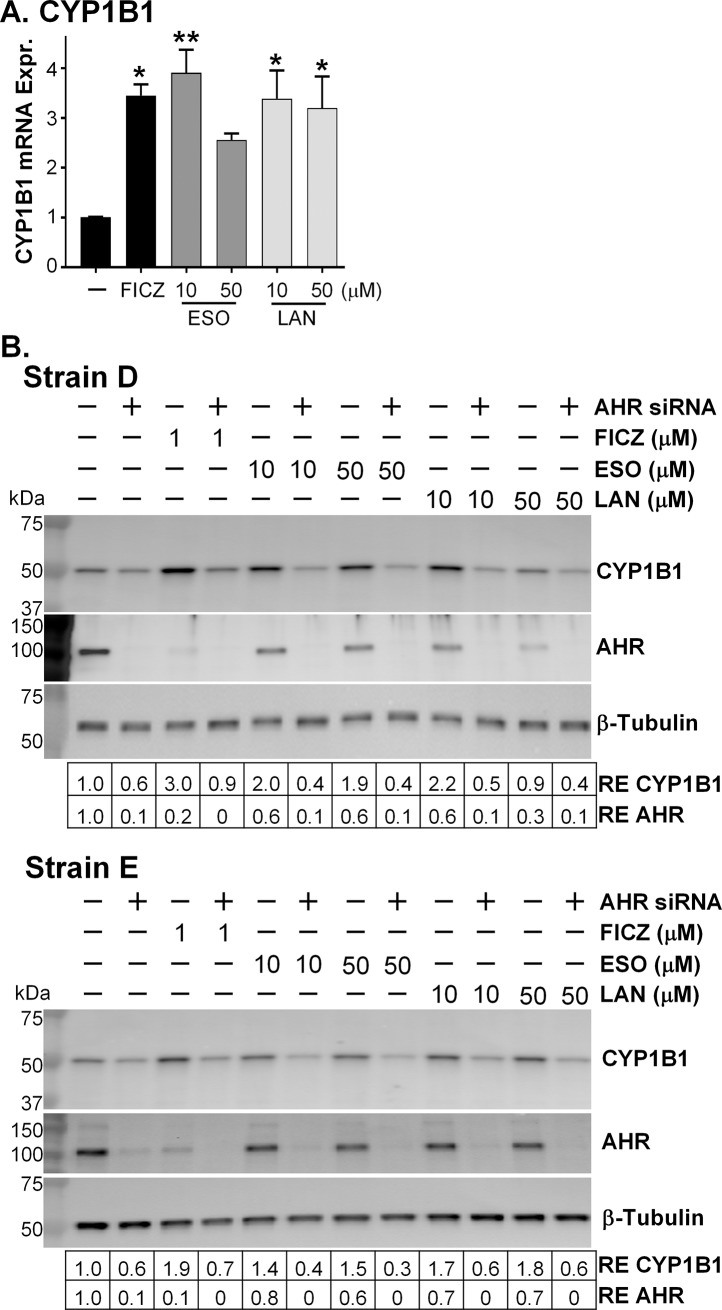
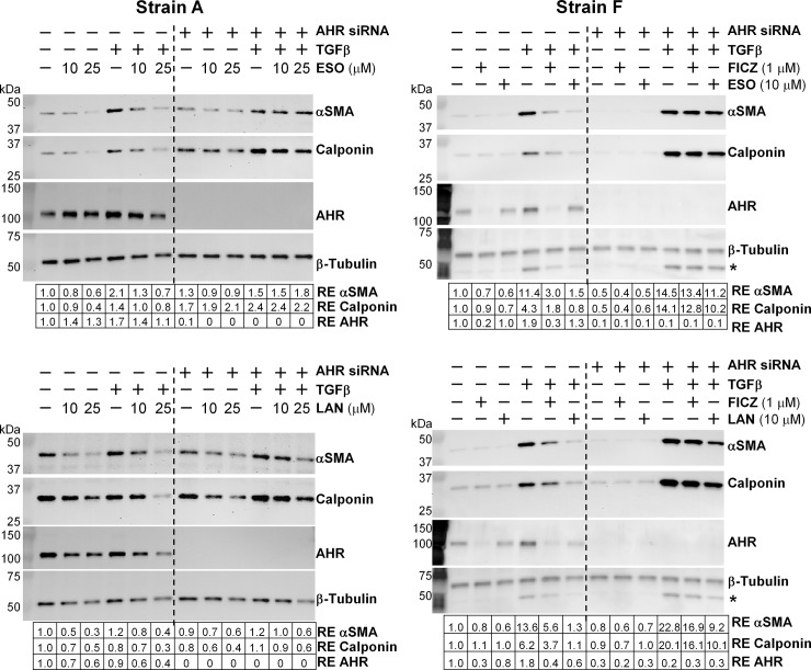
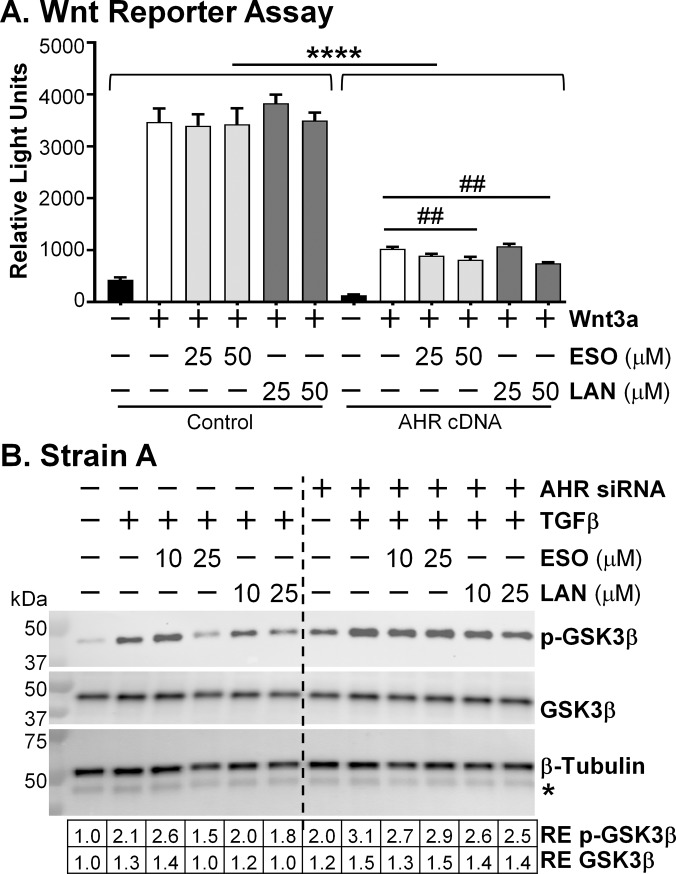
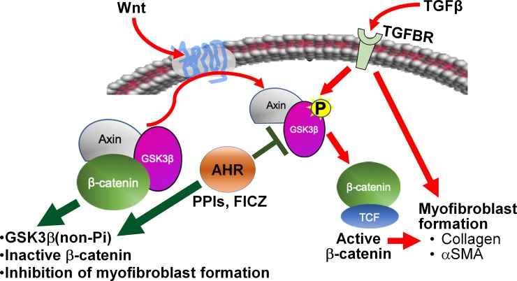
Thyroid eye disease (TED) can lead to scar formation and tissue remodeling in the orbital space. In severe cases, the scarring process leads to sight-threatening pathophysiology. There is no known effective way to prevent scar formation in TED patients, or to reverse scarring once it occurs. In this study, we show that the proton pump inhibitors (PPIs), esomeprazole and lansoprazole, can prevent transforming growth factor beta (TGFβ)-mediated differentiation of TED orbital fibroblasts to myofibroblasts, a critical step in scar formation. Both PPIs prevent TGFβ-induced increases in alpha-smooth muscle actin (αSMA), calponin, and collagen production and reduce TED orbital fibroblast cell proliferation and migration. Esomeprazole and lansoprazole exert these effects through an aryl hydrocarbon receptor (AHR)-dependent pathway that includes reducing β-catenin/Wnt signaling. We conclude that PPIs are potentially useful therapies for preventing or treating TED by reducing the myofibroblast accumulation that occurs in the disease.


Conflict of interest statement

The authors have declared that no competing interests exist.

Figures

Fig 1
Fig 1. PPIs prevent TGFβ-induced myofibroblast marker expression.
A) PPIs were added to TED orbital fibroblasts daily for 4 days. TGFβ (1 ng/mL) was administered after the first 24h of PPI exposure. Cell lysates were collected after the fourth day and protein levels of αSMA and calponin were measured by Western blot. A) Two different representative strains of TED orbital fibroblasts were treated with esomeprazole +/- TGFβ. Esomeprazole led to a dose-dependent prevention in TGFβ-mediated αSMA and calponin increase. RE = relative expression of protein bands compared with first band on the left. * The faint band beneath the β-tubulin band is αSMA that was previously detected. B) The same two representative TED orbital fibroblast strains were treated with lansoprazole +/- TGFβ. Lansoprazole also led to a dose-dependent prevention in TGFβ-mediated αSMA and calponin increase. RE = relative expression of protein bands compared with first band on the left. * The faint band beneath the β-tubulin band is αSMA that was previously detected. Bar graphs depicting the dose-dependent effects of the PPIs on αSMA and calponin expression due to TGFβ are presented beneath the Western blots. N = 4–6 different TED orbital fibroblast strains. ** = p < 0.01; **** = p < 0.0001; significantly different from vehicle treated control. ## = p < 0.01; ### = p < 0.001; #### = p < 0.0001; significantly different from TGFβ alone. Statistics were measured by one-way ANOVA and Tukey’s post hoc analysis.
Fig 2
Fig 2. PPIs prevent the TGFβ-induced myofibroblast marker αSMA expression.
PPIs (25 μM) were added to TED orbital fibroblasts 24h before 1ng/mL TGFβ and then every 24h for the next 72h. TGFβ-induced αSMA expression was measured with fluorescent imaging. Green = αSMA; Blue = DAPI staining of nuclei. Three representative strains are shown. The bar on the images represents 100 μm.
Fig 3
Fig 3. PPIs attenuate TGFβ-induced collagen production.
PPIs were added to TED orbital fibroblasts 24h before 1 ng/mL TGFβ and then every 24h for the next 72h. Supernatants were collected at the end of the 72h and measured for collagen content using collagen slot blots. Data from three representative strains are shown. RE = relative expression; determined by setting each vehicle treated sample in the group of four bands to one. Graphs represent data from images of Strains B-D. N = 3. * = p < 0.05; *** = p < 0.001; significantly different from vehicle treated control. # = p < 0.05; ### = p < 0.001; significantly different from TGFβ alone. Statistics were measured by one-way ANOVA and Tukey’s post hoc analysis.
Fig 4
Fig 4. PPIs attenuate cell associated collagen induced by TGFβ.
PPIs were added to TED orbital fibroblasts 24h before 1ng/mL TGFβ and then every 24h for the next 72h. Cell lysates were collected and measured for collagen 1A1 using Western blotting. Western blots from two representative TED orbital fibroblast strains are shown. A) Orbital fibroblasts treated with esomeprazole (ESO). B) Orbital fibroblasts treated with lansoprazole (LAN). RE = relative expression of protein bands compared with first band on the left. Bar graphs of the mean relative expression for each band depict a dose-dependent reduction in both basal and TGF-induced collagen levels and are shown beneath the Western blots. N = 4 different TED orbital fibroblast strains. ** = p < 0.01; significantly different from vehicle treated control. ## = p < 0.01; significantly different from TGFβ alone. Statistics were measured by one-way ANOVA and Tukey’s post hoc analysis.
Fig 5
Fig 5. PPIs do not reduce TED orbital fibroblast cell viability; however, they do reduce fibroblast proliferation.
A) PPIs (ESO = esomeprazole; LAN = lansoprazole; 50 μM dose) were added to cell cultures every 24h for 96h. TGFβ (1 ng/mL) was added 24h after the first PPI dose. Cell viability in TED orbital fibroblasts was measured using the Alamar Blue assay. PURO = Puromycin, a positive control for cell death. N = 3 different TED orbital fibroblast strains. **** p < 0.0001; significantly different from all other treatments measured with a one-way ANOVA and Tukey’s post hoc analysis. B) Cells were treated with PPIs (ESO = esomeprazole; LAN = lansoprazole; 10 and 50 μM doses) for 2h before some cultures received the addition of 1 ng/mL TGFβ. The PPIs were then added every 24h afterwards for a total of 72h after TGFβ administration. Cellular proliferation was measured with BrdU incorporation. Basal and TGFβ-induced fibroblast proliferation was attenuated by the PPIs. N = 3 different TED orbital fibroblast strains. * p < 0.05; significantly different from vehicle treated control. ## p < 0.01; #### p < 0.0001; significantly different from TGFβ treatment alone. Statistics were measured with a one-way ANOVA and Tukey’s post hoc analysis.
Fig 6
Fig 6. PPIs attenuate TED orbital fibroblast proliferation and migration in a wound healing assay.
Wounds were scratched across confluent TED orbital fibroblasts and bright-field pictures were taken at specific locations across the wound, the area of the wound was determined and set at 0% wound closure. Cells were treated with PPIs for 2h before TGFβ (5 ng/mL) was added. The PPIs were added every 24h afterwards until 120h. Additional pictures were taken at 72 and 120h of the same specific locations imaged directly after the scratch. The wound area was determined using Image J software and is reported as relative to the wound area at time zero. A) Line graphs for two representative strains are shown. ESO = esomeprazole and LAN = lansoprazole. * p < 0.05; ** p < 0.01; *** p < 0.001, **** p < 0.0001; significantly different from TGFβ treatment alone. N = 8 technical replicates. Statistics were measured with a two-way ANOVA and Tukey’s post hoc analysis. B) Representative bright-field pictures of the wounds across the TED orbital fibroblasts in Strain C, demonstrating the prevention of fibroblast migration after TGFβ treatment by esomeprazole (ESO) and lansoprazole (LAN).
Fig 7
Fig 7. PPIs are AHR activators.
A) CYP1B1 mRNA expression after 6h PPI treatment. Cells were treated with an AHR ligand positive control (FICZ) or proton pump inhibitors for 24h before RNA was isolated from cell lysates and analyzed for CYP1B1 mRNA expression using qPCR. ESO = esomeprazole; LAN = lansoprazole. N = 3 different TED orbital fibroblast strains. * p < 0.05; ** p < 0.01; significantly different from vehicle treatment alone. Statistics were measured with a one-way ANOVA and Tukey’s post hoc analysis. B) After reduction of AHR expression with AHR specific siRNA, FICZ, esomeprazole or lansoprazole were added for 24h. Cell lysates were analyzed for CYP1B1, AHR, and β-tubulin protein expression using Western blotting. Two representative TED orbital fibroblast strains are shown, Strain D and Strain E. RE = relative expression of protein bands compared with first band on the left.
Fig 8
Fig 8. PPIs prevent TGFβ-induced myofibroblast marker expression in an AHR-dependent manner.
After reduction of AHR expression with AHR specific siRNA, FICZ or a proton pump inhibitor (ESO = esomeprazole; LAN = lansoprazole) was added every 24h for 96h. After the first 24h, 1 ng/mL TGFβ was added. Cell lysates were collected at the end of 96h and analyzed for αSMA, calponin, AHR, and β-tubulin expression using Western blotting. Two representative strains of different TED orbital fibroblasts are shown, Strain A was used in the left two panels and Strain F was used in the right two panels. RE = relative expression of protein bands compared with first band on the left. * The band beneath the β-tubulin band is αSMA that was previously detected on this membrane.
Fig 9
Fig 9. PPIs reduce Wnt signaling in an AHR-dependent manner.
A) Wnt reporter assay. HEK293FT cells were used in a reporter assay to monitor Wnt signaling in the presence or absence of exogenous AHR expression. This experiment was repeated three times and a representative experiment is shown. N = 3–6 technical replicates. **** p < 0.0001; each control plasmid bar is significantly higher than its respective AHR cDNA plasmid bar. ## p < 0.01; 50 μM PPIs are significantly lower than Wnt treatment alone in the AHR cDNA plasmid group. Statistics were measured with a one-way ANOVA and Tukey’s post hoc analysis. B) Western blot for phospho-GSK3β. After reduction of AHR expression with AHR specific siRNA, esomeprazole or lansoprazole was added every 24h for 96h. After the first 24h, 1 ng/mL TGFβ was added for the remainder of the 72h. Cell lysates were collected and analyzed for phosphorylated GSK3β, total GSK3β, and β-tubulin expression using Western blotting. RE = relative expression of protein bands compared with first band on the left. * The band beneath the β-tubulin band is GSK3β that was previously detected on this membrane.
Fig 10
Fig 10. AHR prevents GSK3β phosphorylation to limit β-catenin/Wnt signaling and block myofibroblast formation.
This is a simplified model of the potential interactions of TGFβ, AHR, and Wnt signaling pathways in TED, and how the PPIs may be signaling through AHR affecting Wnt signaling and preventing TGFβ-myofibroblast formation. TGFβ signaling induces myofibroblast formation through a few different pathways, one of which is the β-catenin/Wnt signaling pathway. TGFβ can directly phosphorylate GSK3β allowing β-catenin to enter the nucleus and promote the transcription of genes important for myofibroblast formation. Signaling through the AHR with PPIs or FICZ inhibits phosphorylation of GSK3β, thereby maintaining GSK3β and β-catenin in the destruction complex retained in the cytoplasm and blocking the transcription of β-catenin upregulated genes. Without β-catenin activated genes, myofibroblast formation is inhibited.

Similar articles

Cited by

References

    1. Bahn RS. Current Insights into the Pathogenesis of Graves' Ophthalmopathy. Horm Metab Res. 2015;47(10):773–8. Epub 2015/09/12. 10.1055/s-0035-1555762 . - DOI - PubMed
    1. Bartalena L. Graves' orbitopathy: imperfect treatments for a rare disease. Eur Thyroid J. 2013;2(4):259–69. Epub 2014/05/02. 10.1159/000356042 - DOI - PMC - PubMed
    1. Douglas RS, Gupta S. The pathophysiology of thyroid eye disease: implications for immunotherapy. Curr Opin Ophthalmol. 2011;22(5):385–90. Epub 2011/07/07. 10.1097/ICU.0b013e3283499446 - DOI - PMC - PubMed
    1. Schluter A, Horstmann M, Diaz-Cano S, Plohn S, Stahr K, Mattheis S, et al. Genetic immunization with mouse thyrotrophin hormone receptor plasmid breaks self-tolerance for a murine model of autoimmune thyroid disease and Graves' orbitopathy. Clin Exp Immunol. 2018;191(3):255–67. Epub 2017/10/24. 10.1111/cei.13075 - DOI - PMC - PubMed
    1. Smith TJ. New advances in understanding thyroid-associated ophthalmopathy and the potential role for insulin-like growth factor-I receptor. F1000Res. 2018;7:134 Epub 2018/05/11. 10.12688/f1000research.12787.1 - DOI - PMC - PubMed

Publication types

MeSH terms