Review
. 2019 Sep 24;74(12):1594-1607.
doi: 10.1016/j.jacc.2019.07.061.

From Focal Lipid Storage to Systemic Inflammation: JACC Review Topic of the Week

Affiliations
Review

From Focal Lipid Storage to Systemic Inflammation: JACC Review Topic of the Week

Peter Libby et al. J Am Coll Cardiol. .

Abstract

Concepts of atherogenesis have evolved considerably with time. Early animal experiments showed that a cholesterol-rich diet could induce fatty lesion formation in arteries. The elucidation of lipoprotein metabolism ultimately led to demonstrating the clinical benefits of lipid lowering. The view of atheromata as bland accumulations of smooth muscle cells that elaborated an extracellular matrix that could entrap lipids then expanded to embrace inflammation as providing pathways that could link risk factors to atherogenesis. The characterization of leukocyte adhesion molecules and their control by proinflammatory cytokines, the ability of chemokines to recruit leukocytes, and the identification of inflammatory cell subtypes in lesions spurred the unraveling of innate and adaptive immune pathways that contribute to atherosclerosis and its thrombotic complications. Such pathophysiologic insights have led to the identification of biomarkers that can define categories of risk and direct therapies and to the development of new treatments.

Keywords: LDL cholesterol; inflammation; smooth muscle cell.


Figures

Central Illustration:
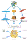
Central Illustration:. Evolution of concepts of the pathogenesis of atherosclerosis.
The diagrams represent the dominant formulation of mechanisms of atherogenesis as they emerged over time (clockwise). In the mid-19th-century Virchow and von Rokitansky fueled a heated controversy regarding the role of incorporated thrombus in atherosclerosis (top pair of diagrams.) The experiments of an Anichkov and many others lead to predominant view of atherosclerosis as primarily a lipid storage disease. This concept prevailed for much of the 20th Century. The pioneering work of Ross and of Benditt in the 1970s emphasized the role of smooth muscle proliferation in lesion formation. The initial formulation of the “response to injury” hypothesis accorded initiating role to endothelial denudation, and did not invoke a role for inflammatory cells. Work in the 1980s and beyond used the evolving tools of immunology to define operation of both innate and adaptive immunity in atherogenesis, coming full circle to Virchow’s older observations implicating inflammation pathways in atherogenesis. Our current synthetic view of atherogenesis (center) encompasses elements of each of these pathogenic processes unraveled through the years.
Figure 1.
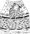
Figure 1.. The atheromatous plaque according to Virchow.
This drawing by Virchow of a human atherosclerotic plaque shows atheromatous foam cells (a’), and a layer of proliferating cells (p) with morphologic evidence of cell division disclosed by mitotic nuclei. The figure depicts a layer of cells (h) beneath an intimal EC lining (i) on the lumenal surface. This depiction from mid 19th century illustrates the lipid core (a, a’), smooth muscle involvement (p), a fibrous cap, and an intact endothelial monolayer. Adapted from (1).
Figure 2.
Figure 2.. A foam cell lesion in a cholesterol-fed rabbit.
This drawing by Anitchkov portrays a foam cell-rich arterial lesion from a cholesterol-fed rabbit. Note the intact endothelial monolayer (End.), the macrophages (Mf.), and the cholesterol laden phagocytes (Chol. Ph.), some with multiple nuclei. Bipolar spindle-shaped cells (likely smooth muscle) reside beneath the cluster of foam cells. Adapted from (5).
Figure 3.
Figure 3.. The ascendancy of smooth muscle proliferation.
This drawing presents the view of Ross and Glomset in the 1970s depicting a desquamative endothelial injury with SMC migration from the media into a growing intimal lesion with mitotic figures indicating division of the SMCs. Ross and Glomset hypothesized a major role for platelet-derived growth factor in stimulating the migration and proliferation of SMCs. This formulation viewed atherogenesis as a bland phenomenon lacking inflammation or leukocytes. Adapted from (14).
Figure 4.
Figure 4.. A simplified view of the operation of innate and adaptive immunity as thought to operate in atherogenesis.
Innate immunity initiates when macrophages (MΦ) recognize PAMPs and DAMPs binding to their pattern recognition receptors. This interaction leads to production of a host of proinflammatory molecules including cytokines (e.g. IL-1-α and -β and TNF) and small molecules such as eicosanoids. Adaptive immune reponses follow the processing of antigens (foreign or autologous) by dendritic cells (DC.) Proteolytic cleavage of protein antigens into peptides within the DC prepares the antigen for presentation on the DC surface bound to major histocompatibility complex (MHC) molecules (human leukocyte antigens, HLA in humans.) Antigen-specific immune cells recognize the nominal nominal antigen in the context of self MHC. T cells can recognize peptide-HLA complexes via specific antigen receptors. Antigen recognition in combination with signals produced by the DC prompts the T cell to activate and differentiate. Activated CD4+ T cells may differentiate into several cell types with different functions. Among them, Th1 cells produce interferon-γ and TNF, are highly proinflammatory, and strongly stimulate macrophage activation and vascular inflammation. Treg, on the other hand, make two anti-inflammatory and immunoregulatory cytokines, namely transforming growth factor-β and IL-10. IL-17 produced by Th17 cells activate granulocytes and stimulates collagen production, as does TGF-β. B cells recognize antigens that ligate their surface-bound immunoglobulins. The most prevalent type of B cell, the B2 cell, receives help from T follicular helper (Tfh) cells and can develop into plasma cells specialized in production of IgG antibodies. In addition, B2 cells produce cytokines that can modulate inflammation. Another B cell subset, the B1 cell, produces IgM antibodies and does not require Tfh cell help. B1 cells largely produce “natural” antibodies encoded by the germline, while the genes that encode IgG antibodies generally undergo somatic mutations to achieve greater affinity for the particular antigen with time. Single-line arrows indicate signaling molecules whereas thick arrows show cell development.
Figure 5.
Figure 5.. Thrombosis begets inflammation.
Platelets not only contribute to thrombus formation through well understood pathways but when activated also release pre-formed proinflammatory mediators from their granules. Thrombin activates platelets to produce the proinflammatory mediators such as RANTES, IL-6, and to exteriorize CD40 Ligand, and also stimulates SMCs to proliferate and activates ECs. Adapted from (47).
Figure 6.
Figure 6.. The expanded cardiovascular continuum.
Risk factors beget atherosclerosis that can cause thrombosis leading to tissue injury such as acute myocardial infarction. The sympathetic stimulation that myocardial infarction engenders can activate the bone marrow to release leukocyte progenitor cells that can reside in the spleen and migrate to the atherosclerotic plaque. The infarcted myocardium also leads release of such pro-inflammatory cytokines as IL-1β, able to propagate inflammation to remote sites, including the atheroma itself. Hence, both systemic and local inflammation can impinge on the prepared soil of the plaque leading to local inflammatory activation exacerbating the inflammatory state. Thus, the cycle of inflammation can perpetuate leading to recurrent events and aggravated atherothrombosis. Adapted from (38).
Figure 7.
Figure 7.. Thrombosis: The ultimate complication of atherosclerosis.
Arterial thrombi cause acute coronary syndromes and many ischemic strokes. The thrombotic potential of a plaque depends on the production of tissue factor by macrophages or SMCs within the intima, the “solid state” of the plaque. Plasminogen activator inhibitor (PAI-1) production by lesional cells in response to inflammatory stimuli inhibits these promoters of fibrinolysis. ECs can express thrombomodulin which can limit thrombosis, but inflammatory stimuli and regulation by KLF-factor 2 can limit thrombomodulin expression by ECs reducing their baseline anti-thrombotic effects. ECs can also produce tissue type and urokinase-type plasminogen activator (tPA and uPA) that can activate the fibrinolytic system. When inflammatory stimuli augment its production, PAI-1 inhibits these promoters of fibrinolysis. Thus an inflammatory state augments the thrombogenicity of the plaque by boosting tissue-factor expression, mutes the intrinsic anticoagulant effect of the normal endothelial monolayer, and combats fibrinolysis (an effect that stabilizes clots). The consequencing of a given plaque disruption, be it fibrous cap rupture as depicted here, or superficial erosion (not shown), depends not only on the solid state of the plaque but also on the fluid phase of blood. Microparticles that bear tissue factor may also contribute to the extension of thrombosis due to plaque disruption. Adapted from (79).

References

    1. Virchow R Cellular Pathology. London: John Churchill, 1858.
    1. Mayerl C, Lukasser M, Sedivy R, Niederegger H, Seiler R, Wick G. Atherosclerosis research from past to present--on the track of two pathologists with opposing views, Carl von Rokitansky and Rudolf Virchow. Virchows Arch 2006;449:96–103. - PubMed
    1. Duguid J Thrombosis as a factor in the pathogenesis of coronary atherosclerosis. J Pathol 1946;58:208–212. - PubMed
    1. Anitchkov N, Chalatow S. On experimental cholesterin steatosis and its significance in the origin of some pathological processes (1913). Reprinted in Arteriosclerosis 1983;3:178–182. - PubMed
    1. Anitchkov N Über die Veränderungen der Kaninchenaorta bei experimenteller Cholesterin-steatose. Beitr Pathol Anat 1913;56:379–404.

Publication types