

Augmented Bladder Tumor Detection Using Deep Learning
- PMID: 31537407
- PMCID: PMC6889816
- DOI: 10.1016/j.eururo.2019.08.032
Augmented Bladder Tumor Detection Using Deep Learning
Abstract
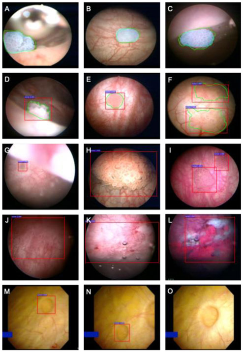
Adequate tumor detection is critical in complete transurethral resection of bladder tumor (TURBT) to reduce cancer recurrence, but up to 20% of bladder tumors are missed by standard white light cystoscopy. Deep learning augmented cystoscopy may improve tumor localization, intraoperative navigation, and surgical resection of bladder cancer. We aimed to develop a deep learning algorithm for augmented cystoscopic detection of bladder cancer. Patients undergoing cystoscopy/TURBT were recruited and white light videos were recorded. Video frames containing histologically confirmed papillary urothelial carcinoma were selected and manually annotated. We constructed CystoNet, an image analysis platform based on convolutional neural networks, for automated bladder tumor detection using a development dataset of 95 patients for algorithm training and five patients for testing. Diagnostic performance of CystoNet was validated prospectively in an additional 54 patients. In the validation dataset, per-frame sensitivity and specificity were 90.9% (95% confidence interval [CI], 90.3-91.6%) and 98.6% (95% CI, 98.5-98.8%), respectively. Per-tumor sensitivity was 90.9% (95% CI, 90.3-91.6%). CystoNet detected 39 of 41 papillary and three of three flat bladder cancers. With high sensitivity and specificity, CystoNet may improve the diagnostic yield of cystoscopy and efficacy of TURBT. PATIENT SUMMARY: Conventional cystoscopy has recognized shortcomings in bladder cancer detection, with implications for recurrence. Cystoscopy augmented with artificial intelligence may improve cancer detection and resection.
Keywords: Bladder cancer; Computer-assisted image analysis; Cystoscopy; Deep learning; Diagnostic imaging.
Copyright © 2019 European Association of Urology. Published by Elsevier B.V. All rights reserved.
Conflict of interest statement
Figures
Comment in
-
Re: Eugene Shkolyar, Xiao Jia, Timothy C. Chang, et al. Augmented Bladder Tumor Detection Using Deep Learning. Eur Urol 2019;76:714-8.Eur Urol. 2020 May;77(5):e133. doi: 10.1016/j.eururo.2019.11.020. Epub 2019 Dec 6. Eur Urol. 2020. PMID: 31818521 No abstract available.
References
-
- Svatek RS, Hollenbeck BK, Holmäng S, et al. The economics of bladder cancer: costs and considerations of caring for this disease. Eur Urol 2014;66:253–62. - PubMed
-
- Witjes JA, Redorta JP, Jacqmin D, et al. Hexaminolevulinate-guided fluorescence cystoscopy in the diagnosis and follow-up of patients with non-muscle-invasive bladder cancer: review of the evidence and recommendations. Eur Urol 2010;57:607–14. - PubMed
-
- Burger M, Grossman HB, Droller M, et al. Photodynamic diagnosis of non-muscle-invasive bladder cancer with hexaminolevulinate cystoscopy: a meta-analysis of detection and recurrence based on raw data. Eur Urol 2013;64:846–54. - PubMed
Publication types
MeSH terms
Grants and funding
LinkOut - more resources
Full Text Sources
Medical
