Review
. 2019 Oct;71(4):539-570.
doi: 10.1124/pr.118.017129.

Novel Therapeutic Approaches Targeting the Renin-Angiotensin System and Associated Peptides in Hypertension and Heart Failure

Affiliations
Review

Novel Therapeutic Approaches Targeting the Renin-Angiotensin System and Associated Peptides in Hypertension and Heart Failure

Lauren B Arendse et al. Pharmacol Rev. 2019 Oct.

Abstract

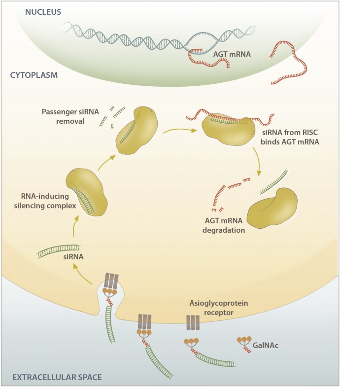
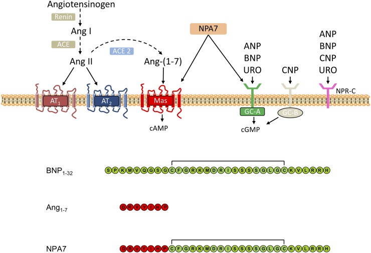
Despite the success of renin-angiotensin system (RAS) blockade by angiotensin-converting enzyme (ACE) inhibitors and angiotensin II type 1 receptor (AT1R) blockers, current therapies for hypertension and related cardiovascular diseases are still inadequate. Identification of additional components of the RAS and associated vasoactive pathways, as well as new structural and functional insights into established targets, have led to novel therapeutic approaches with the potential to provide improved cardiovascular protection and better blood pressure control and/or reduced adverse side effects. The simultaneous modulation of several neurohumoral mediators in key interconnected blood pressure-regulating pathways has been an attractive approach to improve treatment efficacy, and several novel approaches involve combination therapy or dual-acting agents. In addition, increased understanding of the complexity of the RAS has led to novel approaches aimed at upregulating the ACE2/angiotensin-(1-7)/Mas axis to counter-regulate the harmful effects of the ACE/angiotensin II/angiotensin III/AT1R axis. These advances have opened new avenues for the development of novel drugs targeting the RAS to better treat hypertension and heart failure. Here we focus on new therapies in preclinical and early clinical stages of development, including novel small molecule inhibitors and receptor agonists/antagonists, less conventional strategies such as gene therapy to suppress angiotensinogen at the RNA level, recombinant ACE2 protein, and novel bispecific designer peptides.


Figures

None
Graphical abstract
Fig. 1.
Fig. 1.
Outline of the systems involved in blood pressure regulation. Vasoconstrictor and vasodilator peptides are shown in red and blue rectangles, respectively. Vasopeptidases responsible for the production or degradation of vasoactive peptides are shown in colored spheres (ACE, APP, ECE, and NEP). Production of the vasoconstrictor peptides Ang II and ET-1 (red rectangles) in the RAS and endothelin system, respectively, lead to vasoconstriction, aldosterone secretion, and sodium retention. Bradykinin and NPs (ANP, BNP, and CNP) are potent vasodilatory peptides that counter-regulate the effects of Ang II and ET-1. The vasoactive peptides mediate their physiologic effect via a range of receptors (AT1R, AT2R, B1R, B2R, ETAR, ETBR, NPR-A, NPR-B, and NPR-C).
Fig. 2.
Fig. 2.
(A) Angiotensin metabolism. Angiotensin peptides are shown as colored spheres (AGT and Ang metabolites). Peptidases responsible for peptide cleavage are indicated (ACE, ACE2, AP, CHY, DAP, and NEP). Receptors for vasoactive peptides responsible for mediating vasoconstrictive and counteractive vasodilatory responses are indicated in colored rectangles (AT1R, AT2R, and Mas). (B) Schematic showing the counter-regulatory effects of the Ang 1-7/Mas, Ang 1-9/AT2R, and Ang II/AT2R pathways on the Ang II/AT1R pathway. AGT, angiotensinogen; AP, aminopeptidase; CHY, chymase; DAP, dipeptidyl aminopeptidase.
Fig. 3.
Fig. 3.
Bradykinin metabolism. Bradykinin peptides are shown as colored spheres. Peptidases responsible for peptide cleavage are indicated (ACE, ACE2, APP, CPN, DPPIV, and NEP). Bradykinin receptors B1R and B2R are indicated in green rectangles. CPN, carboxypeptidase N; DPP-IV, dipeptidyl peptidase IV.
Fig. 4.
Fig. 4.
A schematic diagram of ACE active sites [Schechter and Berger nomenclature (Schechter and Berger, 1967)] showing the subsite binding pockets accommodating the residues on either side of the ZBG of peptide substrates. ACE domain-specific amino acid residues important for conferring domain selectivity are shown within the relevant subsites of the ACE active site.
Fig. 5.
Fig. 5.
(A) Chemical structures of C-domain–selective inhibitors and the corresponding in vitro inhibition constants for the N and C domains. (B) Overlay of C-domain–selective inhibitors bound to the active site of the C domain from crystal structures [PDB codes 3BKK (kAF), 3BKL (kAW), 2OC2 (RXPA380), and 3L3N (LisW)]. (C) Overlay of crystal structures of ACE N and C domains in complex with lisinopril is shown in yellow (N domain) and green (C domain) (PDB codes 2C6N and 1O86 respectively) and the ACE C domain in complex with LisW in purple (PDB code 3L3N). C-domain unique residues are shown in cyan with corresponding N-domain residues in orange. Cdom, C domain; Ndom, N domain; PDB, Protein Data Bank.
Fig. 6.
Fig. 6.
(A) Chemical structures of C-domain–selective phosphinic tripeptides FI and FII, showing residue positions relative to the zinc binding group together with the in vitro inhibition data for NEP, ECE-1, and ACE N and C domains. (B) FI and FII bound to the active sites of the ACE N and C domains: FI bound to the N domain and C domain in green and cyan, respectively; FII bound to the N domain and C domain shown in yellow and black, respectively. C-domain unique residues within the active site are shown in cyan with corresponding N-domain residues in orange.
Fig. 7.
Fig. 7.
Mode of action of the APA inhibitor prodrug RB150/firibastat on the control of blood pressure in hypertensive rats. After oral administration, the disulfide bridge enables RB150 to cross the blood–brain barrier and to enter the brain. At the opposite, EC33 is not able to enter the brain. In the brain, the disulfide bridge of RB150 is cleaved by brain reductases generating two active molecules of EC33. EC33 subsequently inhibits brain APA activity and blocks the formation of brain Ang III, known to exert, in brain structures (PVN, SON, PPit, NTS, and RVLM), a stimulatory action on the control of blood pressure in hypertensive rats. This results in a blood pressure decrease via a decrease in arginine-vasopressin release and sympathetic neuron activity and an improvement of the baroreflex function. The red dashed lines represent the neuronal angiotensinergic pathways in the adult rat brain. MnPO, median preoptic nucleus; NTS, nucleus of the solitary tract; OVLT, organum vasculosum of the lamina terminalis; PPit, posterior pituitary; PVN, paraventricular nucleus; RVLM, rostral ventrolateral medulla; SFO, subfornical organ; SON, supraoptic nucleus.
Fig. 8.
Fig. 8.
Overview of AGT suppression using siRNAs. siRNAs enter the cell and are incorporated into the RISC in the cytoplasm. The RISC complex with the active guide strand binds the complementary sequence within the target mRNA, resulting in Argonaut 2–mediated cleavage and subsequent AGT mRNA degradation. AGT, angiotensinogen.
Fig. 9.
Fig. 9.
NPA7 is a single peptide entity that coactivates the Mas and pGC-A receptors and their second messengers cAMP and cGMP, respectively. NPA7 incorporates key amino acids from BNP1-32 (a pGC-A activator) and Ang 1-7 (a Mas activator), resulting in a novel bispecific first-in-class bispecific peptide. GC, guanylyl cyclase; URO, urodilatin.

References

    1. Abassi Z, Karram T, Ellaham S, Winaver J, Hoffman A. (2004) Implications of the natriuretic peptide system in the pathogenesis of heart failure: diagnostic and therapeutic importance. Pharmacol Ther 102:223–241. - PubMed
    1. AbdAlla S, Lother H, Abdel-tawab AM, Quitterer U. (2001) The angiotensin II AT2 receptor is an AT1 receptor antagonist. J Biol Chem 276:39721–39726. - PubMed
    1. AbdAlla S, Lother H, Quitterer U. (2000) AT1-receptor heterodimers show enhanced G-protein activation and altered receptor sequestration. Nature 407:94–98. - PubMed
    1. Agah R, Bandi V, Guntupalli KK. (1997) Angioedema: the role of ACE inhibitors and factors associated with poor clinical outcome. Intensive Care Med 23:793–796. - PubMed
    1. Akif M, Schwager SL, Anthony CS, Czarny B, Beau F, Dive V, Sturrock ED, Acharya KR. (2011) Novel mechanism of inhibition of human angiotensin-I-converting enzyme (ACE) by a highly specific phosphinic tripeptide. Biochem J 436:53–59. - PMC - PubMed

Publication types

MeSH terms

Substances