. 2019 Sep 19;9(1):12866.
doi: 10.1038/s41598-019-49177-y.

Characterisation and localisation of the endocannabinoid system components in the adult human testis

Affiliations

Characterisation and localisation of the endocannabinoid system components in the adult human testis

John E Nielsen et al. Sci Rep. .

Erratum in

Abstract

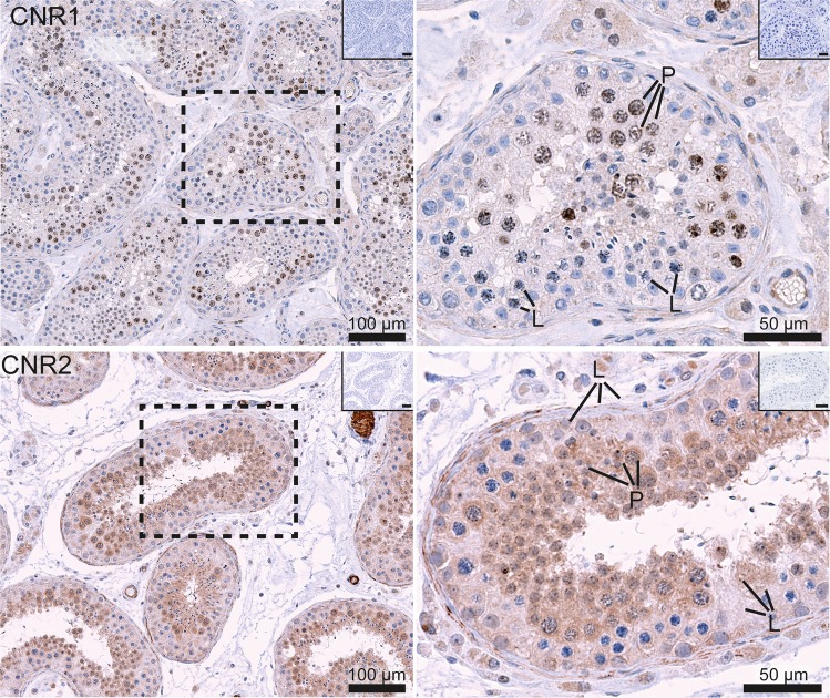
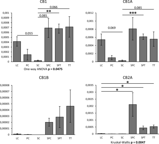
Heavy use of cannabis (marijuana) has been associated with decreased semen quality, which may reflect disruption of the endocannabinoid system (ECS) in the male reproductive tract by exogenous cannabinoids. Components of ECS have been previously described in human spermatozoa and in the rodent testis but there is little information on the ECS expression within the human testis. In this study we characterised the main components of the ECS by immunohistochemistry (IHC) on archived testis tissue samples from 15 patients, and by in silico analysis of existing transcriptome datasets from testicular cell populations. The presence of 2-arachidonoylglycerol (2-AG) in the human testis was confirmed by matrix-assisted laser desorption ionization imaging analysis. Endocannabinoid-synthesising enzymes; diacylglycerol lipase (DAGL) and N-acyl-phosphatidylethanolamine-specific phospholipase D (NAPE-PLD), were detected in germ cells and somatic cells, respectively. The cannabinoid receptors, CNR1 and CNR2 were detected at a low level in post-meiotic germ cells and Leydig- and peritubular cells. Different transcripts encoding distinct receptor isoforms (CB1, CB1A, CB1B and CB2A) were also differentially distributed, mainly in germ cells. The cannabinoid-metabolising enzymes were abundantly present; the α/β-hydrolase domain-containing protein 2 (ABHD2) in all germ cell types, except early spermatocytes, the monoacylglycerol lipase (MGLL) in Sertoli cells, and the fatty acid amide hydrolase (FAAH) in late spermatocytes and post-meiotic germ cells. Our findings are consistent with a direct involvement of the ECS in regulation of human testicular physiology, including spermatogenesis and Leydig cell function. The study provides new evidence supporting observations that recreational cannabis can have possible deleterious effects on human testicular function.


Conflict of interest statement

The authors declare no competing interests.

Figures

Figure 1
Figure 1
Immunohistochemical expression of the endocannabinoid-synthesising enzymes, NAPE-PLD and DAGLA, in the adult human testis. The images on the left show a general expression pattern, and a higher magnification of an area within a stippled line is shown on the right. All pictures show a low magnification image of negative controls in the right upper corner. NAPE-PLD was present in Leydig and Sertoli cells. DAGLA was present predominantly in the nuclei of germ cells, with only weak reaction detected in Sertoli and Leydig cells. L (leptotene) and P (pachytene)  indicate early and late spermatocytes, respectively.
Figure 2
Figure 2
Immunohistochemical expression in human testis tissue of the two canonical EC receptors; CNR1 and CNR2. The images on the left show a general expression pattern, and a higher magnification of an area within a stippled line is shown on the right. All pictures show a low magnification image of negative controls in the right upper corner. CNR1 staining (upper images) is visible within nuclei of primary spermatocytes and in the cytoplasm of Leydig cells. CNR2 (bottom images) is localised in the cytoplasm of all types of germ cells, except early spermatocytes, and in somatic cells, especially in Leydig cells and peritubular cells. L (leptotene) and P (pachytene) indicate early and late spermatocytes, respectively.
Figure 3
Figure 3
Immunohistochemical staining for the EC-degrading enzymes ABHD2, FAAH and MGLL in the human testis. The images on the left show a general expression pattern and the images on the right show a higher magnification of an area within a stippled line. All pictures show a low magnification image of negative controls in the right upper corner. Note that the ABHD2 (top images) is expressed in the cytoplasm of all stages of germ cell maturation, except early spermatocytes. FAAH reaction (middle row) is strongest in post-meiotic germ cells, including late spermatocytes and spermatids. MGLL protein (bottom images) is abundant in Sertoli cells. L (leptotene) and P (pachytene) indicate early and late spermatocytes, respectively.
Figure 4
Figure 4
Expression profiles of transcript isoforms coding for CB1, CB1A, CB1B and CB2A. Quantitative PCR (qPCR) experiments were performed on cDNA from isolated human Leydig cells (n = 4; LC), peritubular cells (n = 2; PC), Sertoli cells (n = 2; SC), pachytene spermatocytes (n = 4; SPC), round spermatids (n = 4; SPT) and total testis tissue (n = 4; TT). Histograms represent mean expression (relative to RPLP0) ± SEM. Following evaluation of normality (Shapiro–Wilk test and D’Agostino’s K-squared test) and of homogeneity of variances (Brown-Forsythe test), global analysis of variances was achieved with One-way ANOVA (CB1 and CB1A) or with Kruskal–Wallis one-way analysis of variance (CB1B and CB2A). Pairwise comparisons were also performed using unpaired Student’s t-test (CB1 and CB1A) or with Mann–Whitney U test (CB1B and CB2A). One, two or three stars denote statistical significance with p < 0.05, p < 0,01 or p < 0.005, respectively. p-values close to significance are also indicated.
Figure 5
Figure 5
Detection of 2-AG in human testis. (A) MALDI imaging with blue colour showing a lipid associated with intra-tubular cells and green colour marking a lipid associated with interstitial cells, while red stain shows recognition of 2-AG dispersed among the tubules. (B) The frozen section after MALDI imaging showing the structure of the testis.
Figure 6
Figure 6
Graphical summary of the findings of this study illustrating the general expression patterns (transcripts and protein combined) of the main components of the endocannabinoid system (ECS) in different cell types of the human adult testis. All germ cell types are depicted with blue nuclei, and the somatic cell types with orange nuclei. The ECS components are grouped into the synthesising enzymes (with the main products shown in parentheses), the receptors and the catabolic enzymes (main substrates in parentheses). Germ cell maturation stages are shown in the upper panel, and the somatic cells at the bottom of the graph. Uncertain expression patterns, with some discrepancy between the transcript and protein levels, are denoted by question marks. The asterisks refer to uncertain subcellular location of the protein (nuclear). The testicular cell types with the greatest activity of the ECS are shaded in pink: the post-meiotic germ cells (upper panel) and Leydig cells (bottom).

Comment in

References

    1. Hembree WC, 3rd, Nahas GG, Zeidenberg P, Huang HF. Changes in human spermatozoa associated with high dose marihuana smoking. Adv. Biosci. 1978;22-23:429–39. - PubMed
    1. Lewis SE, et al. Differences in the endocannabinoid system of sperm from fertile and infertile men. PLoS One. 2012;7:e47704. doi: 10.1371/journal.pone.0047704. - DOI - PMC - PubMed
    1. Gundersen TD, et al. Association between use of marijuana and male reproductive hormones and semen quality: A study among 1,215 healthy young men. Am. J. Epidemiol. 2015;182:473–481. - PubMed
    1. Hsiao P, Clavijo RI. Adverse effects of Cannabis on male reproduction. Eur Urol. Focus. 2018;4:324–328. - PubMed
    1. Daling JR, et al. Association of marijuana use and the incidence of testicular germ cell tumors. Cancer. 2009;115:1215–1223. - PMC - PubMed

Publication types