. 2019 Dec 1:586:113443.
doi: 10.1016/j.ab.2019.113443. Epub 2019 Sep 17.

Perfused murine heart optical transmission spectroscopy using optical catheter and integrating sphere: Effects of ischemia/reperfusion

Affiliations

Perfused murine heart optical transmission spectroscopy using optical catheter and integrating sphere: Effects of ischemia/reperfusion

Tyler M Bauer et al. Anal Biochem. .

Abstract

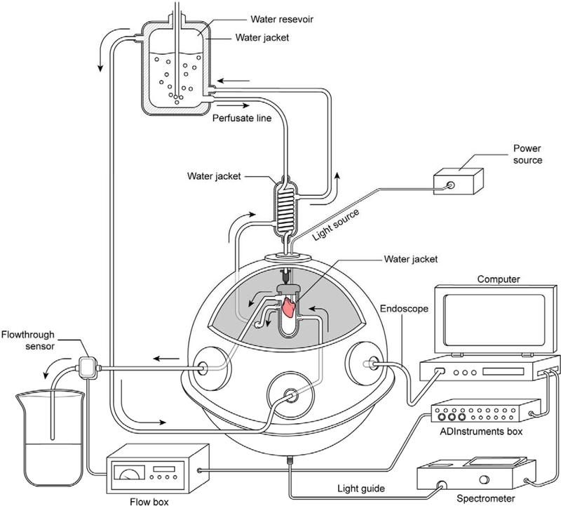
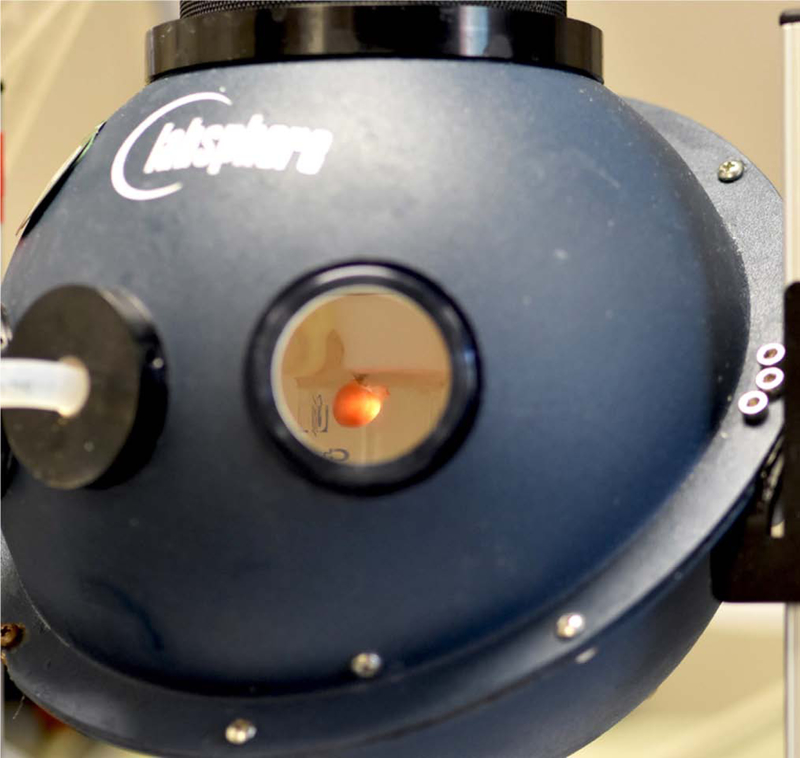
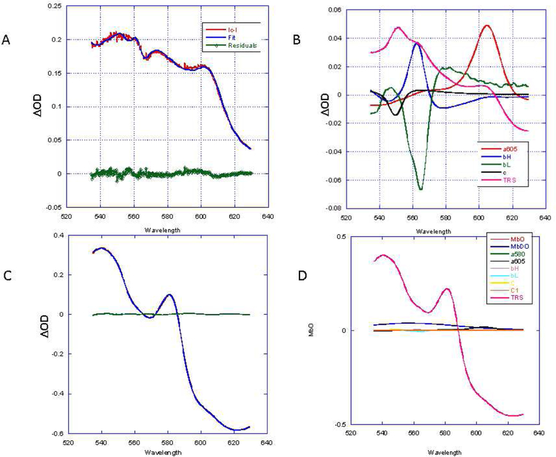
Tissue transmission optical absorption spectroscopy provides dynamic information on metabolism and function. Murine genetic malleability makes it a major model for heart research. The diminutive size of the mouse heart makes optical transmission studies challenging. Using a perfused murine heart center mounted in an integrating sphere for light collection with a ventricular cavity optical catheter as an internal light source provided an effective method of optical data collection in this model. This approach provided high signal to noise optical spectra which when fit with model spectra provided information on tissue oxygenation and redox state. This technique was applied to the study of cardiac ischemia and ischemia reperfusion which generates extreme heart motion, especially during the ischemic contracture. The integrating sphere reduced motion artifacts associated with a fixed optical pickup and methods were developed to compensate for changes in tissue thickness. During ischemia, rapid decreases in myoglobin oxygenation occurred along with increases in cytochrome reduction levels. Surprisingly, when ischemic contracture occurred, myoglobin remained fully deoxygenated, while the cytochromes became more reduced consistent with a further, and critical, reduction of mitochondrial oxygen tension during ischemic contraction. This optical arrangement is an effective method of monitoring murine heart metabolism.

Keywords: Cytochromes; Linear least squares fitting; Mitochondria membrane potential; Myoglobin; Optical pathlength; Oxidative phosphorylation; Oxygen.


Figures

Figure 1:
Figure 1:
Transmission spectroscopy of the perfused murine heart with fixed fiber. Ventricular cavity optical side firing light source with fixed fiber detector collecting light from a fixed position on the beating heart.
Figure 2:
Figure 2:
Integrating sphere light collection system for perfused heart. A) Schematic representation of transmural spectroscopy in the integrating sphere. The side firing catheter is placed in the heart, which is bathed in a sealed water jacketed all glass chamber. The chamber is then placed in the center of an integrating sphere with the light detector on the bottom of the sphere. The bottom of the water jacketed glass chamber is made white to prevent light from striking the detector at the bottom of the sphere directly. The perfusion pressure is maintained at 70 mm Hg by regulated gas pressure over the water jacketed perfusate chamber. The light collected from the heart is directed to a rapid scanning spectrophotometer coupled to a data collecting computer. Coronary flow is monitored as the outflow from the glass chamber and recorded in a computer. An endoscope is inserted in one of the sampling ports of the sphere to permit online visual monitoring of the heart during an experimental procedure. Note the circulating thermoregulated water was maintained at 37 C ° with a temperature regulated circulating bath (not shown for simplicity). B) Image of perfused murine heart with ventricular cavity light source in center mounted position of integrating sphere. The light detector is at the bottom of the integrating sphere collecting all the light transmitted through the heart with little or no geometric selection preference. See Figure 2 for schematic. For the purposes of this picture one of the ports was removed to permit the photography, under normal conditions the sphere is closed to minimize non-reflective surfaces.
Figure 2:
Figure 2:
Integrating sphere light collection system for perfused heart. A) Schematic representation of transmural spectroscopy in the integrating sphere. The side firing catheter is placed in the heart, which is bathed in a sealed water jacketed all glass chamber. The chamber is then placed in the center of an integrating sphere with the light detector on the bottom of the sphere. The bottom of the water jacketed glass chamber is made white to prevent light from striking the detector at the bottom of the sphere directly. The perfusion pressure is maintained at 70 mm Hg by regulated gas pressure over the water jacketed perfusate chamber. The light collected from the heart is directed to a rapid scanning spectrophotometer coupled to a data collecting computer. Coronary flow is monitored as the outflow from the glass chamber and recorded in a computer. An endoscope is inserted in one of the sampling ports of the sphere to permit online visual monitoring of the heart during an experimental procedure. Note the circulating thermoregulated water was maintained at 37 C ° with a temperature regulated circulating bath (not shown for simplicity). B) Image of perfused murine heart with ventricular cavity light source in center mounted position of integrating sphere. The light detector is at the bottom of the integrating sphere collecting all the light transmitted through the heart with little or no geometric selection preference. See Figure 2 for schematic. For the purposes of this picture one of the ports was removed to permit the photography, under normal conditions the sphere is closed to minimize non-reflective surfaces.
Figure 3:
Figure 3:
Spectral analysis of ischemia reperfusion protocol. A) Time course of light transmittance at 700 nm (red) and change in total myoglobin (TM) absorbance (blue) during the ischemia reperfusion protocol. Periods labeled: control (C), ischemia (I), Ischemic Contraction (IC) and Ischemic Reperfusion (IR). IR is divided into an early (e) and late (L) stage. The ΔTM was determined from summing the MbO and MbDO in difference spectra relative to control (C). Examples of the fitting analysis for 4 difference spectra are shown in panels B-E. In each case the overall fitting and residuals is shown in an insert at the top right of the figure. The reference spectral contributions to the fit are presented in the main panel. The color coding of the reference spectra is presented in a key at the upper right corner of the Figure. B) The difference spectrum between C and I. C) The difference spectrum between I and IC. D) The difference spectrum between IC and IRe. E) Difference spectrum between IRe and IRL. Note that the spectral fitting residuals with the individual MbO and MbDO spectra versus the MbO/MbDO difference spectra used in prior studies were slightly different since the myoglobin difference spectrum was not forced on the linear fit and MbO and MbDO spectra were used independently.
Figure 4:
Figure 4:
Fitting of absolute spectrum of steady states in the IR protocol. The fitting solutions for the reference spectra are presented with the raw data and residuals as an insert in the upper left hand of the plot. A: Control. B:Ischemia. C: Ischemic Contraction. D: Reperfusion early. E:Reperfusion Late.
Figure 5.
Figure 5.
Fitting of difference spectra using Tissue Reference Spectrum. A: Difference spectrum of I versus IC with fit and residuals using I TRS. B: Reference spectra contributions to the fit including I TRS. C: Difference spectrum of IRe versus IRL with fit and residuals. D: Reference spectra contributions to the fit including IRe TRS.

References

    1. Keilin D, On cytochrome, a respiratory pigment, common to animals, yeast, and higher plants, Proc.R.Soc.Lond B Biol.Sci, 98 (1925) 312–339.
    1. Wittenberg BA, Wittenberg JB, Transport of oxygen in musle, Anu.Rev.Physiol, 51 (1989) 857–878. - PubMed
    1. Tamura M, Oshino N, Chance B, Silver IA, Optical measurements of intracellular oxygen concentrations of rat heart in vitro, Arch.Biochem.Biophys, 191 (1978) 18–22. - PubMed
    1. Arai AE, Kasserra CE, Territo PR, Gandjbakhche AH, Balaban RS, Myocardial oxygenation in vivo: optical spectroscopy of cytoplasmic myoglobin and mitochondrial cytochromes, Am.J.Physiol, 277 (1999) H683–H697. - PubMed
    1. Heineman FW, Kupriyanov VV, Marshall R, Fralix TA, Balaban RS, Myocardial oxygenation in the isolated working rabbit heart as a function of work, Am.J.Physiol, 262 (1992) H255–H267. - PubMed

Publication types