. 2019 Nov;130(11):2019-2025.
doi: 10.1016/j.clinph.2019.08.021. Epub 2019 Sep 3.

Neonatal infant EEG bursts are altered by prenatal maternal depression and serotonin selective reuptake inhibitor use

Affiliations

Neonatal infant EEG bursts are altered by prenatal maternal depression and serotonin selective reuptake inhibitor use

P G Grieve et al. Clin Neurophysiol. 2019 Nov.

Abstract

Objective: Increasingly, serotonin selective reuptake inhibitor (SSRI) medications are prescribed in pregnancy. These medications pass freely into the developing fetus but little is known about their effect on brain development in humans. In this study we determine if prenatal maternal depression and SSRI medication change the EEG infant delta brush bursts which are an early marker of normal brain maturation.

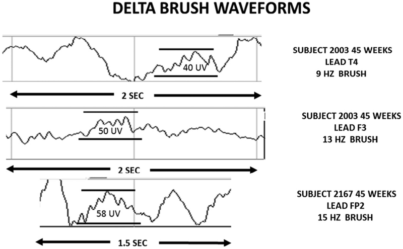
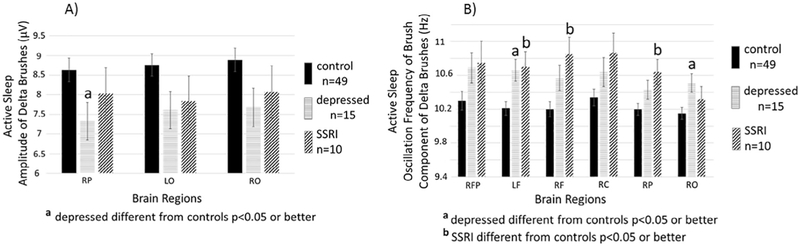
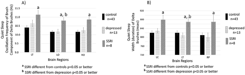
Methods: We measured delta brush bursts from the term infants of three groups of mothers (controls (N = 52), depressed untreated (N = 15), and those taking serotonin SSRI medication (N = 10). High density EEGs were obtained during sleep at an average age of 44 weeks post conceptional age. We measured the rate of occurrence, brush amplitude, oscillation frequency and duration of the bursts.

Results: Compared to infants of control mothers, the parameters of delta brush bursts of the offspring of depressed and SSRI-using mothers are significantly altered: burst amplitude is decreased; the oscillation frequency increased, and the duration increased (SSRI only). These significant differences were found during both sleep states.

Conclusions: Electrocortical bursting activity (i.e. delta brushes) is known to play an important role in early central nervous system (CNS) synaptic formation and function.

Significance: Maternal depression or SSRI use may alter brain function in their offspring.

Keywords: Delta brush; EEG burst; Infant EEG; Prenatal maternal depression; SSRI.


Conflict of interest statement

Declaration of Competing Interest

None of the authors have potential conflicts of interest.

Figures

Fig. 1.
Fig. 1.
Delta brush power spectrum.
Fig. 2.
Fig. 2.
Delta brush EEG waveforms.
Fig. 3.
Fig. 3.
Active Sleep. LEFT PANEL. Shown are means (±SE) of zero to peak amplitudes in microvolts of the high frequency oscillation component of delta brushes detected during active sleep in infants of mothers without depression or SSRIs during pregnancy (control n = 49, solid black bars), infants of mothers who were depressed during pregnancy but did not take SSRIs (depressed n = 15, striped bars), and infants of mothers who took SSRIs during pregnancy (SSRI n = 10, hatched bars). Results are shown for the three brain regions that demonstrated a main effect of group (control, depressed, SSRI) in ANOVAs (see Table 2). Superscript “a” above the bar indicates significant (p < 0.05) post-hoc test (Tukey’s) for the comparison between depressed and control. RP = right parietal, LO = left occipital, RO = right occipital. RIGHT PANEL. Shown are means (±SE) of the central frequency within the high frequency component of delta brushes detected during active sleep infants of mothers without depression or SSRIs during pregnancy (control n = 49, solid black bars), infants of mothers who were depressed during pregnancy but did not take SSRIs (depressed n = 15, striped bars), and infants of mothers who took SSRIs during pregnancy (SSRI n = 10, hatched bars). Results are shown for the six brain regions that demonstrated a main effect of group (control, depressed, SSRI) in ANOVAs (see Table 2). Superscript “a” above the bar indicates significant (p < 0.05) post-hoc test (Tuckey’s) for the comparison between depressed and control and a superscript “b” indicates a significant (p < 0.05) post-hoc test (Tukey’s) for the comparison between SSRI and control. RFP = right frontal polar, LF = left frontal, RF = right frontal, RC = right central, RP = right parietal, RO = right occipital.
Fig. 4.
Fig. 4.
Quiet Sleep. LEFT PANEL. Shown are means (±SE)) of the central frequency within the high frequency component of delta brushes. Detected during quiet sleep for infants of mothers without depression during pregnancy (control n = 43, solid black bars), infants of mothers who were depressed during pregnancy but did not take SSRIs (depressed n = 13, striped bars), and infants of mothers who took SSRIs during pregnancy (SSRI n = 8, hatched bars). Results are shown for the three brain regions that demonstrated a main effect of group (control, depressed, SSRI) in ANOVAs (see Table 2). Superscript “a” above the bar indicates significant (p < 0.05) post-hoc test (Tukey’s) for the comparison between SSRI and control and a superscript “b” indicates a significant (p < 0.05) post-hoc test (Tukey’s) for the comparison between SSRI and depression. LT = left temporal, LO = left occipital, RO = right occipital. RIGHT PANEL. Shown are means (±SE) for the widths (i.e. duration) of delta brushes detected during quiet sleep of infants of mothers without depression or SSRIs during pregnancy (control n = 43, solid black bars), infants of mothers who were depressed during pregnancy but did not take SSRIs (depressed n = 13, striped bars), and infants of mothers who took SSRIs during pregnancy (SSRI n = 8, hatched bars). Results are shown for the three brain regions that demonstrated a main effect of group (control, depressed, SSRI) in ANOVAs (see Table 2). Superscript “a” above the bar indicates significant (p < 0.05) post-hoc test (Tuckey’s) for the comparison between SSRI and control and a superscript “b” indicates a significant (p < 0.05) post-hoc test (Tuckey’s) for the comparison between SSRI and depression. LC = left central, LT = left temporal, RP = right parietal.

References

    1. Akhmetshina D, Zakharov A, Vinokurova D, Nasretdinov A, Valeeva G, Khazipov R. The serotonin reuptake inhibitor citalopram suppresses activity in the neonatal rat barrel cortex in vivo. Brain Res Bul 2016;124:48–54. - PubMed
    1. André M, Lamblin MD, d’Allest AM, Curzi-Dascalova L, Moussalli-Salefranque F, Nguyen Thetich S, et al. Electroencephalography in premature and full-term infants. Developmental features and glossary. Neurophysiol Clin 2010;40(2):59–124. - PubMed
    1. Ansorge MS, Zhou M, Lira A, Hen R, Gingrich JA. Early-life blockade of the 5-HT transporter alters emotional behavior in adult mice. Science 2004;306 (5697):879–81. - PubMed
    1. Arichi T, Whitehead K, Barone G, Pressler R, Padormo F, Edwards AD, et al. Localization of spontaneous bursting neuronal activity in the preterm human brain with simultaneous EEG-fMRI. Elife 6;2017:pii: e27814. - PMC - PubMed
    1. Barnes NM, Sharp T. A review of central 5-HT receptors and their function.Neuropharm 1999;38(8):1083–152. - PubMed

Publication types

MeSH terms

Substances