

Generating and evaluating type I interferon receptor-deficient and feline TMPRSS2-expressing cells for propagating serotype I feline infectious peritonitis virus
- PMID: 31539770
- PMCID: PMC7112123
- DOI: 10.1016/j.virol.2019.08.030
Generating and evaluating type I interferon receptor-deficient and feline TMPRSS2-expressing cells for propagating serotype I feline infectious peritonitis virus
Abstract
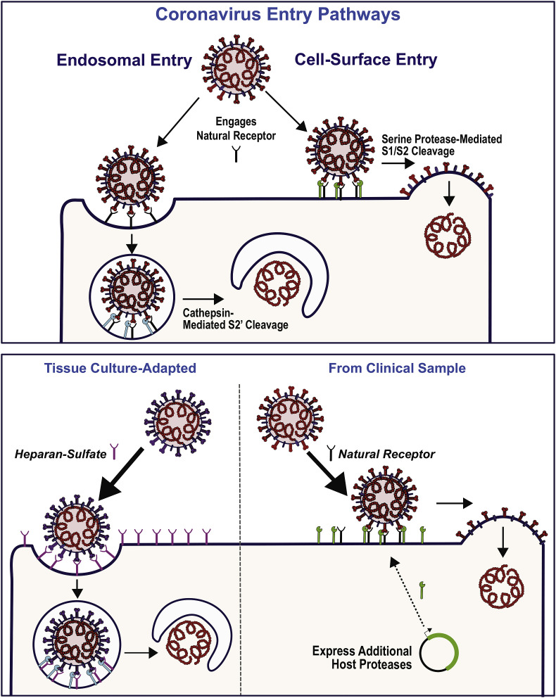
Feline coronavirus infection can progress to a fatal infectious peritonitis, which is a widespread feline disease without an effective vaccine. Generating feline cells with reduced ability to respond to interferon (IFN) is an essential step facilitating isolation of new candidate vaccine strains. Here, we describe the use of Crispr/Cas technology to disrupt type I IFN signaling in two feline cell lines, AK-D and Fcwf-4 CU, and evaluate the replication kinetics of a serotype I feline infectious peritonitis virus (FIPV) within these cells. We report that polyclonal cell populations and a clonal isolate, termed Fcwf-4 IRN, exhibited significantly diminished IFN-responsiveness and allowed FIPV replication kinetics comparable to parental cells. Furthermore, we demonstrate that replication of FIPV is enhanced by ectopic expression of a host serine protease, TMPRSS2, in these cells. We discuss the potential of these cells for isolating new clinical strains and for propagating candidate vaccine strains of FIPV.
Keywords: AK-D cells; Crispr/Cas gene editing; FIPV; Fcwf-4 CU cells; Feline coronavirus; IFNαR-deficient cells; Interferon signaling-deficient cells; TMPRSS2-expressing cells.
Copyright © 2019 Elsevier Inc. All rights reserved.
Figures
Similar articles
-
Characterizing replication kinetics and plaque production of type I feline infectious peritonitis virus in three feline cell lines.Virology. 2018 Dec;525:1-9. doi: 10.1016/j.virol.2018.08.022. Epub 2018 Sep 8. Virology. 2018. PMID: 30205273 Free PMC article.
-
Serotype I and II Feline Coronavirus Replication and Gene Expression Patterns of Feline Cells-Building a Better Understanding of Serotype I FIPV Biology.Viruses. 2022 Jun 22;14(7):1356. doi: 10.3390/v14071356. Viruses. 2022. PMID: 35891338 Free PMC article.
-
TNF-alpha, produced by feline infectious peritonitis virus (FIPV)-infected macrophages, upregulates expression of type II FIPV receptor feline aminopeptidase N in feline macrophages.Virology. 2007 Jul 20;364(1):64-72. doi: 10.1016/j.virol.2007.02.006. Epub 2007 Mar 23. Virology. 2007. PMID: 17382365 Free PMC article.
-
Feline Coronaviruses: Pathogenesis of Feline Infectious Peritonitis.Adv Virus Res. 2016;96:193-218. doi: 10.1016/bs.aivir.2016.08.002. Epub 2016 Aug 31. Adv Virus Res. 2016. PMID: 27712624 Free PMC article. Review.
-
A review of feline infectious peritonitis virus: molecular biology, immunopathogenesis, clinical aspects, and vaccination.Vet Microbiol. 1993 Jul;36(1-2):1-37. doi: 10.1016/0378-1135(93)90126-r. Vet Microbiol. 1993. PMID: 8236772 Free PMC article. Review.
Cited by
-
Development of Feline Ileum- and Colon-Derived Organoids and Their Potential Use to Support Feline Coronavirus Infection.Cells. 2020 Sep 12;9(9):2085. doi: 10.3390/cells9092085. Cells. 2020. PMID: 32932592 Free PMC article.
-
High-throughput viral microneutralization method for feline coronavirus using image cytometry.J Virol Methods. 2020 Dec;286:113979. doi: 10.1016/j.jviromet.2020.113979. Epub 2020 Sep 23. J Virol Methods. 2020. PMID: 32979406 Free PMC article.
-
Placental Expression of ACE2 and TMPRSS2 in Maternal Severe Acute Respiratory Syndrome Coronavirus 2 Infection: Are Placental Defenses Mediated by Fetal Sex?J Infect Dis. 2021 Dec 8;224(Suppl 6):S647-S659. doi: 10.1093/infdis/jiab335. J Infect Dis. 2021. PMID: 34293137 Free PMC article.
-
Endocytic Pathway of Feline Coronavirus for Cell Entry: Differences in Serotype-Dependent Viral Entry Pathway.Pathogens. 2019 Dec 16;8(4):300. doi: 10.3390/pathogens8040300. Pathogens. 2019. PMID: 31888266 Free PMC article.
-
Coronaviruses Associated with the Superfamily Musteloidea.mBio. 2021 Jan 19;12(1):e02873-20. doi: 10.1128/mBio.02873-20. mBio. 2021. PMID: 33468694 Free PMC article. Review.
References
-
- Addie D.D. Feline coronaviral infections. In: Greene C., editor. Infectious Diseases of the Dog and Cat. Saunders; 2011. pp. 92–108.
-
- Bertram S., Dijkman R., Habjan M., Heurich A., Gierer S., Glowacka I., Welsch K., Winkler M., Schneider H., Hofmann-Winkler H., Thiel V., Pohlmann S. TMPRSS2 activates the human coronavirus 229E for cathepsin-independent host cell entry and is expressed in viral target cells in the respiratory epithelium. J. Virol. 2013;87:6150–6160. doi: 10.1128/jvi.03372-12. - DOI - PMC - PubMed
Publication types
MeSH terms
Substances
Grants and funding
LinkOut - more resources
Full Text Sources
Other Literature Sources
