. 2019 Sep 14;24(18):3352.
doi: 10.3390/molecules24183352.

Development of a Fragment-Based Screening Assay for the Focal Adhesion Targeting Domain Using SPR and NMR

Affiliations

Development of a Fragment-Based Screening Assay for the Focal Adhesion Targeting Domain Using SPR and NMR

Carlos Alvarado et al. Molecules. .

Abstract

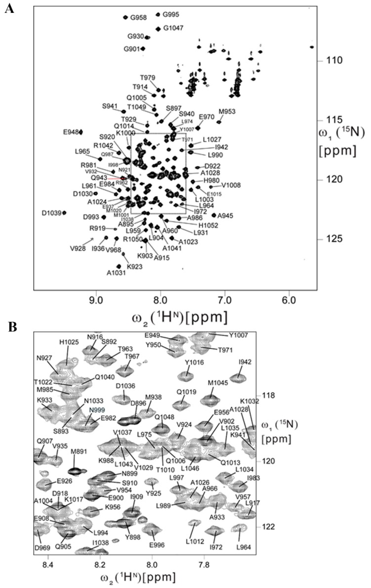
The Focal Adhesion Targeting (FAT) domain of Focal Adhesion Kinase (FAK) is a promising drug target since FAK is overexpressed in many malignancies and promotes cancer cell metastasis. The FAT domain serves as a scaffolding protein, and its interaction with the protein paxillin localizes FAK to focal adhesions. Various studies have highlighted the importance of FAT-paxillin binding in tumor growth, cell invasion, and metastasis. Targeting this interaction through high-throughput screening (HTS) provides a challenge due to the large and complex binding interface. In this report, we describe a novel approach to targeting FAT through fragment-based drug discovery (FBDD). We developed two fragment-based screening assays-a primary SPR assay and a secondary heteronuclear single quantum coherence nuclear magnetic resonance (HSQC-NMR) assay. For SPR, we designed an AviTag construct, optimized SPR buffer conditions, and created mutant controls. For NMR, resonance backbone assignments of the human FAT domain were obtained for the HSQC assay. A 189-compound fragment library from Enamine was screened through our primary SPR assay to demonstrate the feasibility of a FAT-FBDD pipeline, with 19 initial hit compounds. A final total of 11 validated hits were identified after secondary screening on NMR. This screening pipeline is the first FBDD screen of the FAT domain reported and represents a valid method for further drug discovery efforts on this difficult target.

Keywords: FAT domain; focal adhesion kinase; fragment-based drug discovery; nuclear magnetic resonance; surface plasmon resonance.


Conflict of interest statement

The authors declare no conflict of interest.

Figures

Figure 1
Figure 1
Construction and validation of AviTag Focal Adhesion Targeting (FAT). (A) Plasmid map of pET15b AviTag FAT with labeled protein sequence of the insert. The insert contains the 6x HisTag, 6 residue thrombin cleavage site, and the 15 residue AviTag sequence. (B) SDS-PAGE of purified AviTag FAT and western blot of biotinylated AviTag FAT probing with anti-biotin antibody. Biotinylated and non-biotinylated AviTag Maltose Binding Protein (MBP) were used as controls. The SDS-PAGE shows FAT purification at over 90% purity. The western blot shows a band for biotinylated AviTag FAT confirming successful biotinylation. (C) Surface plasmon resonance (SPR) sensorgram showing immobilization of biotinylated AviTag FAT to a SADH streptavidin coated chip. The final immobilization response level of this protein, shown in Channel 1, is at around 7000 RU. Channel 2 and Channel 3 do not have any immobilized protein and demonstrate a consistent baseline providing ideal reference channels.
Figure 2
Figure 2
Development and optimization of SPR assay. (A) SPR sensorgram of AviTag FAT-LD2 binding in various buffers for optimization studies. The buffers listed are as follows-Buffer 1: 20 mM Tris-Cl pH 8.0, 200 mM NaCl, 1% DMSO; Buffer 2: 100 mM Tris-Cl pH 8.0, 200 mM NaCl, 1% DMSO; Buffer 3: 100 mM Tris-Cl pH 8.0, 200 mM NaCl, 1% DMSO, 0.05% Triton-X 100; Buffer 4: 100 mM Tris-Cl pH 8.0, 200 mM NaCl, 1% DMSO, 0.05% Tween-20; Buffer 5: 100 mM Tris-Cl pH 8.0, 200 mM NaCl, 2.5% DMSO, 0.05% Tween-20; Buffer 6: 100 mM Tris-Cl pH 8.0, 200 mM NaCl, 5% DMSO, 0.05% Tween-20. Buffer 6 demonstrated the largest response and best on-rate of all buffers examined and was chosen as the final buffer for all SPR runs moving forward. (B) Comparison between Paxillin-LD2 (50 µM) binding to wild-type (WT) AviTag FAT using a fixed concentration injection (FCI) (top) versus an OneStep injection (bottom). Kinetic fitting of FCI produced a KD of 103.7 ± 0.4 µM and equilibrium fit produced a KD of 102.7 ± 0.2 µM. OneStep injection produced a KD of 88 ± 0.1 µM, very similar to the binding affinity by FCI. (C) Molecular model (PDB ID: IOW8) of the FAT-LD2 binding complex highlighting residues to be mutated as binding controls. (D) SPR sensorgram comparing the binding of LD2 against various FAT constructs, including WT FAT, single mutants I936A, L994E, and double mutant I936E/L994E. WT to mutant selectivity ratios were calculated from these results: I936A-1.5, L994E-3, I936E/L994E-18.
Figure 3
Figure 3
Backbone resonance assignments of human FAT. (A) 2D [15N,1H] HSQC spectrum of human FAT-892 recorded at 750 MHz 1H resonance frequency and 37 °C in about 1.5 h. Resonance assignments are indicated using the one-letter amino acid code and the numbering of the full-length protein. (B) Central spectral region of the 2D [15N,1H] HSQC spectrum of the FAT domain.
Figure 4
Figure 4
Development of 2D heteronuclear single quantum coherence nuclear magnetic resonance (HSQC-NMR) assay. (A) Overlay of 2D HSQC spectra acquired with a 100 µM FAT sample in the presence of varying concentrations of LD2. Two-dimensional HSQC signals represented in blue, green, red, and black are obtained in the presence of 125 µM, 62.5 µM, 31.3 µM of LD2 and 2% DMSO, respectively. Residues displaying notable chemical shift perturbations (CSPs) are indicated with the one letter amino acid code and the numbering of the full-length protein. (B) Molecular model (PDB ID: 1OW8) of the FAT-LD2 binding complex with the CSPs from the LD2 titration highlighted in red. A zoomed in view of both the helix 1-4 and helix 2-3 binding sites with proper amino acid labels indicating that the CSPs line up with the model for LD2 binding.
Figure 5
Figure 5
Pilot SPR fragment screen (A.) Hit plot displaying results of the primary SPR fragment screen with WT AviTag FAT compared to a control compound found through a fluorescence polarization assay. Hits were characterized as having 20% of the control response. Three hits were above the 100% control response, but analysis of the binding curves showed curves that they didn’t reach equilibrium indicating aggregation and were ruled out as hits. The primary screen produced a total of 32 initial hits. (B) Six representative SPR sensorgrams of fragment hits with binding to WT FAT (blue), single mutants I936A (pink), L994E (black), and double mutant I936E/L994E (green). Each binding curve displays their respective KD. Careful visual inspection of the binding curves allowed for a final curated hit count of 19 fragments.
Figure 6
Figure 6
Validation of hits from SPR utilizing 2D HSQC-NMR. (A) Representative HSQC overlays displaying the CSPs of four hits. Compounds 009 and 137 demonstrate multiple CSPs in the helix 2-3 binding site. Compounds 125 and 088 demonstrate a mix of helix 2-3 and 1-4 binding. (B) Venn diagram detailing the number of hits after each screen. The initial library was 189 compounds, with 19 curated hits after SPR, and a final 11 hits after secondary screening by 2D HSQC-NMR.

References

    1. Weiner T.M., Liu E.T., Craven R.J., Cance W.G. Expression of focal adhesion kinase gene and invasive cancer. Lancet. 1993;342:1024–1025. doi: 10.1016/0140-6736(93)92881-S. - DOI - PubMed
    1. Owens L.V., Xu L., Craven R.J., Dent G.A., Weiner T.M., Kornberg L., Liu E.T., Cance W.G. Overexpression of the focal adhesion kinase (p125fak) in invasive human tumors. Cancer Res. 1995;55:2752–2755. - PubMed
    1. Lark A.L., Livasy C.A., Calvo B., Caskey L., Moore D.T., Yang X., Cance W.G. Overexpression of focal adhesion kinase in primary colorectal carcinomas and colorectal liver metastases: Immunohistochemistry and real-time pcr analyses. Clin. Cancer Res. 2003;9:215–222. - PubMed
    1. McLean G.W., Carragher N.O., Avizienyte E., Evans J., Brunton V.G., Frame M.C. The role of focal-adhesion kinase in cancer—A new therapeutic opportunity. Nat. Rev. Cancer. 2005;5:505–515. doi: 10.1038/nrc1647. - DOI - PubMed
    1. Schaller M.D., Borgman C.A., Cobb B.S., Vines R.R., Reynolds A.B., Parsons J.T. Pp125fak a structurally distinctive protein-tyrosine kinase associated with focal adhesions. Proc. Natl. Acad. Sci. USA. 1992;89:5192–5196. doi: 10.1073/pnas.89.11.5192. - DOI - PMC - PubMed

LinkOut - more resources