Review
. 2019 Sep 14;11(9):859.
doi: 10.3390/v11090859.

Recombination in Enteroviruses, a Multi-Step Modular Evolutionary Process

Affiliations
Review

Recombination in Enteroviruses, a Multi-Step Modular Evolutionary Process

Claire Muslin et al. Viruses. .

Abstract

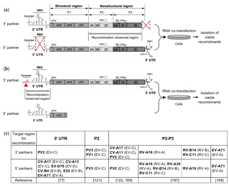
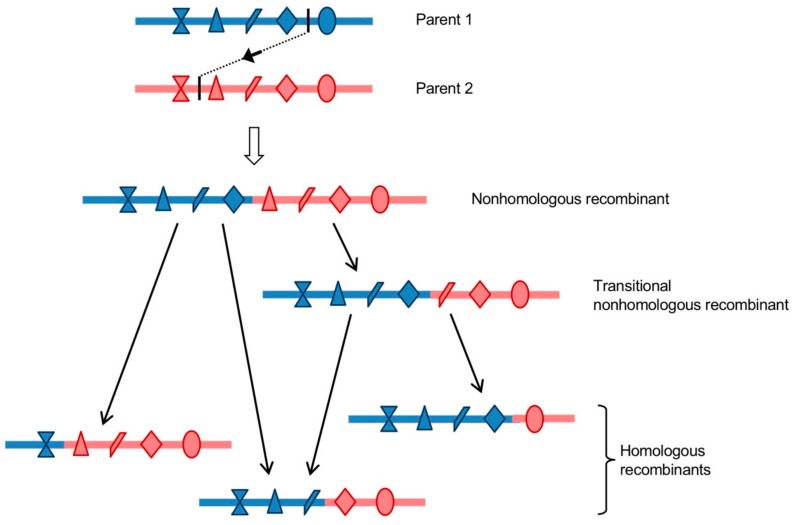
RNA recombination is a major driving force in the evolution and genetic architecture shaping of enteroviruses. In particular, intertypic recombination is implicated in the emergence of most pathogenic circulating vaccine-derived polioviruses, which have caused numerous outbreaks of paralytic poliomyelitis worldwide. Recent experimental studies that relied on recombination cellular systems mimicking natural genetic exchanges between enteroviruses provided new insights into the molecular mechanisms of enterovirus recombination and enabled to define a new model of genetic plasticity for enteroviruses. Homologous intertypic recombinant enteroviruses that were observed in nature would be the final products of a multi-step process, during which precursor nonhomologous recombinant genomes are generated through an initial inter-genomic RNA recombination event and can then evolve into a diversity of fitter homologous recombinant genomes over subsequent intra-genomic rearrangements. Moreover, these experimental studies demonstrated that the enterovirus genome could be defined as a combination of genomic modules that can be preferentially exchanged through recombination, and enabled defining the boundaries of these recombination modules. These results provided the first experimental evidence supporting the theoretical model of enterovirus modular evolution previously elaborated from phylogenetic studies of circulating enterovirus strains. This review summarizes our current knowledge regarding the mechanisms of recombination in enteroviruses and presents a new evolutionary process that may apply to other RNA viruses.

Keywords: RNA virus; emergence; enterovirus; recombination; viral evolution.


Conflict of interest statement

The authors declare no conflict of interest.

Figures

Figure 1
Figure 1
Organization of the genome of poliovirus type 1 (PV1) Mahoney. The poly-adenylated single positive-strand RNA genome is covalently linked to the viral protein VPg (also named 3B) at the 5′ terminus. In addition to the main large open-reading frame (ORF), the majority of the EV-A, EV-B and EV-C genomes, and in particular PV1 genome, contain a second upstream overlapping ORF (uORF). However, PV2 and PV3 genomes do not contain an intact uORF. The coding region is flanked by two untranslated regions (5′ and 3′ UTRs). The 5′ UTR (nucleotides 1 to 743) is magnified to indicate the seven stem-loop structures (I to VII) forming two functional units, the cloverleaf (CL: I) and the internal ribosome entry site (IRES: II-VI). The P1 region encodes the capsid proteins (VP1-4) and the P2 and P3 regions encode the non-structural proteins such as the RNA-dependent RNA polymerase 3D.
Figure 2
Figure 2
Homologous and nonhomologous recombinant genomes. Parental RNA genomes are located in the upper panel of (a), (b) and (c) diagrams. They have a similar genomic structure. The recombination site in each of the parental genomes is represented by a black vertical line, the recombination event is indicated by black dotted line and arrow. The recombination site in the obtained recombinant genome (lower panels) of (a) and (b) diagrams is indicated by a green reversed triangle. (a) Homologous recombination occurs at the same site in both parental genomes, thus the obtained recombinant has the same genomic structure as the parental viruses. (b) and (c) Nonhomologous recombination occurs at different sites in the two parental genomes. (b) a duplication of homologous sequences (hatched) is generated around the recombination site. (c) A deletion of genomic sequence is generated.
Figure 3
Figure 3
Genomic structures of all recombinant circulating vaccine-derived poliovirus lineages characterized so far. A schematic view of the genetic organization of the poliovirus genomes is given in the upper panel (see also Figure 1). The presence of vaccine-derived sequences is indicated (mutated Sabin 1, 2, of 3 sequences) as well as the non-vaccine sequences derived from other species C enteroviruses (EV-Cs). Colors and patterns differentiate EV-C sequences that differed significantly from each other. Non-vaccine sequences showing similarity with those of co-circulating coxsackieviruses A (CV-A11, -A13, -A17) are indicated. Data are modified from [73] (Egypt), [67] (Greece), [68] (Hispaniola), [69] (Philippines), [71,72] (Nigeria), [65,66,70,76] (Madagascar), [120] (Cambodia). The location of the six recombinant hotspots identified by experimental studies of genetic exchanges between poliovirus and enteroviruses is indicated by grey rectangles [77,121,122] (see further in the text).
Figure 4
Figure 4
Mechanisms of replicative and nonreplicative RNA recombination. (a) The replicative mechanism of copy-choice. The replication complex pauses during the synthesis of the complementary strand of the RNA donor (in red) and dissociates from the RNA donor template. Then, the incomplete nascent RNA strand interacts with the acceptor RNA (in blue) where the replication complex reassembles and the synthesis of the complementary strand resumes. The complementary strand is indicated by lighter colors. (b) The nonreplicative mechanism of breakage-ligation. The two parental RNA molecules are degraded, and then the two fragments generated are covalently linked.
Figure 5
Figure 5
Experimental systems of intra- and intertypic recombination between enteroviruses. (a) and (b). Examples of recombination partners designed to target recombination in the P2-P3 region (a) and in the 5′ UTR (b). (a) The 3′ partner is made from the complete enterovirus genome in which the 3′ end of the 3D polymerase and the entire 3′ UTR were deleted. The 5′ partner is made from the enterovirus genome carrying a deletion in the IRES. Red crosses indicate the genomic regions in which deletions were made. Co-transfecting cells with these two defective complementary genomic RNA fragments led to the production of infectious genomes recombinant in the P2-P3 region [122,189]. (b) The 3′ partner is made defective by substitutions in the cloverleaf structure of 5′ UTR. The 5′ partner includes the complete 5′ UTR followed by the N-terminal part of the ppORF. Co-transfecting cells with this pair of defective genomes will generate viable viruses only if a recombination event occurs in the 5′ UTR [77]. (c) List of enterovirus types used for the construction of the 3′ and 5′ partners in the different experimental systems. For each type, the enterovirus species is indicated in brackets.
Figure 6
Figure 6
Model of generation of homologous recombinants from a nonhomologous one. A nonhomologous recombinant genome, displaying a duplication of homologous sequences around the recombination site, is produced by a replicative or nonreplicative recombination mechanism. Icons represent differences between the two homologous parental genomes. During the following replication cycles, the nonhomologous recombinant genome can generate a series of homologous recombinants through one or more rearrangement event(s). Recombination and rearrangement may take place within the initial co-infected (or co-transfected) cell, or in a different cell following re-infection, supposing that the nonhomologous recombinant can be encapsidated [189].
Figure 7
Figure 7
Model of modular evolution of species C enteroviruses. (a) Schematic representation of the enterovirus genomic RNA molecule. Experimental studies of genetic exchanges between poliovirus and enteroviruses led to the identification of six putative intertypic recombination hotspots, indicated by hatched orange rectangles [77,121,122]. (b) Modular recombination process. Each enterovirus species would exist as a pool of genetic material containing a finite set of P1 regions defining different types and a swarm of nonstructural and untranslated regions, divided in functional recombination modules and evolving independently. Each new enterovirus lineage can be considered as a new association of compatible recombination module.

Similar articles

Cited by

References

    1. Zell R., Delwart E., Gorbalenya A.E., Hovi T., King A.M.Q., Knowles N.J., Lindberg A.M., Pallansch M.A., Palmenberg A.C., Reuter G., et al. ICTV Virus Taxonomy Profile: Picornaviridae. J. Gen. Virol. 2017;98:2421–2422. doi: 10.1099/jgv.0.000911. - DOI - PMC - PubMed
    1. Pallansch M., Roos R. Enteroviruses: Polioviruses, coxsackieviruses, echoviruses, and newer enteroviruses. In: Knipe D.M., Howley P.M., editors. Fields Virology. Lippincott Williams and Wilkins; Philadelphia, PA, USA: 2007. pp. 839–893.
    1. Nathanson N., Kew O.M. From emergence to eradication: The epidemiology of poliomyelitis deconstructed. Am. J. Epidemiol. 2010;172:1213–1229. doi: 10.1093/aje/kwq320. - DOI - PMC - PubMed
    1. Rao C.D., Yergolkar P., Shankarappa K.S. Antigenic diversity of enteroviruses associated with nonpolio acute flaccid paralysis, India, 2007–2009. Emerg. Infect. Dis. 2012;18:1833–1840. doi: 10.3201/eid1811.111457. - DOI - PMC - PubMed
    1. Yang T.-T., Huang L.-M., Lu C.-Y., Kao C.-L., Lee W.-T., Lee P.-I., Chen C.-M., Huang F.-Y., Lee C.-Y., Chang L.-Y. Clinical features and factors of unfavorable outcomes for non-polio enterovirus infection of the central nervous system in northern Taiwan, 1994–2003. J. Microbiol. Immunol. Infect. 2005;38:417–424. - PubMed

Publication types

LinkOut - more resources