. 2019 Sep 13;20(18):4530.
doi: 10.3390/ijms20184530.

Immunohistochemical Examination of Trophoblast Syncytialization during Early Placentation in Sheep

Affiliations

Immunohistochemical Examination of Trophoblast Syncytialization during Early Placentation in Sheep

Heewon Seo et al. Int J Mol Sci. .

Abstract

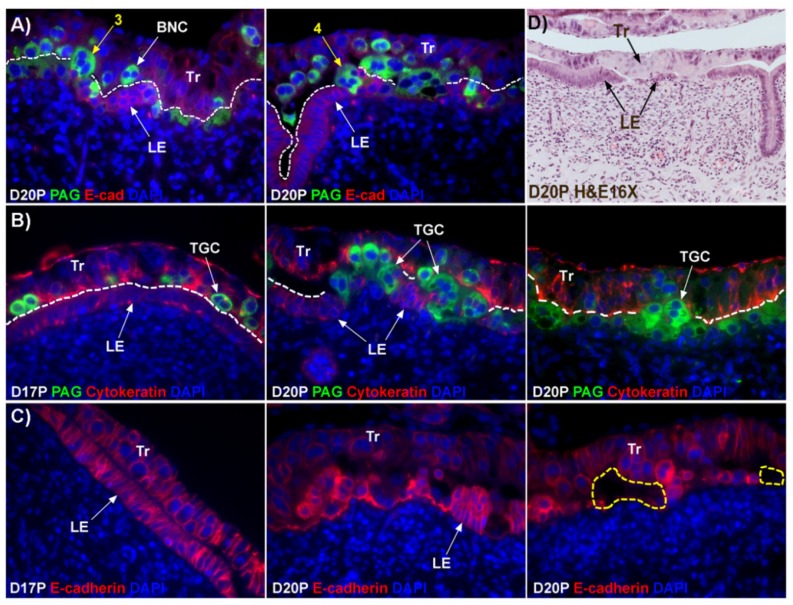
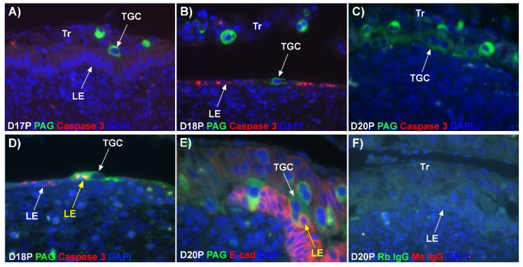
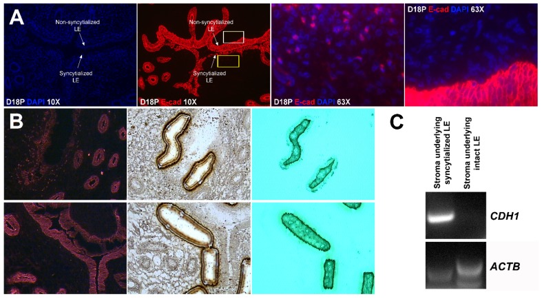
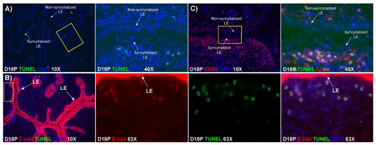
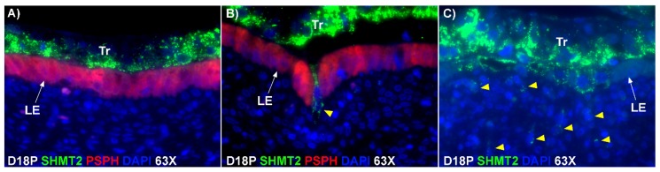
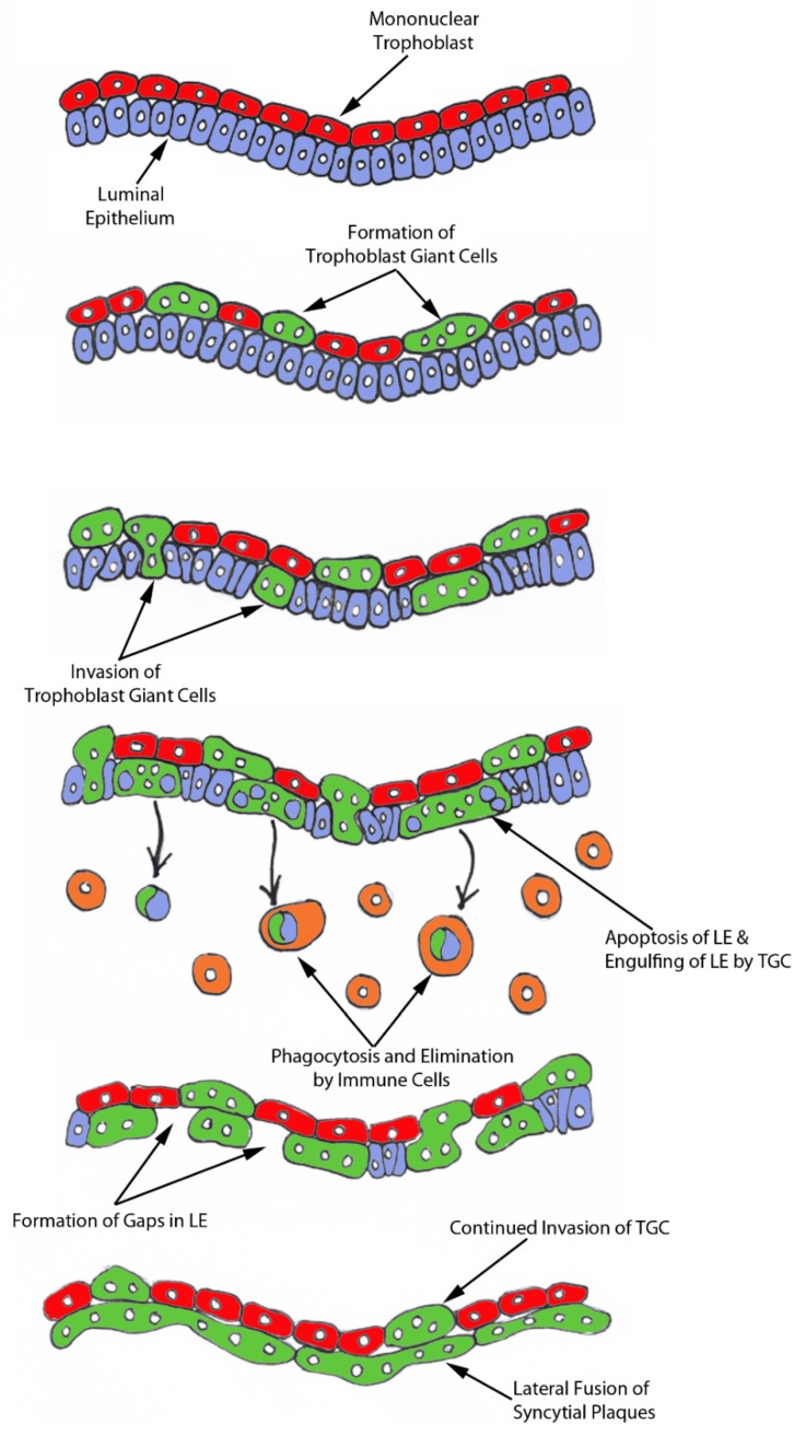
During the peri-implantation period, multinucleated syncytia are formed in the sheep placenta. For over 20 years the scientific consensus has been that during trophoblast syncytialization in sheep, binucleate trophoblast giant cells (BNCs) differentiate from mononuclear trophoblast cells, and individual BNCs fuse with individual luminal epithelial (LE) cells to form trinucleate cells. These trophoblast-LE syncytial plaques then grow through continued BNC migration and fusion. Therefore, LE cells are thought to be incorporated into syncytial plaques. However, these ideas were based on electron microscopy studies, without benefit of molecular markers for BNC and LE cells to support conclusions. The aim of this study was to observe interactions between BNCs and uterine LE cells using immunohistochemical localization for molecular markers for BNCs and uterine LE cells. We performed immunofluorescence staining, laser capture microdissection, and TUNEL staining on the uterine-placental tissues of sheep during early placentation. We observed: (1) syncytial cells containing more than two nuclei within the trophoblast cell layer; (2) depolarized LE cells that express caspase 3 and stain positively for TUNEL; (3) engulfment of caspase 3-positive LE cells by trophoblast giant cells (TGCs) and empty spaces within the LE layer at sites of implantation; (4) rapid enlargement of syncytial plaques; and (5) E-cadherin and TUNEL-positive cells within the uterine stroma underlying degenerating LE was coincident with accumulation of CD45-positive cells at these sites. These data suggest that during early placentation: (1) fusion between trophoblasts is not limited to the formation of BNCs, and the term 'trophoblast giant cell (TGC)' may be appropriate; (2) LE cells undergo apoptosis; (3) apoptotic LE cells are eliminated by TGCs; (4) fusion is not limited to the incorporation of new BNCs but involves the lateral fusion between growing syncytial plaques; and (5) TGCs carry apoptotic LE cells away from the uterine-placental interface for elimination by immune cells within the stroma. These data indicate that uterine LE cells are not incorporated into syncytial plaques, but are engulfed and eliminated, and that early placentation in sheep is more similar to early placentation in humans than is currently understood in that both develop mononucleated cytotrophoblast and multinucleated syncytiotrophoblast layers of entirely placental origin. The elimination of LE cells by sheep TGCs might provide insights into elimination and penetration of LE cells during human embryo implantation.

Keywords: sheep; syncytialization; trophoblast; uterine luminal epithelium.


Conflict of interest statement

The authors declare no conflict of interest.

Figures

Figure 1
Figure 1
Interaction between TGCs and uterine LE cells at implantation sites in sheep. (A) Within the trophoblast layer, there are PAG-stained binucleate cells (BNC) as well as trophoblast cells that contain 3 or 4 nuclei (3 and 4 shown in yellow and termed trophoblast giant cells throughout the manuscript). The dotted line delineates the interface between the trophoblast layer and uterine LE layer. (B) Double immunofluorescence staining for PAG and cytokeratin at implantation sites between day 17 and day 20 of pregnancy. Most TGCs are present within the trophoblast layer on day 17 of pregnancy, but on day 20, there is extensive migration of TGCs into the uterine LE cell layer and replacement of mononuclear uterine LE cells by syncytial cells. (C) E-cadherin (E-cad; red, stains mononuclear trophoblast and LE) immunostaining at implantation sites revealed the development of gaps/voids (yellow dotted line) in the uterine LE layer. (D) H and E staining of a day 20 implantation site in sheep illustrates the absence of sections of uterine LE that are replaced by syncytial cells. Tr, mononuclear trophoblast cells; BNC, binucleate trophoblast cell; TGC, trophoblast giant cell; LE, luminal epithelium; D, day; P, pregnancy. The width of field for the microscopic images of immunofluorescence is 220 μm. The width of the field for the H and E image is 540 μm.
Figure 2
Figure 2
Expression of caspase 3 and internalization of caspase 3-positive LE cells by TGCs at implantation sites of sheep. (AD) Double immunofluorescence staining for PAGs (green, stains TGCs) and caspase 3 (red). The yellow arrow indicates a TGC endocytosing caspase 3-positive LE cells. (E) Immunofluorescence for PAGs (green, stains TGCs) and E-cadherin (E-cad; red, stains mononucleate Tr and LE). The yellow arrow indicates a TGC appearing to physically engulf an LE cell. The dual labelled rabbit (Rb) and mouse (Ms) IgG control is presented in panel (F). Tr, mononuclear trophoblast cells; TGC, trophoblast giant cells; LE, luminal epithelium; D, day; P, pregnancy. The width of field for the microscopic images in panels is 220 μm.
Figure 3
Figure 3
Localization of E-cadherin-positive cells within the uterine stroma during active syncytialization at implantation sites of sheep. (A) Immunofluorescence staining for E-cadherin (E-cad, red) in the endometrium on day 18 of pregnancy. The third panel represents the region indicated by the yellow box in the second panel. The fourth panel represents the region indicated by the white box in the second panel. E-cadherin-positive cells are present within the stroma underlying actively syncytializing uterine LE. The width of field for the microscopic image captured at 10× and 63× is 890 and 140 μm, respectively. (B) Laser capture microdissection (LCM) used to collect cells within the stroma underlying syncytializing LE (top row) and cells within the stroma underlying intact uterine LE (bottom row). Frozen endometrial tissue sections on day 18 of pregnancy stained with E-cadherin immediately before microdissection (top and bottom rows, first panels). The same tissue section is shown with the missing cells after microdissection (top and bottom rows, middle panels). The LCM cells attached to the CapSure Macro LCM Caps (top and bottom rows, third panels). The width of field for the microscopic image is 631 μm. (C) RT-PCR analysis of CDH1 mRNA in the stromal cells captured by LCM. ACTB was used as a positive control. CDH1 mRNA was detected in the LCM cells isolated from stroma underlying actively syncytializing LE.
Figure 4
Figure 4
Apoptosis and elimination of LE cells during syncytialization. (A) TUNEL staining (green) in the endometrium on day 18 of pregnancy. The second panel represents the region indicated by the yellow box in the first panel. TUNEL-positive cells were detected within the stroma underlying syncytialized LE. (B) Double staining for E-cad (red) and TUNEL (green) in the endometrium on day 18 of pregnancy. The second, third, and fourth panels represent the region indicated by the dotted yellow box in the first panel. TUNEL-positive cells co-localized with E-cadherin staining in the stroma. (C) Double staining for CD45 (red) and TUNEL (green) at an implantation site. The second panel represents the region indicated by the yellow box in the first panel. CD45-positive cells were accumulated within the stroma underlying syncytialized LE, and these CD45-positive cells were closely associated with TUNEL-positive cells within the stroma. The width of field for the microscopic image captured at 10×, 40×, and 63× is 890, 220, and 140 μm, respectively.
Figure 5
Figure 5
TGCs invade into the uterine stroma during syncytialization. (A,B) Double immunofluorescence staining for SHMT2 (serine hydroxymethyltransferase 2; green) and PSPH (phosphoserine phosphatase; red) at implantation sites on day 18 of pregnancy. SHMT2-positive trophoblast cells are shown penetrating the PSPH-stained LE layer (yellow arrow head). (C) Immunofluorescence staining for SHMT2 at implantation sites on day 18 of pregnancy. SHMT2-positive trophoblast cells were detected within the uterine stroma by day 18 of gestation (yellow arrow heads). Tr, trophoblast cells; LE, luminal epithelium; D, day; P, pregnancy. The width of field for the microscopic images is 140 μm.
Figure 6
Figure 6
A working hypothesis for the syncytialization of the sheep placenta. Mononuclear trophoblast cells fuse with one-another to become multinucleated TGCs. Large numbers of TGCs migrate to insert themselves between the uterine LE cells that are simultaneously undergoing apoptosis. TGCs then engulf LE cells and carry them to the stroma for elimination by immune cells. The remaining TGCs then fuse with each other to form an extensive trophoblast syncytial layer that fills spaces left by removal of uterine LE and form the interface between caruncles and cotyledons in placentomes of the functional synepitheliochorial placenta of sheep.

Similar articles

Cited by

References

    1. Wintenberger-Torres S., Flechon J.E. Ultrastructural evolution of the trophoblast cells of the pre-implantation sheep blastocyst from day 8 to day 18. J. Anat. 1974;118:143–153. - PMC - PubMed
    1. Rowson L.E., Moor R.M. Development of the sheep conceptus during the first fourteen days. J. Anat. 1966;100:777–785. - PMC - PubMed
    1. Farin C.E., Imakawa K., Roberts R.M. In situ localization of mRNA for the interferon, ovine trophoblast protein-1, during early embryonic development of the sheep. Mol. Endocrinol. 1989;3:1099–1107. doi: 10.1210/mend-3-7-1099. - DOI - PubMed
    1. Guillomot M., Michel C., Gaye P., Charlier N., Trojan J., Martal J. Cellular localization of an embryonic interferon, ovine trophoblastin and its mRNA in sheep embryos during early pregnancy. Biol. Cell. 1990;68:205–211. doi: 10.1016/0248-4900(90)90309-Q. - DOI - PubMed
    1. Godkin J.D., Bazer F.W., Thatcher W.W., Roberts R.M. Proteins released by cultured day 15–16 conceptuses prolong luteal maintenance when introduced into the uterine lumen of cyclic ewes. J. Reprod. Fertil. 1984;71:57–64. doi: 10.1530/jrf.0.0710057. - DOI - PubMed

MeSH terms