. 2019 Sep 13;7(9):352.
doi: 10.3390/microorganisms7090352.

The Architecture of Monospecific Microalgae Biofilms

Affiliations

The Architecture of Monospecific Microalgae Biofilms

Andrea Fanesi et al. Microorganisms. .

Abstract

Microalgae biofilms have been proposed as an alternative to suspended cultures in commercial and biotechnological fields. However, little is known about their architecture that may strongly impact biofilm behavior, bioprocess stability, and productivity. In order to unravel the architecture of microalgae biofilms, four species of commercial interest were cultivated in microplates and characterized using a combination of confocal laser scanning microscopy and FTIR spectroscopy. In all the species, the biofilm biovolume and thickness increased over time and reached a plateau after seven days; however, the final biomass reached was very different. The roughness decreased during maturation, reflecting cell division and voids filling. The extracellular polymeric substances content of the matrix remained constant in some species, and increased over time in some others. Vertical profiles showed that young biofilms presented a maximum cell density at 20 μm above the substratum co-localized with matrix components. In mature biofilms, the maximum density of cells moved at a greater distance from the substratum (30-40 μm), whereas the maximum coverage of matrix components remained in a deeper layer. Carbohydrates and lipids were the main macromolecules changing during biofilm maturation. Our results revealed that the architecture of microalgae biofilms is species-specific. However, time similarly affects the structural and biochemical parameters.

Keywords: FTIR spectroscopy; architecture; biofilm; confocal laser scanning microscopy; microalgae.


Conflict of interest statement

The authors declare no conflict of interest. The funders had no role in the design of the study; in the collection, analyses, or interpretation of data; in the writing of the manuscript, or in the decision to publish the results.

Figures

Figure 1
Figure 1
Representative three-dimensional reconstructions of the microalgae biofilms (a) and maximum intensity projection (b) of cells (red signal), lectins (green signal), and dextran (green signal) signals of the biofilms at day 11. In panel (a), day 0 represents the inoculum, while days 2, 7, and 11 are the days at which the biofilms were stained and analyzed by ATR-FTIR spectroscopy. The brightness of the images was adjusted for better visualization. The image’s size in (a,b) is 638 × 638 μm. The XY ticks interval in (a) is 100 μm, and the scale bar in (b) is 100 μm.
Figure 2
Figure 2
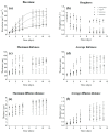
Dynamics of the structural parameters obtained from the z-stacks acquired at the confocal laser scanning microscopy (CLSM): biovolume (a), roughness (b), maximum thickness (c), average thickness (d), maximum diffusion distance (e), and average diffusion distance (f). The results are reported as the mean and standard deviation of 12 independent biological replicates. The fitting of the logistic model is also presented for the biovolume.
Figure 3
Figure 3
Structural parameters characterizing the biofilm matrix at days 2, 7, and 11. Volume of binding lectins ((a); specifically binding to glycoconjugates), lectin-to-cell ratio (b), volume of dextran ((c); used to stain the matrix non-specifically), and dextran-to-cell ratio (d). The results are reported as the mean and standard deviation of at least four independent biological replicates. Bars with different letters represent statistically different means (p < 0.05), as determined by pair-wise comparisons after two-way ANOVA.
Figure 4
Figure 4
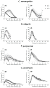
Vertical profiles of cells (a,c,e,g) and extracellular polymeric substances (EPS) (b,d,f,h) coverage of four different monospecific microalgae biofilms after 2, 7, and 11 days of maturation. The vertical profiles are reported as the percentage of coverage of cells or of EPS obtained from the z-stacks acquired at the confocal laser scanning microscopy (CLSM). The vertical profiles are reported as the mean and standard deviation of at least four independent biological replicates.
Figure 5
Figure 5
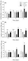
Macromolecular composition of the biofilms after 2, 7, and 11 days of maturation. Panel (a) depicts the carbohydrate-to-protein ratio, panel (b) depicts the lipid-to-protein ratio, and panel (c) represents the carbohydrate-to-lipid ratio. The results are reported as the mean and standard deviation of at least four independent biological replicates. Bars with different letters represent statistically different means (p < 0.05) as determined by pair-wise comparisons after the two-way ANOVA.

References

    1. Jorquera O., Kiperstok A., Sales E.A., Embiruçu M., Ghirardi M.L. Comparative energy life-cycle analyses of microalgal biomass production in open ponds and photobioreactors. Bioresour. Technol. 2010;101:1406–1413. doi: 10.1016/j.biortech.2009.09.038. - DOI - PubMed
    1. Ozkan A., Kinney K., Katz L., Berberoglu H. Reduction of water and energy requirement of algae cultivation using an algae biofilm photobioreactor. Bioresour. Technol. 2012;114:542–548. doi: 10.1016/j.biortech.2012.03.055. - DOI - PubMed
    1. Berner F., Heimann K., Sheehan M. Microalgal biofilms for biomass production. J. Appl. Phycol. 2015;27:1793–1804. doi: 10.1007/s10811-014-0489-x. - DOI
    1. Slade R., Bauen A. Micro-algae cultivation for biofuels: Cost, energy balance, environmental impacts and future prospects. Biomass Bioenerg. 2013;53:29–38. doi: 10.1016/j.biombioe.2012.12.019. - DOI
    1. Di Pippo F., Ellwood N.T.W., Gismondi A., Bruno L., Rossi F., Magni P., De Philippis R. Characterization of exopolysaccharides produced by seven biofilm-forming cyanobacterial strains for biotechnological applications. J. Appl. Phycol. 2013;25:1697–1708. doi: 10.1007/s10811-013-0028-1. - DOI

LinkOut - more resources